International Journal of Spine Research
Biomechanical etiology of the so-called Idiopathic Scoliosis-New classification; Rules of therapy and causal prophylaxis
T Karski*
Cite this as
Karski T (2019) Biomechanical etiology of the so-called Idiopathic Scoliosis-New classification; Rules of therapy and causal prophylaxis. Int J Spine Res 1(1): 012-016. DOI: 10.17352/ijsr.000003The biomechanical etiology of the so-called idiopathic scoliosis [Adolescent Idiopathic Scoliosis (AIS)] is the subject of the author’s research from 1984. The beginning of observation and search about etiology of “idiopathic scoliosis” was in Finland in 1984 in Invalid Foundation Hospital during scholarship stay.
In the period 1984 till 2019 in Lublin, Poland were observed many children with scoliosis and was given all information about etiology, classification, new therapy and causal prophylaxis already in years 1995 - 2007.
The etiology of AIS is strict biomechanical and is connected with asymmetry of hips movement and next with function. Scoliosis develop because of permanent standing ‘at ease’ on the right leg in “C” 2nd / A etiopathological group (epg) and “S” 2nd / B epg type and because of special properties of gait in situation of maximal limited adduction, internal rotation and very often also extension of right hip in 1 st epg and in 3 rd epg type of scoliosis [1-28].
Material
In the years 1984 – 2018, more than 2500 patients with scoliosis have been observed and treated and in this group were children (80%). The age of children and youth patients was 4 to 25. In 20 % there were older patients 50 – 70 years old, coming because of spinal pain.
In all these cases of scoliosis was found the same etiological factors -limited movement of right hip in various forms of the restriction and various types of scoliosis.
Classification
“The model of hip movements” (T. Karski - described in 2006) explains the new classification of scoliosis. When movements of hips are equal, so-called idiopathic scoliosis never develops. Exist no biomechanical pathological influence acting on the spine. The growth of spine is proper.
When movements of hips left to right are asymmetrical - there is an input to develop scoliosis in the three groups and four types. The asymmetry of movement of hips is one of symptoms of “Sydnrome of Contracture” (SofC) according to Prof. Hans Mau (Tubingen, Germany – in original “Siebenersyndrom” in English “Seven Contractures Syndrome”). From 2006 we speak in Lublin, about “Sydnrome of Contracture and Deformities” (SofCD) because to seven contractures I add the varus deformity of shanks in newborn and babies as eighths deformity.
There are following groups and types of scoliosis:
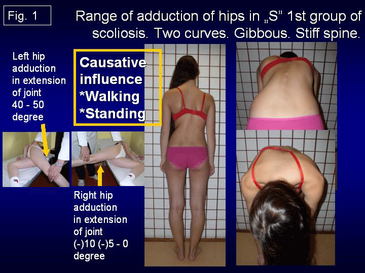
(1) Scoliosis 3D - “S” 1st etiopathological group (epg) [Figure 1] - double curve. Stiff spine. Rib hump on the right side of the thorax. Connection with gait and permanent standing „at ease’ on the right leg.
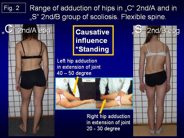
(2a) Scoliosis 1D or 2D - “C” 2nd/A epg [Figure 2] – one curve – lumbar left convex. Spine flexible. Connection with permanent standing „at ease’ on the right leg.
(2b) Scoliosis “S” 2nd/B epg [Figure 2] – two curves, (2D or 3D). Connection with permanent standing ‘at ease’ on right leg and additionally with laxity of joints or / and harmful previous exercises.
In these second 2nd/A and 2nd/B types of scoliosis - the spine is flexible.
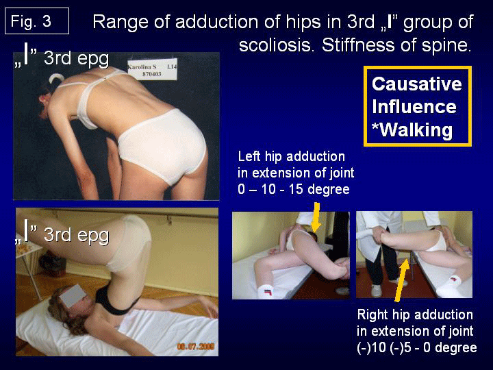
(3) Scoliosis 2D or 3D - “I” 3rd epg [Figure 3]. Deformity has the form of a stiff spine. No curves or small ones. The cause is gait only. Such “spine deformity” was till 2004 never included and classified as “scoliosis”.
The questions and the authors answers to the problem of scoliosis based on biomechanical etiology [Literature 1-28].
In research of etiology of the so-called idiopathic scoliosis everybody who will answer the question: what is “the etiology?” - must answer the all below presented questions about scoliosis. Answering only some / few questions is not enough and is not the answer for question about “the etiology”.
The questions are following:
1/ The etiology - of course - the subject of whole discussion of many researches, many orthopedic institutions?
2/ Why girls have more frequent scoliosis?
3/ Why we observe the lumbar left convex curve?
4/ Why we observe the thoracic right convex curve?
5/ When and why we observe one or two curves scoliosis?
6/ Why the rib hump is on the right side of thorax?
7/ In which age start to develop scoliosis?
8/ What kind of classification is proper?
9/ Why is the rapid progression of scoliosis in growth acceleration period?
10/ In which type of scoliosis we observe the progresses of deformity?
11/ Which type of scoliosis does not progress?
12/ Why the blind children do not have scoliosis?
13/ Is there any influence of CNS in development of scoliosis?
14/ What kind of therapy – conservative or operative should be used in treatment?
15/ Are extension and strengthening exercises in therapy of scoliosis correct?
16/ What kinds of rehabilitation exercises should be applied?
17/ Is corset treatment proper. In which cases should be apply?
18/ Is causative prophylaxis possible?
My answers and comments:
For all these questions only “biomechanical etiology of the so-called scoliosis” give full and proper answers.
1/ The etiology - is biomechanical, connected with asymmetry of the anatomy of body of children and with the asymmetry of movements of hips. All these asymmetries are signs of a “Syndrome of Contractures” (SofC) according Prof. Hans Mau (original in German “Siebenersyndrom” - see Literature) or “Syndrome of Contractures and Deformities [(SofCD), 2006 – T. Karski].
Because of these “asymmetries” – it exist: a/ asymmetry of loading during gait, b/ asymmetry of time of standing on left / right leg – more on the right(!), c/ asymmetry in development and growth of spine, d/ in result scoliosis in three ethiopathological groups and four types.
2/ Why girls have more frequent scoliosis? Answer - SofCD – appears mostly in girls. Girls are more sensible for forces during pregnancy and more frequently they have “Syndrome of Contractures and Deformities”.
3/ Why lumbar left convex curve? Answer: The SofCD is mostly “left sided” (90 % - 95 % of pregnancies - Prof. Jan Oleszczuk / Lublin and all gynecologists). Permanent standing on the right leg is due “contracture” more stable (!) and such form of standing is taken by all scoliosis children. Primary is only “functional change of spine axis”, but after 10 – 12 years of such standing appears fix lumbar left convex scoliosis.
4/ Why thoracic scoliosis / curve is right convex? Answer: The SofCD is mostly “left sided” as told above. Permanent standing on the right leg makes lumbar left convex scoliosis and secondary right convex thoracic curve in 2nd / B ethiopathological group (epg). Some cases in “S” 2nd / B epg scoliosis are “kifoscoliosis / kiphoscoliosis”. Important remark: in I-epg group both curves – lumbar left convex and thoracic right convex develop at the same time – please look the next points.
5/ Why there can be one curve or two curves scoliosis? The answer: one curve scoliosis is “C” lumbar left convex deformity is in 2nd / A epg group connected with standing ‘at ease’ on the right leg. The scoliosis in form of “S” is in 1st epg group - spine is stiff and also is in “S” 2nd / B epg group - spine is flexible. These both types of scoliosis are double curve deformities.
6/ Why the rib hump is on the right side? The answer: The SofCD is mostly left sided. In “S” 1 st epg group permanent standing on the right leg - this leg due to “contracture” is more stable, triggers the lumbar left convex and secondary right convex scoliosis. Due to walking and permanent rotation distortion in inter-vertebral joints a rib hump develops on the right side of thorax. Important remark: a/ the adduction and internal rotation movement in right the hip in 1st epg group of scoliosis is maximally limited, b/ during gait “this absent movement of right hip” is produce and transmitted as compensatory movement to the pelvis and to the spine, c/ because of this appears distortions procedure in inter-vertebral joints by every step, d/ in result appears stiffness of spine.
This rotation deformity develops in three clinical stages: a/ flat back,
b/ disappearance of processi spinosi, c/ in some cases appears a lordotic deformity in thoracic part of spine. Some cases in “S” 1st epg scoliosis we called “lordo-scoliosis”. The lordo-scoliosis are in result of: a/ etiological factors plus b/ incorrect therapy, extensions exercises.
7/ In which year of child’s life starts to develop scoliosis? Every type of scoliosis starts to develop when the child starts to “stand” and “walk” – at the age of two – three years.
The “S” scoliosis in 1st epg we can observe at the age of 4 - 6. The “C” 2nd/A epg scoliosis and “S” 2/B epg we can observe at the age of 10-12.
8/ What kind of classification is proper? The proper classification are: three groups and four types of scoliosis. All groups in this “new classification” are connected with “the special model of hips movements” (T. Karski, 2006):
1st epg –“S” scoliosis with stiffness of spine - connection with gait and with permanent standing ‘at ease’ on right leg,
2nd/A “C” scoliosis - connection with permanent standing ‘at ease’ on right leg,
2nd/B “S” scoliosis - connection with permanent standing ‘at ease’ on right leg – plus laxity of joints and incorrect exercises
3rd epg “I” scoliosis – small curves or none, small gibbous or none – only stiffness of spine. This group of scoliosis is connected with gait.
9/ Why is there a rapid progression of scoliosis in the period of accelerated growth of the child? Answer: bones grow - even 10 or more centimeter per year in some children. Contracted soft tissue in the region of the right hip does not grow and its influence become to be bigger (!), the deformity progress.
10/ Which type of scoliosis progresses? The progression is in the 1st epg “S” scoliosis, because in this group the difference in movement of hips is maximal. In right hips exist abduction contracture or adduction in straight position of joint is limited to 0 (zero) degree.
11/ Which type of scoliosis does not progress? The “C” 2nd/A epg, “S” 2nd/B epg scoliosis and 3rd epg type of deformity are without progression. We must remember – the causes in this group is only “permanent standing ‘at ease’ on the right leg. The range of deformity depend to the “cumulative time of standing” on the right leg. In scoliosis 3rd epg – patients notice the problem only because of pain in the adult age.
12/ Why blind children do not have scoliosis? The gait of blind children protects before scoliosis – they walk without lifting of legs and every step is very careful. They stand also carefully – symmetrical on both legs (observation of ophthalmologist).
13/ Is there any influence of CNS in the development of scoliosis? Yes – there are only indirect influences in children with Minimal Brain Dysfunction (MBD) or with Attention Deficit Hyperactivity Disorder (ADHA) – [according author – MBD and ADHD is equal]:
a/ extension contracture of the trunk in small children – because of spastic (semi spastic) contracture of trunk extensors,
b/ anterior tilt of pelvis – because of spastic (semi spastic) contracture of m. rectus (part of m. quadriceps) both sides,
c/ “laxity” of joints – because of changed properties of collagen.
14/ What kind of therapy – conservative or operative should be applied in treatment? Answer: only conservative therapy. In material from the 1995 – 2009, only 13 % children need surgery and there were children previously treated by wrong, incorrect exercises. In years 2010 – 2019 the number of children needing surgery in my material is maximally low – 3 %.
15/ Are extension exercises correct? [Figures 4a-c] No – such exercises are wrong - they cause “iatrogenic deformity” - bigger curves, bigger rib hump, stiffer spine. We should stop in all countries such incorrect therapy.
16/ What kinds of rehabilitation exercises should be applied? [Figures 5a-d] Only – stretching exercises – giving symmetry of movements and next symmetry of growth and development of pelvis and spine are correct. As the first aim – we should restore the “full movement of hips”, next symmetry of “function of trunk muscles”. When the child has full, symmetrical movements of both hips - it is no more asymmetry of loading of body left / right side during walking and no more “permanent standing ‘at ease’ on right leg” – but “symmetrical time of standing left / right leg”. In such situation never develops scoliosis.
17/ Corset treatment – yes ? no ? I had to use the corset in 20 % of children in “S” 1st epg scoliosis and in 5% - 10% of children in “S” 2nd / B epg scoliosis in years 1995 – 2009. Now this percent is lower.
18/ Is causative prophylaxis possible? Yes, the causative prophylaxis should be introduced in all countries. Exercises leading to symmetry of movements and symmetry of function of hips and spine are important in prophylaxis and therapy. All exercises removing abduction contracture or only “too small range of adduction” of the right hip, removing flexion contractures of both hips, removing external rotation contracture of right hip and extension contracture of whole spine belong to these prophylactics methods. Flexion exercises for spine should be introduced already in small children at the age of 3 - 5. Sport forms like karate, taekwondo, aikido, kung fu, fulfils all awaiting for “causative prophylaxis”.
Also is very important to inform parents of small patients about position of standing – all children should stand ‘at ease’ only on the left leg.
Here, is my moral obligation to inform readers, that “flexion exercises” in therapy of scoliosis in Poland in years 1960 – 1970 had introduce Professor Stefan Malawski from Warsaw. He saw beneficial results after such therapy, however in this time was not found the etiology of scoliosis.
Conclusions
1/ In all years of observations (T. Karski, 1984 – 2019), the biomechanical etiology of the so-called idiopathic scoliosis was confirmed.
2/ Development of scoliosis and the types of spine deformity are connected with pathological “model of hips movements” (T. Karski, 2006) and function – “standing ‘at ease’ on the right leg” and “walking”.
3/ Restricted range of movements in the right hip is connected with the “Syndrome of Contractures and Deformities” according Prof. Hans Mau.
4/ Every type of scoliosis starts to develop at the age of 2-3.
5/ There are three groups and four types of scoliosis:
(A) “S” scoliosis 1st epg, 3D. Causative influence: standing and gait,
(B1) “C” scoliosis 2nd / A epg, 1D. Causative influence: standing.
(B2) “S” scoliosis 2nd / B epg, 1D or 2D. Causative influence: standing, plus, - laxity of joints and/or incorrect exercises in previous therapy,
(C) “I” scoliosis 3rd epg, 2D or 3D. Clinically only stiffness of the spine. Causative influence: gait. The clinical symptom of this deformity are: sport problems at a young age and “permanent pain” in adults.
6/ The proper therapy of scoliosis – are only stretching exercises help to obtain full movements of the right hip, the proper position of the pelvis and full movement of the spine.
7/ The causal prophylaxis of scoliosis is possible and should be introduced in every country.
8/ The rules in prophylaxis – are – standing ‘at ease’ on the left leg, sitting relax, sleeping in embryo position, active participation in sport - especially in karate, taekwondo, aikido, kung fu and other similarly.
I would like to express my many thanks to Honorata Menet for correction of the article.
- Burwell G, Dangerfield PH, Lowe T, Margulies J (2000) Spine. Etiology of Adolescent Idiopathic Scoliosis: Current Trends and Relevance to New Treatment Approaches. 14: 324.
- Green NE, Griffin PP (1982) Hip dysplasia associated with abduction contracture of the contralateral hip. J Bone Joint Surg Am 268: 1273-1281. Link: http://bit.ly/2LM4xFV
- Hensinger RN (1979) Congenital dislocation of the hip. Clinical Symp 31: 270.
- Howorth B (1977) The etiology of the congenital dislocation of the hip, Clin Orthop 29: 164-179.
- Karski T (2002) Etiology of the so-called “idiopathic scoliosis”. Biomechanical explanation of spine deformity. Two 272 groups of development of scoliosis. New rehabilitation treatment. Possibility of prophylactics. Studies in Health Technology and Informatics 91: 37-46. Link: http://bit.ly/2NWnnwu
- Karski T, Kalakucki J, Karski J (2006) Syndrome of contractures" (according to Mau) with the abduction contracture of the right hip as causative factor for development of the so-called idiopathic scoliosis. Stud Health Technol Inform 123: 34-39. Link: http://bit.ly/2Y23XKu
- Karski T (2010) Explanation of biomechanical etiology of the so-called idiopathic scoliosis (1995 – 2007). New 276 clinical and radiological classification. Locomotor System 17: 26-42. Link: http://bit.ly/2SelE4s
- Karski T (2011) Biomechanical Etiology of The So-Called Idiopathic Scoliosis (1995 – 2007) – Connection with 279 “Syndrome of Contractures” – Fundamental Information for Paediatricians in Program of Early Prophylactics / 280 Journal of US-China Medical Science, USA. 8: 281.
- Tomasz K (2010) Factores biomechanicos en la etiologia de las escoliosis dinominadas idiopaticas. Nueva clasificación. Nuevos test clínicos y nuevo tratamiento conservador y profilaxis. Dialnet 39: 13-143. Link: http://bit.ly/2XWu9Gw
- Tomasz K (2010) Biomechanical Etiology of the So-called Idiopathic Scoliosis (1995-2007). New Classification: Three Groups and Four Types in the New Classification. J Nov Physiother 14: 286. Link: http://bit.ly/32mOhAT
- Tomasz K (2013) Biomechanical Etiology of the So-called Idiopathic Scoliosis (1995-2007). Three Groups and Four Types in the New Classification. J Nov Physiother S2: 289-290. Link: http://bit.ly/2S6j4xn
- Jacek K, Karski T (2013) So-Called Idiopathic Scoliosis. Diagnosis. Tests Examples of Children Incorrect 291 Treated. New Therapy by Stretching Exercises and Results, Journal of Novel Physiotherapies, OMICS Publishing 292 Group, USA.
- Tomasz K (2014) Biomechanical Aetiology of the So-Called Idiopathic Scoliosis. New Classification (1995 – 2007) in Connection with “Model of Hips Movements. Global Journal of Medical Research 14. 12. Link: http://bit.ly/2XZ1X5R
- Tomasz K (2014) Biomechanical Etiology of the So-called Idiopathic Scoliosis (1995 – 2007) - Connection with „Syndrome of Contractures” – Fundamental Information for Pediatricians in Program of Early Prophylactics. Surgical Science 5: 33-38. Link: http://bit.ly/2NOxNhN
- Tomasz K, Jacek K (2015) Syndrome of Contractures and Deformities” according to Prof. Hans Mau as Primary Cause of Hip, Neck, Shank and Spine Deformities in Babies, Youth and Adults. American Research Journal of Medicine and Surgery 1: 26-35. Link: http://bit.ly/2XX6y8H
- Tomasz K, Karski J (2015) Biomechanical Etiology of the So-called Idiopathic Scoliosis: Classification and Dates in History of Research. Principles of Causal Prophylaxis, Indications to New Therapy. Canadian Open Medical Science & Medicine Journal 1: 1-16. Link: http://bit.ly/2YMtXqD
- Karski T, Karski J (2016) Bóle krzyża – problem neurologiczno-ortopedyczny. Objawy, przyczyny, leczenie i profilaktyka Postępy Neurologii Praktycznej, Wydawnictwo Czelej. 9-16. Link: http://bit.ly/2JtPZt2
- Jacek K, Tomasz K (2016) Imperfect hips” As a Problem at an Older Age. Early and Late Prophylactic Management before Arthrosis. Jacobs Journal of Physiotherapy and Exercises / USA / Texas. 1: 015.
- Tomasz K (2018) Biomechanical Aetiology of the So-called Adolescent Idiopathic Scoliosis (AIS). Lublin Classification (1995-2007). Causative Influences Connected with “Gait” and “Standing ‘at ease’ on the Right Leg”. Journal of Orthopaedics and Bone Research (USA), Scholarena 10. Link: http://bit.ly/2Y55hww
- Mau H (1979) Zur Ätiopathogenese von Skoliose, Hüftdysplasie und Schiefhals im Säuglinsalter. Zeitschrift f. 294 Orthop 5: 601-605.
- Mau H (1982) Die Atiopatogenese der Skoliose, Bücherei des Orthopäden, Band 33, Enke Verlag Stuttgart 1-296.
- Mau Hans – personal information and letter.
- Normelly H (1985) Asymmetric rib growth as an aetiological factor in idiopathic scoliosis in adolescent girls, 298 Stockholm 1-103.
- Stokes IAF (1999) Studies in Technology and Informatics, Research into Spinal Deformities 2. 59: 1-385.
- Sevastik J, Diab K (1997) Studies in Technology and Informatics, Research into Spinal Deformities 1. 37: 1-509.
- Sevastik John – personal information.
- Thom Harald – personal information.
- Link: www.ortopedia.karski.lublin.pl
Article Alerts
Subscribe to our articles alerts and stay tuned.
 This work is licensed under a Creative Commons Attribution 4.0 International License.


 Save to Mendeley
