International Journal of Radiology and Radiation Oncology
Autologous flap reconstruction as a unique opportunity for weight loss and breast cancer risk reduction: A case report
Brian P Dickinson1-4*, Ayushi Patel1,5, Judy Pham1, Nikkie Vu-Huynh1, Monica B Vu1, Heather Macdonald3,6, Abigail Madans3,6, Merry Tetef8, Kevin Lin3, Jennifer Overstreet3, Lucia Vu1 and Peter Ashjian2-4,7
2Providence Tarzana Medical Center, Tarzana, CA, USA
3Hoag Hospital Presbyterian, Newport Beach, CA, USA
4Glendale Adventist Medical Center, Glendale, CA, USA
5University of South Florida Morsani College of Medicine, Tampa, FL, USA
6University of Southern California, Keck School of Medicine. Los Angeles, CA, USA
7Inc, Glendale, CA, USA
8University of California Los Angeles, USA
Cite this as
Dickinson BP, Patel A, Pham J, Huynh NV, Vu MB, et al. (2020) Autologous flap reconstruction as a unique opportunity for weight loss and breast cancer risk reduction: A case report. Int J Radiol Radiat Oncol 6(1): 017-021. DOI: 10.17352/ijrro.000040Objective: The purpose of this paper is to discuss how autologous flap reconstruction following mastectomy can serve as a unique opportunity for patients with breast cancer to reduce their risk of recurrence through weight reduction in the pre-operative, intraoperative and postoperative phases of healing.
Background: Autologous mastectomy reconstruction with tissue from the lower abdomen is common. Patients, with breast cancer, who test positive for the BRCA gene are known to have a survival benefit from prophylactic mastectomy. It is also known that satisfaction for autologous breast reconstruction is high in the patient population with a high body mass index (BMI). This population has the unique opportunity to further reduce their risk of breast cancer through weight loss.
Methods: Prior to surgery, our patients are instructed to consume a high protein diet intake on the magnitude of 1-2 grams per/kg of body weight which was on average 80-100 grams of protein per day. A case report chart review was conducted on a breast cancer patient who underwent autologous breast reconstruction with deep inferior epigastric artery perforator (DIEP) flaps. The patient had a high body mass index on the initial presentation of their breast cancer diagnosis. After the bilateral mastectomies, the patient lost additional weight after the completion of autologous flap reconstruction.
Results: Our patient with a high body mass index, who underwent bilateral mastectomy with autologous flap reconstruction, was followed between 2018 and 2020. Aesthetic outcome and weight loss following reconstruction was evaluated at various time points. Weight loss, as documented in chart review and patient report, was examined as well as before and after photographs post autologous flap reconstruction.
Conclusions: Bilateral mastectomy is a common procedure and typical among patients with BRCA positivity or other strong family history of breast cancer. Although mastectomy reduces the risk of recurrence, the risk reduction is not zero. Breast cancer patients often wish to complete all possible modalities to reduce their risk of recurrence. We have found that weight loss is often a controllable risk factor that patients often choose to pursue.
Introduction
Plastic and reconstructive surgeons, via oncoplastic surgery, have become integral in the oncologic care for patients with breast cancer to evolve the standard of breast cancer care with the goal of eradicating cancer and optimizing aesthetic outcomes [1,2]. Plastic and reconstructive breast surgeons also have the unique opportunity to reduce future breast cancer risk in patients with a high BMI following mastectomy with autologous flap reconstruction.
Prior studies have determined that reconstruction satisfaction is high in patients with an elevated body mass index who choose to undergo autologous flap reconstruction. When patients present to us and request autologous reconstruction, we emphasize that their high body mass index predisposes them to a higher rate of complications post-operatively. To mitigate the risk of complications, we initiate a diet high in protein to increase albumin and prealbumin levels. The diet is not restrictive in any way; it is supplemented with counting grams of protein intake and eliminating carbohydrates in the evening meals. We simply tell patients that we use the rule of 20s – “A can of tuna fish is 25 grams, a chicken breast the size of their palm is 25 grams, three eggs is 25 grams, and a protein shake at night is 30 grams.” Compliance with this simple routine daily can help patients exceed 100 grams of protein per day. We have found that when patients attain this level of protein intake, there is little additional room for empty calories, leading to weight loss prior to surgery. This initiates a functional cascade of positive behaviors with infinitely beneficial outcomes.
We encounter many patients in our practice who receive a new diagnosis of cancer and have often been healthy throughout their lives. Patients may indicate they never smoked, never consumed alcohol, and ate reasonably throughout the duration of their lives. Other patients test positive for genetic mutations or have a strong family history. One of the easily and readily reversible risk factors for breast cancer in the highly motivated patient is weight loss. We have found with the right education and guidance, weight loss can not only be an empowerment to fight breast cancer, but can be a re-introduction to a healthy perspective and new setpoint on health and fitness.
Materials and methods
A retrospective chart review was conducted on a patient who underwent bilateral autologous free flap reconstruction procedure and was followed between 2018 to 2020 by the plastic and reconstructive authors (B.D., P.A.). The patient’s weights, self-reports of waist circumference, and photographs were obtained. Satisfaction was assessed subjectively in the chart review.
Results
A 54-year-old female with a strong family history of breast cancer presented with suspicious microcalcifications in her right breast which were biopsied and determined to be Atypical Ductal Hyperplasia (ADH). Her mother died of breast cancer at the age of 38 and her maternal aunt developed breast cancer at age 78. Given her history, she underwent genetic screening and additional imaging. Her genetic testing identified her to have a variant of undetermined significance in the RAD51C gene. Although she was aware this was a negative test, she stated her strong intention to undergo bilateral mastectomy. The initial oncoplastic reconstructive plan included right breast lumpectomy with oncoplastic reconstruction and left breast reduction mastopexy for symmetry. This would allow nipple-sparing reconstruction through second stage mastectomy and eventual autologous flap reconstruction.
Through the bilateral MRI imaging as part of her work-up, a suspicious lesion was identified on the left breast in the upper outer quadrant. The lesion was 1.1 cm in diameter at the 1:30 position, 9 cm from the nipple. Biopsy of the mass returned as a left breast infiltrating ductal carcinoma ER/PR positive and Her-2/Neu negative. Given her left breast cancer, right breast ADH, and strong family history of breast cancer, the patient desired bilateral mastectomies with immediate autologous free tissue transfer reconstruction.
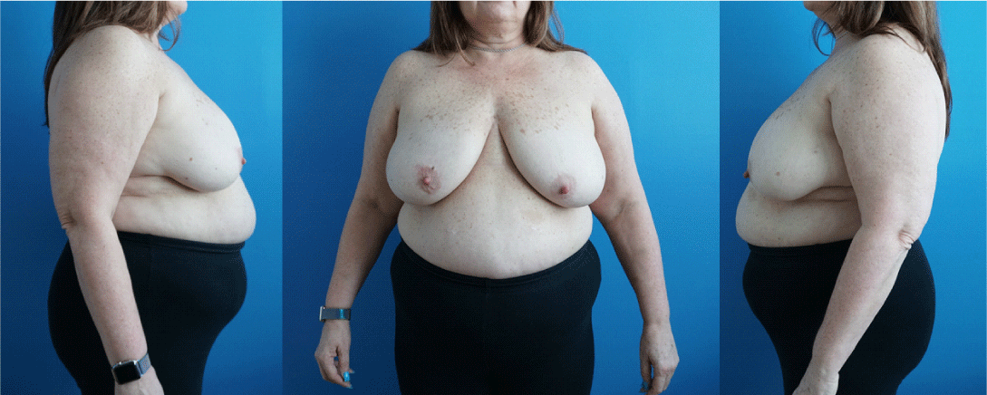
The patient was 5’6” tall and 224 lb (Body Mass Index = 36.2 kg/m2) , with a chest circumference of 39 inches, right breast diameter of 14 inches, and left breast diameter of 15 inches. The right sternal notch to nipple distance was 29 cm, and the left sternal notch to nipple distance was 30 cm. The right nipple to fold distance was 12 cm and the left nipple to fold distance was 12 cm. The breast demonstrated Regnault’s bilateral grade II ptosis (Figure 1).
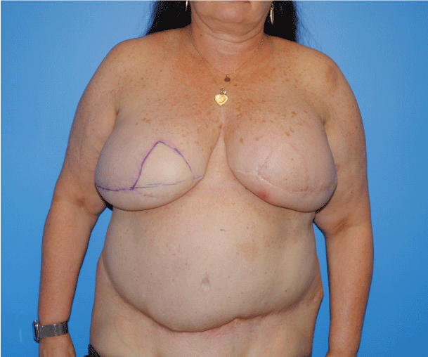
The patient had a high BMI; an incision pattern was drawn with two options. Given her desire for bilateral mastectomy and immediate reconstruction, nipple preservation would not be possible due to the long sternal notch to nipple distance. The patient was in agreement with removal of the nipple-areola complex from an oncologic perspective due to her strong family history of breast cancer. Two incision patterns were drawn–one was a wise pattern closure and the other preserved the skin along the inframammary fold [Figure 2]. It was explained to the patient that there would be a higher degree of wound complications and skin breakdown because of the larger abdominal flaps and tension on the T-junction of the wounds. Given the evolving nature of the patient’s workup, we wished to wait to decide intra-operatively which skin pattern would be conducive to optimal healing and aesthetic success and would not delay any possible radiation therapy.
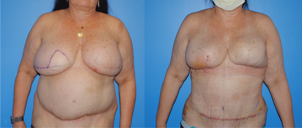
Intraoperatively, the frozen section yielded a positive sentinel lymph node biopsy. Given there would be a high probability of adjuvant radiation therapy, we maintained the skin along the inframammary fold to optimize wound healing and kept the flaps large in anticipation of radiation contraction post-operatively. When the breast wounds healed appropriately well at the six-week mark, the patient initiated left breast radiation in a timely fashion based on tumor board discussion. She received 50 Gy in 25 fractions to the left chest wall and flap, including a separate supraclavicular field. By the end of her radiation course, she had developed just minimal irritation in the inferior breast. She did have dry desquamation in the supraclavicular area, but no desquamation in the flap region. The left-sided flap tolerated radiation well without significant asymmetry [Figure 3].
The patient had a previous sleeve gastrectomy with intention and motivation to lose weight post-operatively. As part of a second stage reconstruction, the patient chose to undergo reduction of her bilateral flap reconstruction. Typically, revision on radiated flaps does not occur until nine months to one year after completion of radiation therapy. As a component of second stage reconstruction, the abdominal donor site is revised. This procedure involves abdominal scar revision to correct any height asymmetry and suction-assisted lipectomy of the abdominal donor site and revision of “dog ears’’ to make this area more symmetric. At this time, patients often choose to pursue additional aesthetic liposuction. Suction-assisted lipectomy can be a powerful weight-loss tool, as fat is removed and metabolic demands increase for optimal healing.
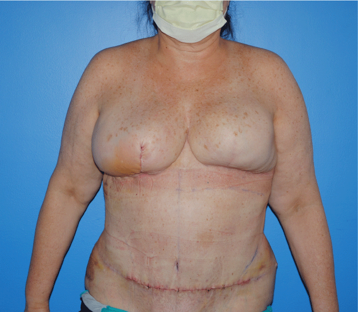
During the DIEP flap reconstruction, patients with a high body mass index commonly have some degree of abdominal wound complication such as delayed wound healing, seroma, or wound dehiscence. We counsel patients preoperatively that this is likely, but can be corrected at a second stage of the reconstruction. After revision of the abdominal donor site, the abdominal contour had improved and any indications of previously delayed wound healing or seroma were no longer apparent [Figure 4].
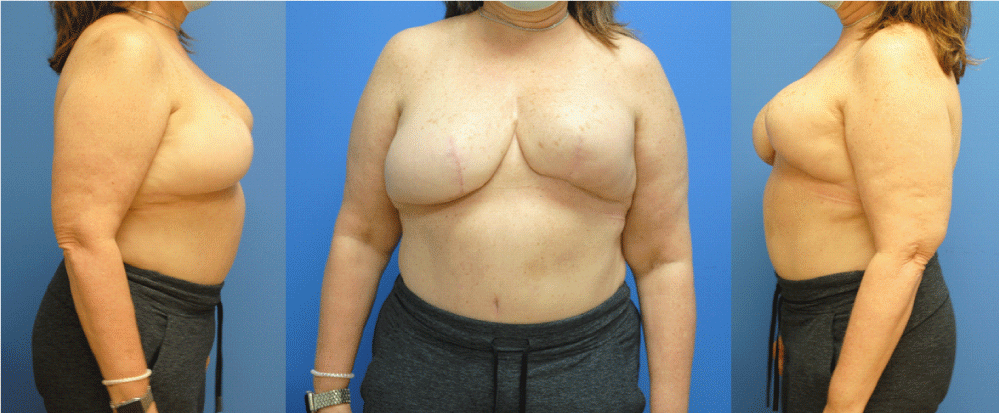
Radiation is always a possibility following immediate reconstruction. In our case, the autologous flap reconstruction had tolerated radiation well. Asymmetry consistent with radiation included an elevated inframammary fold and tightened skin envelope [Figure 5]. Correction of the asymmetry consisted of contralateral right breast mastopexy/reduction and raising of the right inframammary fold [Figure 6]. Over the course of her surgical cancer treatment and reconstruction, this patient lost 45 lbs and reduced her BMI from 36.2 to 28.9, changing her BMI classification from obese to overweight.
Discussion
We have encountered many patients in our practices who have a high body mass index and who choose to undergo autologous flap reconstruction. Our experience is similar to that encountered nationally where patients with a high body mass index have a high satisfaction rate with autologous flap reconstruction [3-5]. This may be accounted for by the ability to match the pre-operative breast size. The largest silicone implant available is 800 cc while some mastectomy specimens in patients with high BMI exceed 1000 grams. Autologous flap reconstruction also gives patients with a higher BMIs the ability to change their overall body shape and lose weight. We commonly encounter many patients who have been given a new diagnosis of breast cancer and state, “I did everything right my whole life... I don’t smoke, I don’t drink alcohol, etc.” and often a feeling of powerlessness or “unluckiness” ensues. We have found in a subset of patients with a high BMI that safe and educated weight reduction preoperatively and postoperatively empowers patients regarding their future breast cancer risks, body image, and overall health. New analysis from Harvard of the Pooling Project of Prospective Studies of Diet and Cancer (DCPP) demonstrates a direct relationship between the amount of weight lost and the reduction of breast cancer risk. In this study, women over fifty who sustained weight loss of greater than 20 lbs had a 26% reduction in breast cancer risk. The analysis excludes women on HRT [5]. The effects of obesity on breast cancer treatment include high recurrence rates, poorer aesthetic outcomes, increased surgical and radiation therapy complications, and decreased effectiveness of chemotherapy and endocrine therapy [6-8].
Wang determined in his meta-analysis of over 50,000 patients from 20 cohort studies, that for every 1 kg/m2 increment increase of BMI, the risk of breast cancer lymph node metastasis increased as linear dose-response reaction. [9] Chlebowski determined that there was a lower risk of breast cancer in post-menopausal women who lose weight. [10] Blackburn determined in the Women’s Intervention Nutrition Study that risk of breast cancer recurrence was reduced 24% in women who lost an average of 6 lbs. with a low fat diet intervention over those who had regular care. [11] The advantage of multiple-staged procedures for breast cancer reconstruction is that the metabolic rate increases as the body attempts to heal the reconstruction. Furthermore, the protein diet preparation for these surgeries facilitates weight loss which are consistent with associations to reduce breast cancer risk.
Pre-operative increases in daily protein intake helps patients develop nutritional habits that aid in healing and recovery post-operatively, when metabolic demands are high. While autologous flap reconstruction does not necessitate entry into the abdominal or thoracic cavity, there are increased, surgery-associated metabolic demands on the integumentary system and a significant volume and surface area of wounds that require healing. In addition, we have encountered many patients who experience early satiety following DIEP flap reconstruction secondary to the tightening of the abdominal domain. This early satiety, increased metabolic demand, and proper nutrition can help reduce breast cancer risk in patients with an elevated BMI.
We have encountered obstacles to our premise of autologous reconstruction in patients with a high BMI. Lumpectomy and oncoplastic reconstruction can yield excellent results with a functional breast. Lumpectomy and radiation can be the endpoint of treatment, and mastectomy can be saved for recurrence if it occurs. Weight loss can be initiated after the lumpectomy to reduce risk of recurrence. However, lumpectomy in patients with a high BMI is also not without complications, and similar principles that we discuss below should be utilized to ensure radiation is initiated in a timely fashion. Fisher, et al. believe that autologous reconstruction in obese patients leads to a higher rate of complications with autologous flap reconstruction [5]. It is well known that many surgical and anesthetic complications occur as a result of obesity or elevated body mass index. We understand this to be true, and it became our impetus to advocating the high protein diet to minimize complications in these patients. Our goal was to achieve albumin levels of 4.0 g/dL. In general, we would attempt to educate our patients to consume 1.5-2 grams of protein per kg of body weight.
As we initiated our protein nutrition plan prior to surgery, we found many patients lost weight prior to surgery and often HgA1c levels returned closer to normal in the process. To contrast Fisher’s points, we try to avoid the free TRAM or MS free TRAM in obese patients and focus on DIEP flaps to minimize donor site morbidity. This often requires the presence of a superficial epigastric vein for backup outflow and appropriate perforator anatomy. The importance of high BMI and associated complications should also focus on minimizing mastectomy skin flap necrosis and wound healing of the reconstructed breast. This healing is time-sensitive in order to ensure that the breast cancer patient can complete their oncologic therapy which may include adjuvant chemotherapy or radiation therapy. Patients with a higher BMI often have ptotic breasts, so a Wise-pattern mastectomy is often beneficial aesthetically. If done in an immediate reconstructive setting, the chances of partial- or full-thickness skin necrosis is high as the weight of the flap rests against a skin flap that lacks a normal venous dependent drainage. If done for prophylactic mastectomy, the skin necrosis that ensues can be addressed with hyperbaric oxygen therapy or can be revised at a later date without consequence. For patients with larger cancers (>5cm) or positive lymph nodes, there is a higher likelihood of adjuvant chemotherapy or radiation, so the selection of a skin pattern that minimizes wound complications is paramount. In our patient, we maintained the inframammary fold skin and kept the autologous flap larger to tolerate radiation.
Our case highlights another issue that commonly occurs in the breast reconstruction process. Larger flaps with a robust blood supply can tolerate radiation well. During an immediate reconstruction, sentinel lymph node biopsies can return positive which may direct radiation therapy postoperatively. When this occurs, we often make more conservative decisions so that we do not negatively affect radiation timing postoperatively. In this case, we maintained the skin along the inframammary fold instead of completing a Wise pattern which would remove the venous drainage and place heavy autologous flaps against the repair. Second, we kept the flaps larger. We have found that larger bulky flaps tolerate radiation well and often do not require a second stage for symmetry. If a symmetry operation is necessary, the contralateral non-radiated side can usually be reduced or lifted. In our patient’s case, we proceeded with delayed bilateral reductions of the autologous flaps and completed the Wise pattern reduction because she desired to have a smaller breast size. At this second stage the Wise pattern mastectomy skin flaps have been delayed and exhibit more robust healing, and in this case, healed well.
Summary
Autologous mastectomy reconstruction represents a unique opportunity for the plastic and reconstructive surgery team to reduce breast cancer risk in patients with a high body mass index. Autologous flap mastectomy reconstruction offers patients a durable, natural-appearing reconstruction with an improved body appearance and self-image. The autologous mastectomy flap reconstruction in combination with appropriate nutrition and exercise can help minimize breast cancer risk.
- Silverstein MJ, Mai T, Savalia N, Vaince F, Guerra L (2014) Oncoplastic breast conservation surgery: the new paradigm. J Surg Oncol 110: 82-89. Link: https://bit.ly/2R32Q8g
- Dickinson BP, Holmes D, Vu-Huynh N, Vu MB, Snyder L, et al. (2020) Autologous mastectomy reconstruction: Communication among the breast surgery team to maximize aesthetic and oncologic outcome. Breast Journal. Link: https://bit.ly/333s1NR
- Frost MH, Schaid DJ, Sellers TA, Slezak JM, Arnold PG, et al. (2000) Long-term satisfaction and psychological and social function following bilateral prophylactic mastectomy. JAMA 284: 319-324. Link: https://bit.ly/2ZeDIzV
- Cho EH, Shammas RL, Glener AD, Greenup RA, Hwang ES, et al. (2017) The Impact of Autologous Breast Reconstruction on Body Mass Index Patterns in Breast Cancer Patients: A Propensity-Matched Analysis. Plast Reconstr Surg 140: 1121-1131. Link: https://bit.ly/3bCQKfB
- Garvey PB, Villa MT, Rozanski AT, Liu J, Robb GL, et al. (2012) The advantages of free abdominal-based flaps over implants for breast reconstruction in obese patients. Plast Reconstr Surg 130: 991-1000. Link: https://bit.ly/3jMuEKu
- Fischer JP, Nelson JA, Sieber B, Cleveland E, Kovach SJ, et al. (2013) Free tissue transfer in the obese patient: an outcome and cost analysis in 1258 consecutive abdominally based reconstructions. Plast Reconstr Surg 131: 681e-92e. Link: https://bit.ly/3h44P6X
- Teras LR, Patel AV, Wang M, Yaun SS, Anderson K, et al. (2019) Sustained weight loss and risk of breast cancer in women ≥50 years: a pooled analysis of prospective data. J Natl Cancer Inst. Link: https://bit.ly/331x0i4
- Lee K, Kruper L, Dieli-Conwright CM, Mortimer JE (2019) The Impact of Obesity on Breast Cancer Diagnosis and reatment. Current Oncology Reports 21: 41. Link: https://bit.ly/324eWnU
- Chlebowski RT, Luo J, Anderson GL, Barrington W, Reding K, et al. (2019) Weight loss and breast cancer incidence in postmenopausal women. Cancer 125: 205-212. Link: https://bit.ly/2F7IIQl
- Wang J, Cai Y, Yu F, Ping Z, Liu L (2020) Body mass index increases the lymph node metastasis risk of breast cancer: a dose-response meta-analysis with 52904 subjects from 20 cohort studies. BMC Cancer 20: 601. Link: https://bit.ly/2Rae0bF
- Blackburn GL, Wang KA (2007) Dietary fat reduction and breast cancer outcome: results from the Women's Intervention Nutrition Study (WINS). Am J Clin Nutr 86: s878-s881. Link: https://bit.ly/33l7Qv0
Article Alerts
Subscribe to our articles alerts and stay tuned.
 This work is licensed under a Creative Commons Attribution 4.0 International License.


 Save to Mendeley
