International Journal of Radiology and Radiation Oncology
Hyperprogression after immunotherapy in HNC: literature review and our experience
Nerina Denaro*
Cite this as
Denaro N (2018) Hyperprogression after immunotherapy in HNC: literature review and our experience. Int J Radiol Radiat Oncol 4(1): 001-002. DOI: 10.17352/ijrro.000026Background
Checkpoint inhibitors demonstrate salutary anticancer effects, including long-term remissions. PD-L1 expression/amplification, high mutational burden, and mismatch repair deficiency correlate with response. Champiat et al for the first time described a small subset of patients that could actually have tumor growth accelerated when given PD1/PDL1-targeting agents [1].
Hyperprogression may be defined as accelerated progression outpaces the expected rate of growth induced by the therapies.
Champiat suggests that a subset of patients with disease progression have a course that is more deleterious than they might have had with other therapies, or even in the absence of therapy.
Champiat et al used the tumor growth rate as a measure for either response or hyperprogression.
The tumor growth rate prior (“reference”) and upon (“experimental”) anti-PD-1/PD-L1 therapy was compared to identify patients with accelerated tumor growth. In particular hyperprogression in the French was defined as a RECIST progression at the first evaluation and as a ≥2-fold increase of the TGR between the REF and the EXP periods [1].
In the study the number of hyperprogressor was about 9% of cases overall. Since several patients progressed clinically prior to an imaging assessment, the number of hyperprogression events in their patient cohort could have been significantly higher [1].
In recurrent/metastatic head and neck squamous cell carcinoma Saâda-Bouzid E et al retrospectively compared tumor growth kinectics (TGK) on immunotherapy and TGK on last treatment in patients treated with PD-1/PD-L1 inhibitors in four French centers. The definition of hyperprogression slightly differed compared with Champiat’s definition. In this study on 34 patients the TGK ratio was calculated considering ratio of the slope of tumor growth before treatment with PD-1/PD-L1 and the slope of tumor growth on treatment. Hyperprogression was defined as a TGK ratio ≥ 2. Hyperprogression was observed in 29% of patients with R/M HNSCC treated with anti-PD-L1/PD-1 agents and correlated with a shorter progression free survival PFS. It occurred in 39% of patients with at least a locoregional recurrence and 9% of patients with exclusively distant metastases [2].
Investigating on these phenomena Kato et al evidenced that some mutations can explain this behaviour [3].
Hyper-progressors harbored MDM2/4 or EGFR alterations, which independently correlated with time-to-treatment failure <2 months, suggesting the need for caution in the presence of these genomic profiles [3].
Description
Male 69 years old (15/5/1948) heavy smokers high amount of alcohol consumption.
From September 2016 dysphagia, weight loss of 10 Kg (>8% ), clinical evidence of base of tongue neoplasm. Histology confirmed squamous cell carcinoma moderately differentiated, grading G2, keratinising.
October 2016 CT scan showed extension through pyriform sinus, bilateral neck nodes.
November 2016 Enteral tube placement
Exclusive Radiotherapy (IMRT) from 3/1/2017 to 21/2/2017 Total dose 68Gy daily dose 2Gy. Patient did not receive concurrent platinum therapy because of rapid impairment of performance status. However nevertheless acute mucosyte during RT, after treatment with nutritional and psychological support patients performance status improved to ECOG value 1.
April 2017 CT scan assessment evidenced complete response: no pathological contrast enhancement accumulation in oro-hypo pharyngeal region.
ENT evaluation doubt of persistence at glossoepiglottic vallecula, MRI required.
19May 2017 MRI irregular enhancement in base of tongue region with longitudinal extension of 4 cm thickness 1 cm. No neck node involvement.
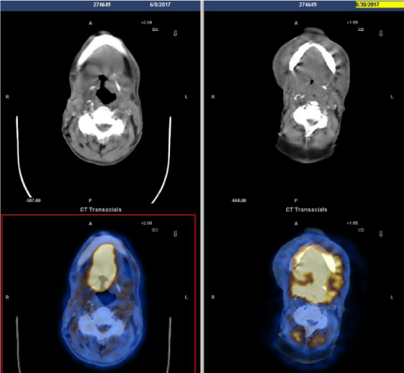
Patient received a basal (8th June 2017)TCPET scan FDG uptake (SUV max 20.6) at right tongue diameter 6.5 x 3.5 x 4 cm , right laterocervical node level II (SUV max 6.7)
At the multidisciplinary meeting on 22nd of May 2017 patient was referred to Oncology department to start first line treatment.
Patient informed about study protocol accepted to be enrolled and after screening procedure received investigational arm treatment.
Treatment started on 6th of July 2017 with a combination of anti PD1 and anti CTLA4 .Patient completed the first cycle and started the second cycle (4 doses of anti PD1 every 14 days and 2 doses of antiCTLA4 every 6weeks)
During treatment, patient accessed to emergency department because of fistulisation of lateral neck node, he underwent antibiotics with modest benefit. He received one cycle more of combination of anti PD1 and anti CTLA4 and then first imaging assessment.
Patient underwent PET scan reassessment (30th August 2017) with evidence of intense FDG uptake at base of tongue oral floor and sovraglottic larynx. The simultaneous presence of flogistic phenomena may alter the interpretation of the current overall imaging reassessment. MRI required but progressive deterioration of clinical PS with death on 14th of October 2017 (Figure 1, Table 1 and 2).
Conclusion
Hyperprogression might be considered the higher adverse event of anti PD1/antiCTLA4 for treating HNSCC, because it means not only no response but a startling progression.
The fact that hyperprogressor usually are those with local recurrence alarm even more because in HNSCC local progression corresponds with symptoms increasing (affecting the phonation respiration swallowing for the site of the tumour).
- Champiat S, Dercle L, Ammari S, Massard C, Hollebecque A, et al. (2017) Hyperprogressive Disease Is a New Pattern of Progression in Cancer Patients Treated by Anti-PD-1/PD-L1. Clin Cancer Res 23: 1920-1928. Link : https://goo.gl/3RC3bW
- Saâda-Bouzid E, Defaucheux C, Karabajakian A, Coloma VP, Servois V, et al. (2017) Hyperprogression during anti-PD-1/PD-L1 therapy in patients with recurrent and/or metastatic head and neck squamous cell carcinoma. Ann Oncol 28: 1605-1611. Link : https://goo.gl/yz9DBR
- Kato S, Goodman A, Walavalkar V, Barkauskas DA, Sharabi A, et al. (2017) Hyperprogressors after Immunotherapy: Analysis of Genomic Alterations Associated with Accelerated Growth Rate. Clin Cancer Res 23: 4242-4250. Link : https://goo.gl/FZqTMk
Article Alerts
Subscribe to our articles alerts and stay tuned.
 This work is licensed under a Creative Commons Attribution 4.0 International License.


 Save to Mendeley
