International Journal of Radiology and Radiation Oncology
Cardiac Computed Tomography Coronary Angiography Post Non-diagnostic or Equivocal Myocardial Perfusion SPECT
Abdullah Al-harbi, Mervat M Aboulkheir and Ahmed Fathala*
Cite this as
Al-harbi A, Aboulkheir MM, Fathala A (2017) Cardiac Computed Tomography Coronary Angiography Post Non-diagnostic or Equivocal Myocardial Perfusion SPECT. Int J Radiol Radiat Oncol 3(1): 015-020. DOI: 10.17352/ijrro.000024Background: Myocardial perfusion single- photon emission computed tomography (MP-SPECT) plays a key role in the management of patients with known or suspected coronary artery disease (CAD). However, Infrequently, MP-SPECT is non-diagnostic or equivocal. Computed Tomography coronary angiography (CTCA) has high diagnostic accuracy and excellent negative predicted value .The goal of the study is to investigate the diagnostic value of CTCA after non-diagnostic, equivocal, and or borderline abnormal MP-SPECT.
Methods: 64 patients were with suspected CAD were enrolled in this study, the mean age was 51 ± 24 years, 36 (56%) male and 28 (44%) female. MP-SPECT result was classified into 3 categories: Probably normal or probably abnormal, Abnormal function and normal myocardial perfusion, and equivocal or non-diagnostic.
Results: Probably normal/ abnormal MP-SPECT: 23 (36%) patients, abnormal function with normal MP-SPECT: 10 patients (16%), equivocal and /or non-diagnostic: 31 (48%) patients. CCTA: Normal Coronary arteries: 46 (72%) patients, Mild stenosis 5 (8%) patients, Moderate stenosis: 7(11%) patients, and more than 75%: 6 (9%) patients. Patients with normal coronary and mild stenosis contributed to total of 51(80%) patients, Patients with moderate and severe stenosis 13 (20%). Obstructive CAD was found in 2 (9%), 5 (50%) and 6 (19%) in patients with probably normal or probably abnormal MP-SPECT, abnormal function and normal perfusion, and equivocal and non-diagnostic MP-SPECT respectively.
Conclusion: Non-diagnostic, equivocal, or mildly abnormal MP-SPECT is infrequent but it causes a management dilemma. CTCA is essential in such clinical circumstances because the majority of such patients have normal coronary artery but few patients may require conventional coronary angiography.
Background
Myocardial perfusion imaging with single- photon emission computed tomography (SPECT) is an important imaging modality in the management of patients with cardiovascular disease. Myocardial perfusion SPECT (MP-SPECT) plays a key role in the establishing diagnosis, prognosis, assessment of therapy and intervention, and myocardial viability [1-3]. The accuracy of the interpreting and reporting MP-SPECT studies, either by visual interpretation or with assistance of computer software packages is dependent on the overall quality of the study and how consistent and predictable is the tracer distribution for patient’s specific disease state. MP-SPECT is a complex process, artifacts and pitfalls can arise at any stage in MP-SPECT process, and can be grouped into issues related to the instrument, issues related to the patients, attenuation artifacts, tracer kinetics/distribution artifacts, and processing related artifacts [4]. The interpreting physicians should develop understanding of all possible MPS artifacts and pitfalls, and develop a systematic approach to correct them and how to incorporate their influence into the final interpretation of the study. The American Society of Nuclear Cardiology (ASNC) is strongly in favor of the standardization of MP-SPECT reporting [5]. The final impression is the most critical part of the MP-SPECT report. The final report must states clearly, whether the study is normal or abnormal. However, in some cases with interpretive uncertainty, multi category reporting of MPS has been proposed, 5- category system include: normal, probably normal, equivocal, probably abnormal, and abnormal [6]. The use of multi category in the final impression results in incorporating non-perfusion and clinical information, and therefore enhances risk stratification of MPS compared with a dichotomous normal/abnormal approach. Non diagnostic study such as inadequate stress or poor quality images is another possibility in final conclusion of the MP-SPECT reporting. As per ASNC guidelines, equivocal category should be used less than 10% of all studies interpretation. Equivocal, non-diagnostic, probably normal or probably abnormal MPS poses a management dilemma and difficult clinical decision, in fact many referring physicians are not in favoring probably normal or probably abnormal due to lack of certainty and all of these patients need further work up for diagnosis of coronary artery disease (CAD).Computed Tomography coronary angiography (CTCA) represents the most rapidly developed imaging modality in cardiac imaging with high diagnostic accuracy and excellent negative predicted value 96 to 99% [7-10]. As per American college of cardiologists and other major Societies appropriateness criteria, CTCA is appropriate and indicated in such cases of nondiagnostic or non-interpretable MP-SPECT [11]. The goal of the study is to investigate the diagnostic value and performance of CTCA after non-diagnostic, equivocal, probably normal or probably abnormal MPS.
Methods
Study population
64 patients were with suspected CAD were enrolled in this study, the mean age was 51 ± 24 years, (36 (56%) male and 28 (44%) female). All patients underwent both MPS and CCTA within 35 days during the period of January 2013 to May 2015. SPECT result was classified into 3 categories:
1. Probably normal or probably abnormal: Patients with anterior and/or inferior wall defect most likely related to attenuation artifact related to soft tissue attenuation. Attenuation artifacts was not corrected by attenuation correction or prone imaging, these patients were refereed for CCTA as they continue to have some symptoms or their referring physician wanted to have more robust confirmatory test. Probably normal MPS was defined as number of segments with abnormal perfusion ≥3 with stress score and rest segmental score =1-1. Probably abnormal MPS was defined as number of segments with abnormal perfusion more than 2 with segmental stress score =2 [6].
2. Abnormal function and mildly abnormal perfusion: Patients with mild heterogeneous perfusion, or mild fixed perfusion defect, dilated cardiomyopathy, patients with wall motion abnormalities, abnormal left ventricular function, and patients with left bundle branch block.
3. Equivocal and non-diagnostic: This category includes inadequate exercise (non-diagnostic), uncorrected patients motion, and other technical issues, Equivocal MPI is defined as MPI with Segmental stress score of 2 to 3 [6].
All patients within one of the above categories underwent CTCA after MP-SPECT, the average time between MP-SPECT and CCTA was 35 days. Most of the follow up CTCA was requested based on referring physician request or based on our recommendation. Our recommendation to perform CT was mainly after non-diagnostic, equivocal SPECT. In patients with abnormal function and normal perfusion, and patients with probably normal or probably abnormal, CCTA was recommended after discussion with the referring physician on case by case basis. The exclusion criteria include: patients with known CAD based on prior abnormal SPECT, prior abnormal conventional coronary angiogram, patients with prior intervention such as PCI, or post-CABG. Patient was excluded if there were any contraindications to cardiac CT, such as impaired renal function. The study was approved by our local hospital ethics committee.
SPECT acquisition and analysis
Patients underwent rest-stress myocardial perfusion studies with either a separate-day protocol or a same-day stress-rest sequence. The choice of tracer and same-day or separate-day protocol was based on logistic requirements. The rest dose in patients who underwent a separate-day rest-stress protocol was 10 to 370 Megabecqurel (MBq) of either technetium-99 (Tc-99m) sestamibi or tetrofosmin. The stress dose in patients who underwent the rest-stress same-day protocol was 1100 MBq mCi of either (Tc99m) sestamibi or tetrofosmin (estimated patient’s radiation dose 8 to 12 mSv). Tc-99m sestamibi or Tc-99m Tetrofosmin was injected during peak pharmacological vasodilatation with adenosine (140μg/kg/min), or dipyridamole. Single photon computed tomography (SPECT) imaging was started 30 minutes following pharmacological vasodilatation. Rest SPECT myocardial perfusion imaging was initiated at approximately 60 minutes following injection. SPECT imaging was performed with line source attenuation correction at 900 dual-detector gamma camera (Cardio MD, Philips Medical System, Milpitas, California) equipped with attenuation correction and truncation compensation. The acquisition parameters and post processing were performed according to the most recent guidelines of the ASNC for nuclear cardiology procedures [12].
All images were reoriented in short, vertical, and horizontal views utilizing Auto SPECT (Cedars-Sinai Medical Center, Los Angeles, California) for visual interpretation by an experienced nuclear medicine physician. The reader was not biased by clinical information. Stress and rest perfusion images were scored using 17 tomographic segments, which included 6 segments each for the basal and midventricular slices, and 4 segments for the apical short-axis slices. The final segment is located on the most apical part of the left ventricle [13]. Finally; gated short-axis images were processed with quantitative SPECT software to measure the ejection fraction. In the visual analysis the 17 segments were scored for perfusion defects on a 4-point system (0 = normal; 1 = mild; 2 = moderate; and 3 = severe) for both the stress and rest images. From this analysis, ischemia was defined as a change in segmental score between stress and rest. Segments with no change between stress and rest were classified as nonreversible. Summed stress and rest scores were calculated by summing the 17 segmental scores in each imaged set. Utilizing the summed difference score (SDS) in measuring defect reversibility was calculated from the difference between the summed stress and rest scores. A SDS lower than 2 was considered nonischemic, 2 to 7 was considered as mild ischemia, and greater than 7 as considered as moderate to severe ischemia. The reader made the final determination of an abnormal SPECT study by comparing both the perfusion and functional data. The perfusion defects represented by the perfusion scores at stress and rest were used to form the interpretation of the MP-SPECT studies using the 5 levels of certainty: (1) normal; (2) probably normal; (3) equivocal; (4) probably abnormal; and (5) definitely abnormal, based simply on the various combinations of perfusion scores as it has been described previously.
Computed tomographic coronary angiography and image analysis
Patients, without contraindications, received metoprolol targeting a heart rate of ≤65 bpm and nitroglycerin 0.8 mg sublingually before image acquisition. A bolus tracking technique was used to calculate the time interval between intravenous contrast (Visipaque 320, GE Healthcare; Milwaukee, Wisconsin, USA) infusion and image acquisition. Final images were acquired with a triphasic protocol (100% contrast, 40%/60% contrast/saline, and 40 cc saline). The contrast volume and infusion rate (5 to 6 cc/s) were individualized according to scan time and patient body habitus. Prospective or Retrospective ECG-gated data sets were acquired with the GE high definition CT (GE Healthcare; Milwaukee, Wisconsin, USA) with 64×0.625 mm slice collimation and a gantry rotation of 350 msec (mA=300 to 800, kV=120). Pitch (0.16 to 0.24) was individualized to the patient’s heart rate. The CCCTA data sets were reconstructed with an increment of 0.4 mm using the cardiac phase with the least cardiac motion [14].
CTA image analysis
Images were processed using the GE Advantage Volume Share Workstation (GE Healthcare, Milwaukee, Wisconsin, USA) and visually interpreted by 2 expert observers blinded to all clinical data. A 17 segment model of the coronary arteries and 4 point grading score (normal, mild [<50%], moderate [50% to 69%], severe [≥70%]) was used for the evaluation of coronary stenosis, obstructive CAD was defined as coronary diameter stenosis ≥50%.
Statical analysis
Continues variable with normal distribution were expressed as mean (standard deviation). Variable with Skewed distributions were expressed as median. Categorical variable were expressed as frequency (percentage).
Results
64 patients were with suspected CAD were enrolled in this study, the mean age was 51 ± 24 years, (36 (56%) male and 28 (44%) female). Table 1 shows patients characteristics.
SPECT Findings
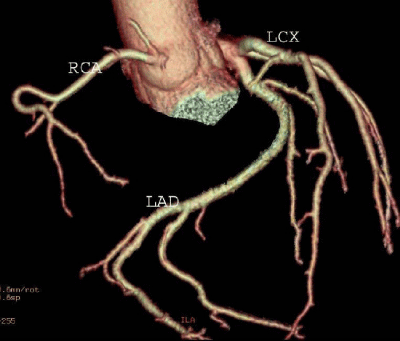
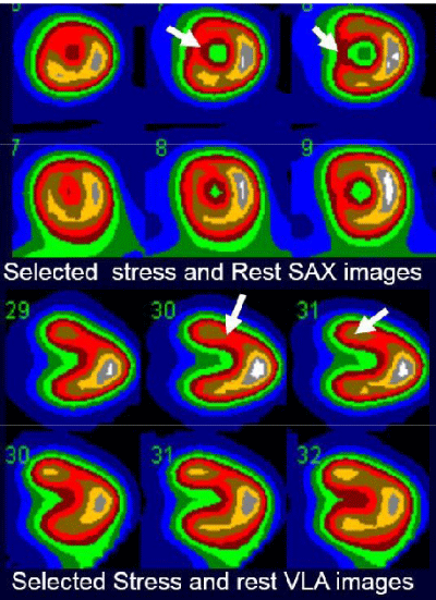
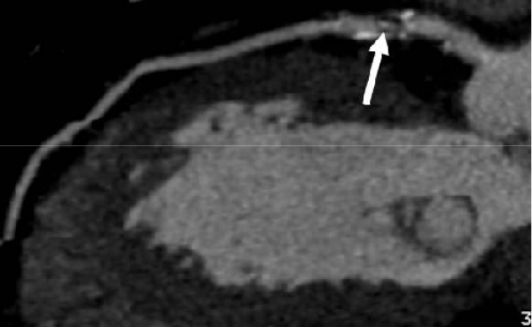
1. Probably normal or probably abnormal SPECT: 23 (36%) patients, Patients with anterior and/or inferior wall defect most likely related to attenuation artifact related to soft tissue attenuation. Attenuation artifacts were not corrected by attenuation correction or prone imaging (Figure 1a,b).
2. Abnormal function and mildly abnormal perfusion: 10 patients (16%), such as mild heterogeneous perfusion or mild fixed or reversible perfusion defect, dilated cardiomyopathy, patients with segmental wall motion abnormalities, abnormal left ventricular function, and patients with left bundle branch block (Figure 2a,b).
3. Equivocal and non-diagnostic: 31 (48%) patients, this category include inadequate exercise (non-diagnostic), uncorrected patients motion, and other technical issues.
CCTA findings
1. Normal Coronary arteries: 46 ( 72%)patients
2. Mild stenosis , less than 50% stenosis : 5 (8%) patients
3. Moderate stenosis , more than 50% stenosis but less than 70 stenosis: 7(11%) patients
4. Severe stenosis, more than 75%: 6 (9%) Patients.
Relationship between CCTA and SPECT
Patients with normal coronary and mild stenosis contributed to total of 51(80%) patients of the study population and considered to have nonobstructive CAD. Patients with moderate stenosis 7 (11%) and severe stenosis 6 (9%) patients contributed to total 13 (20%) patients and considered to have obstructed CAD. The relationship between SPECT and obstructive and non-obstructive CAD as follow:
1. Patients with probably normal or probably abnormal SPECT: only 2 ( 9%) patients out of 23 had obstructive CAD
2. Patients with Abnormal function and mildly abnormal perfusion: 5 (50%) patients out of 10 had obstructive CAD.
3. Patients with Equivocal and non-diagnostic SPECT: 6 (19%) patients out of 32 had obstructive CAD.
Discussion
The main findings
CTCA is the next diagnostic test of choice in patients with equivocal, non-diagnostic, or portably normal and probably normal MPS, 80% of such patients will typically have normal coronary artery or nonobstructive CAD (less than 50% luminal stenosis). Although, MP-SPECT is the traditionally gate keeper before conventional coronary angiography (CCA), it is not an optimum strategy in such clinical circumstances due to very low specificity of MP-SPECT in such cases [15]. However, patients with abnormal MP-SPECT and obstructive CAD, CCA is the appropriate next step to confirm the diagnosis and for possible intervention, in patients with normal CTCA no additional test required, CTCA ha a negative predicted value of 96 to 99%.
Interpretation of MP-SPECT
MP-SPECT must be reported by qualified and certified physician in nuclear cardiology and or nuclear medicine. The preferred report should follow the most recent guidelines published by major relevant societies: e.g. ASNC or society of nuclear Medicine (SNM) [5]. Although all component of MP-SPECT report are important, the final impression is the most important part. The final impression must state clearly, the study is normal or abnormal. Other categories such as non-diagnostic, equivocal, probably normal or probably abnormal must be used very infrequently as much as possible, less than 10%. We consider these categories are uncertain and in most circumstances further work up is mandatory required, these patients typically referred to CCA Before availability and widespread of CTCA. CCA of many patients was typically normal which subsequently decreased the MPS specificity and exposed patients to invasive test with potential complications and higher cost. CTCA is noninvasive test with added value of coronary artery calcification assessment and calculation of coronary calcium score for CAD risk stratification, and can assess cardiac function and structure if needed. For many practical and clinical purposes, we believe MPS final impression should be reported into one of these three categories, Normal; abnormal and, third category includes non-diagnostic, equivocal, probably normal, or probably abnormal. From author experiences referring physician are not accepting the category of probably normal or probably normal.
Non-diagnostic or equivocal MPS
Both non-diagnostic and equivocal MP-SPECT has same uncertainty in patient management and clinical decision. In fact, referring physician rely very much on our recommendation. The interpreting physician must be aware of all MP-SPECT process; there is certain true artifact such as patient’s motion gating [16]. These must minimized in preparation for and during the study, recognized and corrected if they are present. Some other factors related to the patients and cardiac abnormalities such as balanced ischemia, dilated cardiomyopathy, hypertrophic cardiomyopathy, and LBBB are more probably classified as interpretation pitfalls. It is essential for MP-SPECT reader to be aware of these factors, to limit them whenever possible, and to and recognize them when they arise in clinical situations.
Relationship between MPS and CTCA
Based on our 3 MPS results categories, CTCA was diagnostic in all patients; however the distribution of abnormalities varies among the 3 categories. Patients with probably normal or probably abnormal have only 9% obstructive CAD, indicating the perfusion abnormalities in these patients most likely related to soft tissue attenuation and true perfusion defect, but approximately 50% of patients with abnormal LVEF, cardiomyopathy, or patients with LBBB have obstructive CAD. This observation is important and indicates in such patients before nonischemic cardiomyopathy diagnosed, further evaluation of CAD with CTCA or CCA must be considered. Only 19% of patients with non-diagnostic or equivocal MP-SPECT have obstructive CAD. The significant variable CTACA results in the three MPS categories suggests further work up to diagnose or exclude CAD but with different disease prevalence.
Complementary role of CTCA
CTCA in such clinical circumstances offers several benefits, because majority of such patients (80%) have normal coronary arteries and CTCA obviate any further work up. In addition, Coronary calcium score can be acquired as a component of CTCA; coronary artery calcium score has major prognostic value and CAD risk stratification. Finally, left ventricular function and other cardiac structure may be evaluated if needed in certain clinical situations. We believe that certain patients may benefit from CTCA directly without MPS, such as patients without prior history of CAD e.g. prior PCI of CABG, patients with newly diagnosed heart failure, patients with LBBB and young patients with atypical chest pain [17,18]. The essential role of MPS in patients with known CAD is very well established, MPS is the key in management of CAD in diagnosis, prognosis, risk stratification, assessment of therapy and assessment of myocardial viability.
Hybrid imaging versus sequential imaging
Hybrid imaging is simultaneous imaging of both coronary anatomy and physiology with SPECT or positron emission tomography (PET) and CTCA. One study found that, using invasive coronary angiography as a gold standard, the hybrid technique had a higher positive predictive value than either technique alone (81%, for CTCA, 86% for PET, and 100% for hybrid imaging) and similarly higher specificity (87%, 91%, and 100%) [19]. The added value of simultaneous assessment of physiology and anatomy is well established. However, before widespread acceptance it will be essential to identify which patients subsets testing strategies can be enhanced by the use of technique. Sequential imaging techniques with MP-SPECT and CTCA is an alternative approach, in this approach, as in our study, Equivocal, non-diagnostic or borderline MP-SPECT may be further investigated by CTCA as a complementary test. Similarly nondiagnostic CTCA, uncertainty of coronary stenosis or none- valuable coronary artery due to sever calcification or motion artifacts on CTCA can be further evaluated by MP-SPECT. The advantages of the sequential approach include: minimization of radiation exposure, reduction of unnecessarily invasive coronary angiography and revascularization after CTCA or equivocal or nondiagnostic MP-SPECT, minimizes misdiagnosis such as false positive or false negative MP-SPECT, e.g. balanced ischemia of left main disease [20].
Study limitations
Our study has some important limitations, one of the major limitation is relatively small number particularly in subsets categorization of MP-SPECT to non-diagnostic, equivocal or borderline ( probably normal or probably abnormal), definitely study with higher number is more representative, however patients selection and exclusion in such retrospective study limit total patient population number. Other limitation include no follow up with CCA which considered as the gold standard test for evaluation of coronary artery anatomy, classification of CTCA as normal or mildly abnormal versus obstructive based on well-established guidelines of CTCA reporting.
Conclusion
Non-diagnostic, equivocal, or probably normal or probably abnormal MPS l is infrequent final impression of MPS report nevertheless it causes a management dilemma. CTCA is essential in such clinical circumstances the majority of such patients have normal coronary artery based on CTCA with excellent negative predictive value. Some patients will require further evaluation of CAD with gold standard CCA. The main advantages of CTCA in such clinical circumstances include avoidance of unnecessarily invasive test and additional cost. The sequential approach of both MPS and CTCA seems to highly suitable in patients with nondiagnostic or borderline normal MP-SPECT.
- Hachamovitch R, Hayes SW, Friedman JD, Berman DS (2004) Stress myocardial perfusion SPECT is clinically effective and cost-effective in risk-stratification of patients with a high likelihood of CAD but no known CAD. J Am Coll Cardiol 43: 200–208. Link: https://goo.gl/vst0ja
- Elhendy A, Sozzi FB, van Domburg RT, Bax JJ, Geleijnse ML, et al. (2000) Accuracy of exercise stress technetium 99m sestamibi SPECT imaging in the evaluation of the extent and location of coronary artery disease in patients with an earlier myocardial infarction. J Nucl Cardiol 7: 432-438. Link: https://goo.gl/ZVR4zy
- Schelbert HR, Wisenberg G, Phelps ME, Gould KL, Henze E, et al. (1982) Noninvasive assessment of coronary stenoses by myocardial imaging during pharmacologic coronary vasodilation. VI. Detection of coronary artery disease in human beings with intravenous N-13 ammonia and positron computed tomography. Am J Cardiol 49: 1197-1207. Link: https://goo.gl/n43nxK
- Burrell S, MacDonald A (2006) Artifacts and pitfalls in myocardial perfusion imaging. J Nucl Med Technol 34: 193-211. Link: https://goo.gl/S7ffTX
- Hendel RC, Wackers FJ, Berman DS, Ficaro E, Depuey EG, et al. (2006) American Society of Nuclear Cardiology consensus statement: Reporting of radionuclide myocardial perfusion imaging studies. J Nucl Cardiol 13: e152-156. Link: https://goo.gl/qQ4On6
- Abidov A, Hachamovitch R, Hayes SW, Friedman JD, Cohen I, et al. (2009) Are shades of gray prognostically useful in reporting myocardial perfusion single-photon emission computed tomography? Circ Cardiovasc Imaging 2: 290-298. Link: https://goo.gl/lBoh7n
- Achenbach S, Giesler T, Ropers D, Ulzheimer S, Derlien H, et al. (2001) Detection of coronary artery stenoses by contrast-enhanced, retrospectively electrocardiographically-gated, multislicespiral computed tomography. Circulation 103: 2535–2538. Link: https://goo.gl/oRNgGN
- Nieman K, Cademartiri F, Lemos PA, Raaijmakers R, Pattynama PM, et al. (2002) Reliable noninvasive coronary angiography with fast submillimeter multislice spiral computed tomography. Circulation 106: 2051–2054. Link: https://goo.gl/xeYFVJ
- Kopp AF, Schroeder S, Kuettner A, Baumbach A, Georg C, et al. (2002) Non-invasive coronary angiographywith high resolution multidetector-row computed tomography. Results in 102 patients. Eur Heart J 23: 1714–1725. Link: https://goo.gl/8AaQNp
- Leschka S, Alkadhi H, Plass A, Desbiolles L, Grünenfelder J, et al. (2005) Accuracy of MSCT coronary angiographywith 64-slice technology: first experience. Eur Heart J 15: 1482–1487. Link: https://goo.gl/NPd8Om
- Wolk MJ, Bailey SR, Doherty JU, Douglas PS, Hendel RC, et al. (2014) ACCF/AHA/ASE/ASNC/HFSA/HRS/SCAI/SCCT/SCMR/STS 2013 multimodality appropriate use criteria for the detection and risk assessment of stable ischemic heart disease: a report of the American College of Cardiology Foundation Appropriate Use Criteria Task Force, American Heart Association, American Society of Echocardiography, American Society of Nuclear Cardiology, Heart Failure Society of America, Heart Rhythm Society, Society for Cardiovascular Angiography and Interventions, Society of Cardiovascular Computed Tomography, Society for Cardiovascular Magnetic Resonance, and Society of Thoracic Surgeons. J Card Fail 20: 65-90. Link: https://goo.gl/jgb7XP
- Van Train KF, Areeda J, Garcia EV, Cooke CD, Maddahi J, et al. (1993) Quantitative same-day rest-stress technetium-99m-sestamibi SPECT: definition and validation of stress normal limits and criteria for abnormality. J Nucl Med 34: 1494-1502. Link: https://goo.gl/eHOiB6
- Cerqueira MD, Weissman NJ, Dilsizian V, Jacobs AK, Kaul S et al. (2002) Standardized myocardial segmentation and nomenclature for tomographic imaging of the heart: A statement for healthcare professionals from the Cardiac Imaging Committee of the Council on Clinical Cardiology of the American Heart Association. J Nucl Cardiol 9: 240-245. Link: https://goo.gl/O0Bebh
- Raff GL, Gallagher MJ, O’Neill WW, Goldstein JA (2005) Diagnostic accuracy of noninvasive coronary angiography using 64-slice spiral computed tomography. J Am Coll Cardiol 46: 552-557. Link: https://goo.gl/W6c79Y
- Pilz G, Bernhardt P, Klos M, Ali E, Wild M, et al. (2006) Clinical implication of adenosine-stress cardiac magnetic resonance imaging as potential gatekeeper prior to invasive examination in patients with AHA/ACC class II indication for coronary angiography. Clin Res Cardiol 95: 531-538. Link: https://goo.gl/WfkSbC
- Burrell S, MacDonald A (2006) Artifacts and pitfalls in myocardial perfusion imaging. J Nucl Med Technol 34: 193-1211. Link: https://goo.gl/bewzOp
- Ghostine S, Caussin C, Daoud B, Habis M, Perrier E, et al. (2006) Non-invasive detection of coronary artery disease in patients with left bundle branch block using 64-slice computed tomography. J Am Coll Cardiol 48: 1929-1934. Link: https://goo.gl/ZlfHk3
- Budoff MJ, Jacob B, Rasouli ML, Yu D, Chang RS, et al. (2011) Comparison of electron beam computed tomography and technetium stress testing in differentiating cause of dilated versus ischemic cardiomyopathy. J Nucl Cardiol 18: 407-420. Link: https://goo.gl/sbzDIL
- Kajander S, Joutsiniemi E, Saraste M, Pietilä M, Ukkonen H, et al. Cardiac positron emission tomography/computed tomography imaging accurately detects anatomically and functionally significant coronary artery disease. Circulation 122: 603-613. Link: https://goo.gl/5AwwcY
- Tamarappoo B, Hachamovitch R (2011) Myocardial Perfusion Imaging Versus CT Coronary Angiography: When to Use Which? J Nucl Med 52: 1079–1086. Link: https://goo.gl/sQuVCc
Article Alerts
Subscribe to our articles alerts and stay tuned.
 This work is licensed under a Creative Commons Attribution 4.0 International License.


 Save to Mendeley
