International Journal of Radiology and Radiation Oncology
Giant Fibrolipoma Mimicking Subcutaneous Liposarcoma in Shoulder
Özkan Özen1, Abdullah Çelik2, Alptekin Tosun3*, Kürşad Aytekin4 and Cem Zeki Esenyel5
2Assistant Professor, Department of Cardiovascular Surgery, Faculty of Medicine, Giresun University, Turkey
3Associate Professor, Department of Radiology, Faculty of Medicine, Giresun University, Turkey
4Assistant Professor, Department of Orthopaedic & Trauma Surgery, Faculty of Medicine, Giresun University, Turkey
5Professor, Department of Orthopaedic & Trauma Surgery, Faculty of Medicine, Giresun University, Turkey
Cite this as
Özen Ö, Çelik A, Tosun A, Aytekin K, Esenyel CZ (2017) Giant Fibrolipoma Mimicking Subcutaneous Liposarcoma in Shoulder. Int J Radiol Radiat Oncol 3(1): 013-014. DOI: 10.17352/ijrro.000023Lipomas are the most common lesions in subcutaneous tissues. Fibrolipomas are a rare subtype of lipomas. A patient with the mass growth on the left shoulder within 1 year applied to hospital and a large sized fixed mass was detected on the localization. The patient underwent to contrast enhanced MRI for diagnosis. The lesion was heterogeneous isointense according to fatty tissue on both T1- and T2-weighted imaging and also had hypointense band shaped areas. Fat saturated series revealed suppression and post contrast series showed enhancement in band like hypointense areas within the lesion. The patient underwent to surgery with preliminary diagnosis of liposarcoma. The lesion was diagnosed as fibrolipoma after pathological examination. In this report, we present a case of fibrolipoma mimicking liposarcoma in MRI examination.
Introduction
Lipomas are probably the most common lesions in subcutaneous tissues and may present in every location of the body. Lipomas have different subtypes and fibrolipoma is a part of this group [1]. Histopathologically, it contains broad bands of mature fat cells and fibrous connective tissue [2]. Fibrolipomas are most commonly determined in buccal mucosa and mouth [3]. In this report, we aim to present a large size subcutaneous fibrolipoma on the shoulder mimicking liposarcoma in US and MRI with clinical-surgical-radiological findings.
Case Report
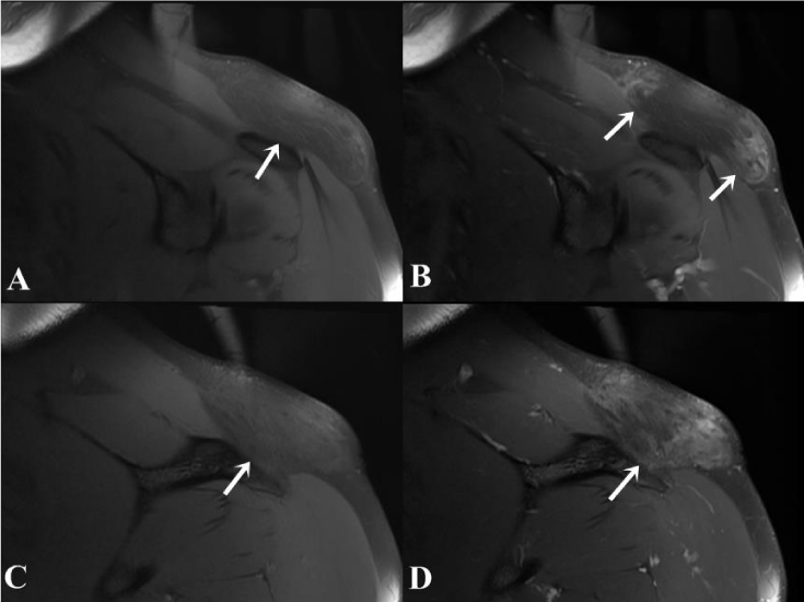
A 35-year-old male patient admitted to hospital with the mass growth on the left shoulder within 1 year. The patient complained of pain spreading from the lesion location to the left arm for the last 3 months. Physical examination revealed a rubbery and fixed lesion within 8x7 cm diameter on the left shoulder. A mild erythema was established on the skin without any varicose dilatation. US examination showed a large heterogeneous isoechoic lesion according to fatty tissue in the subcutaneous location. The solid lesion was in distinguishable from the adjacent muscle-fatty plans. MRI study revealed a solid lesion on the left shoulder within 27x68x92 mm diameter that had heterogeneous isoechoic character according to the fatty tissue on both T1- and T2-weighted imaging and fat suppressed technique showed suppression. The lesion had band shaped hypointense content. After the contrast material administration, fat saturated T1-weighted imaging demonstrated band shaped hypointense areas within the lesion were enhanced. The borders of the lesion were indistinguishable from normal fatty tissue in some regions (Figures 1,2). No evidence of any lesion was found in the adjacent bone tissue. The preliminary diagnosis of liposarcoma was considered after the imaging findings. The patient was operated under general anesthesia. In surgery, the lesion had no capsule and the borders were unable to separate from the surrounding fatty tissue. The mass with adjacent normal fatty tissue within 4x8x10 cm area were wide excised. The skin was primary sutured and closed. Pathological examination result was uncapsuled fibrolipoma. The lesion weight was 928 gram. At follow-up, there was no evidence of residual-recurrence in the US control at the 6 months and patient complaints disappeared.
Discussion
Lipomas are the most common benign mass lesions in subcutaneous fatty tissue. The etiology of lipomas and fibrolipomas is unclear. Benign lipomatous tumors are classified into subtypes according to their clinical histological and cytogenetic features. Fibrolipomas are one of the least common subtypes of lipomas [1,3]. Fibrolipomas are involving fibrous tissue at a significant level intermingled in the fatty lobes [1].
In the literature, fibrolipoma locations have been described in the oral cavity, esophagus and parathyroid gland and in other regions. However, there are few cases describe the radiological findings of subcutaneous fibrolipomas [4]. In our case, we performed US as the first step radiological examination; although, we used IV contrast enhanced MRI in diagnosis due to inadequacy of US.
The radiologic features of subcutaneous location of fibrolipomas defined in the literature are heterogeneous intensity on both T1- and T2-weighted imaging according to fatty tissue, presence of suppressed areas in fat saturation, well bordered or thin capsule occurrence and presence of enhancing areas on post-contrast series [1,4-6]. In one of these cases, calcification within the lesion is present in the foci [6]. MRI findings of a fibrolipoma in the nuchal region are defined as just submucosal fatty tissue thickening [7]. Our case differs from the literature in that undistinguished from the surrounding normal subcutaneous fatty tissue in some areas of the lesion borders. Lipomas are usually encapsulated lesions whereas fibrolipomas may be uncapsuled [3]. In our case, MRI was unable to demonstrate the borders of the lesion in some areas. In surgery, the lesion was inconvenient to separate from surrounding normal fatty tissue. These findings describe the formation of the lesion without capsule. Liposarcoma, spindle cell lipomas, pleomorphic lipomas are in the differential diagnosis of fibrolipomas. Classically, MRI findings of lipomas are isointensity with fat in MR sequences and suppressed in fat-suppressed techniques, and usually do not retain contrast material. MRI findings of spindle cell lipomas are not characteristic.
MRI features of well-differential liposarcoma are wide lipomatous content more than 75% and non-lipomatous component in extended septa more than 2 mm diameter with irregular appearance and/or nodular foci. After contrast material administration, the extended septa demonstrate evident or moderate enhancement [1]. In our case, MRI revealed enhanced thick septas larger than 2 mm and nodular foci. The radiological preliminary diagnosis considered liposarcoma due to imaging findings.
In conclusion, MRI findings of fibrolipoma may mimic liposarcoma rarely as in our case.
- Nishio J, Ideta S, Aoki M, Hamasaki M, Nabeshima K, et al. (2013) Fibrolipoma of the ring finger: MR imaging and histological correlation. In Vivo 27: 541-544. Link: https://goo.gl/sA5aK7
- Shin SJ (2013) Subcutaneous fibrolipoma on the back. J Craniofac Surg 24: 1051-1053. Link: https://goo.gl/xgXOTw
- Iwase M, Saida N, Tanaka Y (2016) Fibrolipoma of the Buccal Mucosa: A Case Report and Review of the Literature. Case Rep Pathol 2016: 5060964. Link: https://goo.gl/ECKCdo
- Kajihara M, Sugawara Y, Sakayama K, Abe Y, Miki H, et al. (2006) Subcutaneous fibrolipoma in the back. Radiat Med 24: 520-524. Link: https://goo.gl/0cSosJ
- Alsaleh N, Amanguno H (2015) Nuchal Fibroma: A rare entity of neck masses. Gulf J Oncolog 1: 10-12. Link: https://goo.gl/RYn0AG
- Akinkunmi M, Balogun B, Fadeyibi I, Benebo A, Awosanya G, et al. (2010) Giant fibrolipoma of the thigh in a Nigerian woman: A case report. Internet J Radiol 12: 1 Link: https://goo.gl/x2itA1
- Bayne DR, Combes J, Pandya A (2016) Nuchal fibrolipoma gives rugby players the 'hump'. ANZ J Surg 86: 416-418. Link: https://goo.gl/hq0442
Article Alerts
Subscribe to our articles alerts and stay tuned.
 This work is licensed under a Creative Commons Attribution 4.0 International License.


 Save to Mendeley
