International Journal of Radiology and Radiation Oncology
Contribution of Tc-99m RBC SPECT/CT in Intrahepatic and Peritoneal Splenosis Mimicking Malignancy
Kevser Oksuzoglu1, Rabia Ergelen2, Tunc Ones1, Sabahat Inanir1 and Tanju Yusuf Erdil1
2Department of Radiology, Marmara University Pendik Training and Research Hospital, Istanbul, Turkey
Cite this as
Oksuzoglu K, Ergelen R, Ones T, Inanir S, Erdil TY (2017) Contribution of Tc-99m RBC SPECT/CT in Intrahepatic and Peritoneal Splenosis Mimicking Malignancy. Int J Radiol Radiat Oncol 3(1): 007-009. DOI: 10.17352/ijrro.000021Introduction
Splenosis is the autoimplantation of ectopic spleen tissue resulting heterotopic auto-transplantation and implantation of splenic tissue after splenic ruptur caused by trauma or splenectomy. It is a benign condition that often mimics malignancy in healthy patients or peritoneal metastases in cancer patients. We report a case of diffuse peritoneal splenosis mimicking neoplastic lesions.
Case Presentation
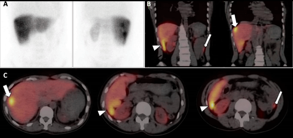
A 41-year-old woman with a history of abdominal pain was referred to ultrasonography (US). US was negative and computerized tomography (CT) was performed for further investigation. CT images disclosed the absent of spleen and a well-defined hypodense lesion compared to the liver, located near the liver capsule with a maximum diameter of 22 mm in the segment 8. In addition, multiple nodular formations were seen near the segment 6 of the liver. During the portal phase, the lesions were hyperdens compared to the liver. On MRI imaging, the lesions were hypointense on T1-weighted images and hyperintense on T2-weighted images. After contrast media administration (Gd+), all lesions showed heterogeneous contrast enhancement on the arterial phase, and homogeneous enhancement on the portal phase images (Figure 1). Peritoneal carcinomatosis was suspected but the patient had no complaints of fever, night sweats or weight loss, which suggests malignancy. Her history included splenectomy 20 years previously due to the traumatic rupture of the spleen during an accident. She went on to have 99mTc-labeled heat-damaged red blood cell (RBC) scintigraphy because of prior splenectomy. One gr of stannous ion (Sn+2) in the form of pyrophosphate was injected intravenously to the patient. 20 min later, a blood sample (approximately 8 cc) was obtained, 20 mCi of NaTc99mO4 was added, the sample was incubated at room temperature for 20 min then the sample was re-injected to the patient. Planar and hybrid single photon emission tomography/computed tomography images (SPECT/CT) were obtained. Planar images showed suspicious visual moderate activity uptake in the right upper quadrant and mild activity uptake in the left upper quadrant of the abdomen (Figure 2A). Hybrid SPECT/CT images provided the accurate localisation of the lesions in the liver (Figure 2B,C, thick arrow) and the other mesenteric and peritoneal ones (in the Morrison’s pouch and left paracolic gutter - Figure 2B,C, arrow head and thin arrow, respectively). These lesions were unchanged for ≥ 5 months compared to prior exams and did not demonstrate any changes in a second MRI session which performed 5 months later. The diagnosis of intrahepatic and peritoneal splenosis was confirmed without invasive diagnostic techniques.
Discussion
Splenosis is a benign, usually asymptomatic condition involving autotransplantation of ectopic splenic tissue that occurs commonly after splenic rupture caused by traumatic rupture of the spleen or splenectomy [1]. Splenosis may develop anywhere within the abdominal and pelvic cavity, being the most common location, even in the thorax when the diaphragm is damaged [2]. The most frequent locations include the greater omentum, small-bowel serosa, parietal peritoneum, and undersurface of the diaphragm [3]. Splenosis in the abdominal or pelvic cavity is thought to occur in as many as 65% of cases of splenic rupture. The average time between the inciting trauma and abdominal or pelvic splenosis is 10 years, although splenosis has been found to occur in as few as five months after trauma [1]. Although abdominal splenosis is frequently asymptomatic, it can present with hemorrhage, pain secondary to infarction or torsion, or obstruction of the intestinal or urinary tract [1,4]. They can be confused with other entities including peritoneal carcinomatosis, endometriosis, renal cancer, abdominal lymphomas, metastatic disease and hepatic adenomas [5-12]. Sonographic and radiological findings are not specific in splenosis, so ultrasound, CT, and MRI show limited value in the diagnostic management of abdominal splenosis [7]. Superparamagnetic iron oxide (SPIO)-enhanced MRI allows a confident characterisation of splenic tissue; but this technique is expensive [13]. Scintigraphic agents localize in the reticuloendothelial system (liver, spleen, bone marrow) and scintigraphy is specific to asses the phagocytosis function. 99mTc-labeled heat-damaged RBC, Indium 111-labeled platelets and Tc-99m sulphur colloid scintigraphy are scintigraphic modalities in diagnosing splenosis. 99mTc-labeled heat-damaged RBC scintigraphy and Indium 111-labeled platelets scintigraphy are more sensitive and specific for the diagnosis of splenic sequestration and phagocytosis than Tc-99m sulphur colloid scanning because of their better signal-to-background ratio, and their specificity for splenic tissue [1,5]. 99mTc-labeled heat-damaged RBC scintigraphy is actually considered as the ‘‘gold standard’’ test to establish the diagnosis of splenosis [14]. The performance of 99mTc-labelled heat-damaged RBC SPECT/CT allows the non-invasive diagnosis of this entity and avoids more aggressive diagnostic techniques but for some untypical cases, tissue sampling for pathologic diagnosis is still necessary [14]. In a recent comprehensive study, Ekmekçi et al reported that Tc-99m RBC SPECT/CT has a high specificity in the detection of accessory spleens/splenosis [15]. There were no cases suggesting false positivity have been reported, but we do not have readily available statistical results in this topic in the literature [15]. CT and MRI are highly sensitive in the detection of an accessory spleen in the usual locations; although, in the presence of multiple splenosis in different areas, detection of all lesions with a single injection of contrast agent and a single shot may be challenging. It is one of the advantages of Tc-99m RBC SPECT/CT that many other areas could be scanned with a single injection [16]. In this case report, the history of splenectomy together with the anatomical distribution of these lesions and the hybrid SPECT/CT images suggested this diagnosis. This nodules were unchanged for ≥ 5 months compared to prior exams, did not demonstrate any changes and the diagnosis of intrahepatic and peritoneal splenosis was confirmed without invasive diagnostic techniques. Additionally, this case report shows that hybrid SPECT/CT imaging has added clinical value over planar/SPECT imaging alone primarily due to more precise anatomical lesion localisation in splenosis.
Conclusion
Splenosis should be part of the differential diagnosis when faced with newly discovered lesions, solitary or multiple, anywhere in the peritoneal, pelvic and thoracic cavity, in patients with a history of abdominal trauma or splenectomy even with a history of neoplasia. Correct identification is essential because misinterpretation can have a significant impact on patient management. 99mTc-labelled heat-damaged RBC scintigraphy with SPECT/CT imaging should be considered prior to avoiding any further unnecessary surgery or chemotherapy.
- Fremont RD, Rice TW (2007) Splenosis: a review. South Med J 100: 589-593. Link: https://goo.gl/KhYXMa
- Bertolotto M, Quaia E, Zappetti R, Cester G, Turoldo A (2009) Differential diagnosis between splenic nodules and peritoneal metastases with contrast-enhanced ultrasound based on signal intensity characteristics during the late phase. Radiol Med 114: 42-51. Link: https://goo.gl/5dDg4G
- Gurses B, Kabakci N, Aksit HZ, Yencilek F, Kovanlikaya A, et al. (2007) Cystic splenosis mimicking a renal mass: a case report and review of the literature. Australas Radiol 51: B52–B55. Link: https://goo.gl/JRgxmv
- Sikov WM, Schiffman FJ, Weaver M, Dyckman J, Shulman R, et al. (2000) Splenosis presenting as occult gastrointestinal bleeding. Am J Hematol 65: 56-61. Link: https://goo.gl/p8ea36
- Tsitouridis I, Michaelides M, Sotiriadis C, Arvaniti Mary (2010) CT and MRI of intraperitoneal splenosis. Diagn Interv Radiol 16: 145-149. Link: https://goo.gl/UvbGjR
- Vercher-Conejero JL, Bello-Arqués P, Pelegrí-Martínez L, Hervás-Benito I, Loaiza-Góngora JL, et al. (2011) Esplenosis intraabdominal: una entidad frecuentemente infradiagnosticada. Rev Esp Med Nuc 30: 97-100. Link: https://goo.gl/AUe65i
- Ksiadzyna D, Peña AS (2011) Abdominal splenosis. Rev Esp Enferm Dig 103: 421-426. Link: https://goo.gl/EVROQj
- Levy AD, Shaw JC, Sobin LH (2009) Secondary tumors and tumorlike lesions of the peritoneal cavity: Imaging features with pathologic correlation. Radiographics 29: 347-373. Link: https://goo.gl/EqVDPh
- Kiser JW, Fagien M, Clore FF (1996) Splenosis mimicking a left renal mass. AJR 167: 1508-1509. Link: https://goo.gl/C0JRzL
- Schenkein DP, Ahmed E (1995) Case records of the Massachusetts General Hospital. Weekly clinic-pathological exercises: Case 29-1995 A 65-year-old man with mediastinal Hodgkin's disease and a pelvic mass. N Engl J Med 333: 784-791. Link: https://goo.gl/GuEiDW
- Yildiz AE, Ariyurek MO, Karcaaltincaba M (2013) Splenic anomalies of shape, size, and location: Pictorial essay. ScientificWorldJournal 2013: 321810. Link: https://goo.gl/RlDrUQ
- Gruen DR, Gollub MJ (1997) Intrahepatic splenosis mimicking hepatic adenoma. AJR Am J Roentgenol 168: 725-726. Link: https://goo.gl/1Ytftb
- Kim SH, Lee JM, Han JK, Lee JY, Kang WJ, et al. (2006) MDCT and superparamagnetic iron oxide (SPIO)-enhanced MR findings of intrapancreatic accessory spleen in seven patients. Eur Radiol 16: 1887–1897. Link: https://goo.gl/upn6BN
- Grande M, Lapecorella M, Ianora AA, Longo S, Rubini G (2008) Intrahepatic and widely distributed intraabdominal splenosis: multidetector CT, US and scintigraphic findings. Intern Emerg Med 3: 265-267. Link: https://goo.gl/VuOzRO
- Ekmekçi Ş, Diz-Küçükkaya R, Türkmen C, Adalet I (2015) Selective Spleen Scintigraphy in the Evaluation of Accessory Spleen/Splenosis in Splenectomized/Nonsplenectomized Patients and the Contribution of SPECT Imaging. Mol Imaging Radionucl Ther 24: 1-7. Link: https://goo.gl/1hhFiQ
- Yammine JN, Yatim A, Barbari A (2003) Radionuclide imaging in thoracic splenosis and a review of the literature. Clin Nucl Med 28: 121–123. Link: https://goo.gl/eG97Qo
Article Alerts
Subscribe to our articles alerts and stay tuned.
 This work is licensed under a Creative Commons Attribution 4.0 International License.


 Save to Mendeley
