International Journal of Radiology and Radiation Oncology
Successful Treatment of a Bilateral Lower Palpebral MALT Lymphoma with Rapid Arc: Description of a New Technique
Issam Lalya1*, Laila Baddouh2, Keltoum Dahmani2, Noha Zaghba1, Khalid Andaloussi1, Mohamed Elmarjany1, Khalid Hadadi1, Hassan Sifat1 and Hamid Mansouri1
2Department of Radio physics, Military Teaching Hospital Mohammed V, University Mohammed V Souissi, Rabat, Morocco
Cite this as
Lalya I, Baddouh L, Dahmani K, Zaghba N, Andaloussi K, et al. (2016) Successful Treatment of a Bilateral Lower Palpebral MALT Lymphoma with Rapid Arc: Description of a New Technique. Int J Radiol Radiat Oncol 2(1): 021-022. DOI: 10.17352/ijrro.000015Clinical Image
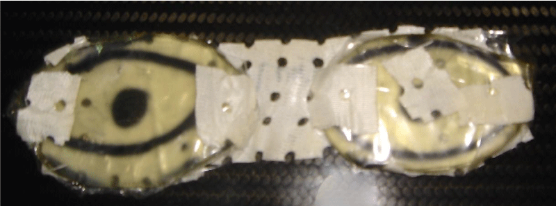
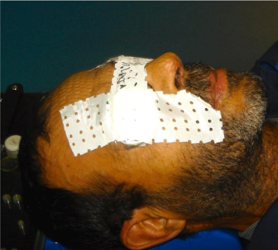
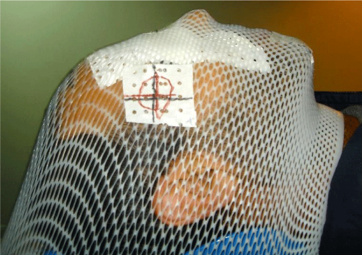
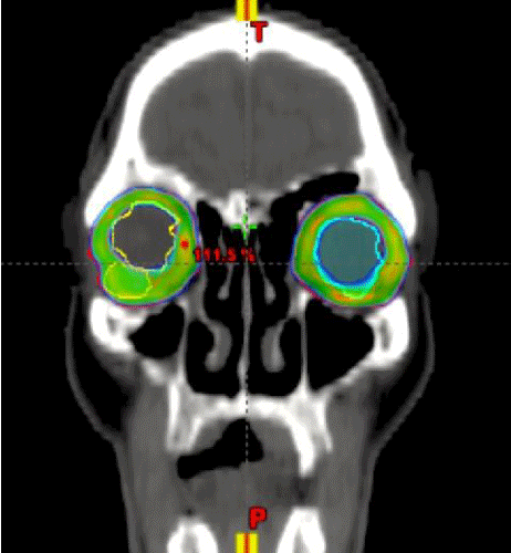
Primary MALT lymphoma of the eyelids is a rare disease; chronic infection by Chlamydophila psittaci has been identified as a possible causative agent, but other pathogens may be implicated such Hepatitis C virus and Helicobacter Pylori. This tumor is considered to have an indolent natural history and a favorable prognosis. Radiation therapy with moderate doses is the corner stone of treatment offering excellent local control with low complications rate. We report herein the radiation therapy treatment plan of a 65-year-old man presented to our department with a primary bilateral lower palpebral MALT lymphoma. CT scan of 2.5-mm slice thickness in supine position was realized. A bolus of 1cm adapted to closed eyes (glasses-like) were placed and fixed to palpebral surface (Figures 1,2), before immobilization with standard thermoplastic mask (Figure 3). The clinical target volume included the tumor, lachrymal glands and the entire orbit. The planning target volume was obtained by automatic margin of 5 mm around the clinical target volume. Optic nerves, eyeballs and lens were contoured as organs at risk. The prescribed dose was 16 Gy delivered in 5 daily fractions of 3.2 Gy (Biological effective dose: 36 Gy). RapidArc plan (Varian Medical Systems) was performed using the Eclipse software (Varian Medical Systems). A maximum dose rate of 600 MU/min and 6 MV photon beams were selected. Two coplanar arcs of 360° were delivered with opposite rotation (clockwise and counter-clockwise). For each arc the field size and the collimator rotation were determined by the automatic tool from Eclipse to encompass the planning target volume. Optimization was carried out by the progressive resolution optimizer version 3 (PRO 3), to have an optimal coverage of the target volume (high priority) while reducing dose to organs at risk as low as possible. Dose calculation was performed using the analytic anisotropic algorithm (AAA). The use of Rapidarc allowed the optimal coverage of target volume by isodose 95%, with perfect protection of eyeballs and lens (Figure 4). The treatment was delivered by a Clinac iX Linear accelerator (Varian Medical Systems). In order to verify the correct positioning we performed a cone beam CT (CBCT) before each treatment fraction. At the fifth fraction of radiation therapy, we noted a complete clinical response, which we reported in a previous article as a clinical image [1]. To the best of our knowledge this is the first report describing the use of RapidArc in this location.
Authors’ contributions
IL and LB invented and realized the treatment plan. IL wrote the manuscript. All authors read and approved the final manuscript. HM revised the intellectual content and approved the final manuscript.
Article Alerts
Subscribe to our articles alerts and stay tuned.
 This work is licensed under a Creative Commons Attribution 4.0 International License.


 Save to Mendeley
