International Journal of Radiology and Radiation Oncology
Effectiveness of Radiotherapy in Heterotopic Ossification
Yasemin Benderli Cihan* and Alaettin Arslan
Cite this as
Cihan YB, Arslan A (2015) Effectiveness of Radiotherapy in Heterotopic Ossification. Int J Radiol Radiat Oncol 1(1): 029-032. DOI: 10.17352/ijrro.000008Objective: Heterotopic ossification (HO) is a biological process characterized by de novo bone formation in tissues which should not undergo ossification in normal condition. Frequently, it is a complication that occurs after head injuries, spinal cord injuries, cerebrovascular events, burns, fractures, dislocations and joint replacements. Etiopathogenesis is unclear in HO. Clinical symptoms and signs become apparent at late phase. Limitation and pain occur by advancing disease. The diagnosis and management are of importance. Non-steroidal anti-inflammatory drugs (NSAIDs) and radiotherapy (RT) are preferred in prophylaxis. The aim of present study was to assess safety and effectiveness of radiation therapy in preventing HO.
Material and method: Data of patients received RT with a diagnosis of HO between 2005 and 2013 were retrospectively reviewed.
Findings: Overall 12 patients aged 20-72 years, including 8 men and 4 women, received RT for HO prophylaxis. HO incidence was highest in hip joint (n=7); followed by elbow (n=3) and knee joints (n=2). Of these patients, 7 received postoperative RT while 5 received RT for HO prophylaxis. RT was given in a single fraction at a dose of 7 Gy (6-8 Gy). Patients received postoperative RT was also given indomethacin (75 mg/day) over 15-20 days. No RT-related complication was observed in any of the patients. Imaging modalities were used in follow-up. In addition, Broker’s Grading Scale was used for the assessment of lower extremities whereas Hastings and Graham Grading Scale was used for upper extremities. Median follow-up was 25.3 months (range: 2-69 months). No radiological HO recurrence was observed in any of the patients received postoperative RT.
Conclusion: RT in combination with NSAIDs is a safe and effective therapeutic modality in the prophylaxis of patients at high-risk for HO development.
Introduction
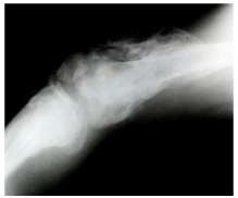
Heterotopic ossification is progressive but self-limiting de novo trabecular bone formation occurring at skin, subcutaneous tissue, and skeletal muscle and periarticular tissues in which ossification doesn’t occur in normal conditions. This ectopic bone tissue, induced by osteoplastic activity, is an extra-articular bone localized to adjacent areas of joint and joint capsule is generally intact [1,2]. It involves large joints such as hip, elbow and knee (Figure 1). It generally appears 1-4 months after trauma, but it may also occur a few years after trauma. Clinic symptoms and signs become apparent at late phase [3-5]. HO is classified according to Broker’s Grading Scale scoring system in lower extremities and Hastings and Graham Grading Scale scoring system in upper extremities [6].
In general, trauma increases incidence of heterotopic ossification. Its incidence is 11-76% after head injury, 18-37% after spinal cord injury, 2-7% after total hip prosthesis, 1-3% after burns, and 0.5-1.2% after cerebrovascular events [7].
The diagnosis and management is of importance in HO which is frequently observed in orthopedic patients and those undergoing neurological rehabilitation. Treatment options include physical therapy to protect range of motion, drug therapy (etidronate, non-steroidal anti-inflammatory drugs), radiotherapy, and resection of ectopic bone tissue in joints with severe dysfunction [1,3,5,6].
Prophylaxis is more important than treatment in heterotopic ossification. Early mobilization is of importance in these patients. Risk factors should be eliminated in the development of heterotopic ossification. In addition, radiotherapy and NSAIDs have major role in the prophylaxis of heterotopic ossification [5-9].
The aim of this study was to assess etiology, clinical signs, and diagnostic and therapeutic options in patients received RT for heterotopic ossification.
Material and Method
Demographic characteristics of patients
We retrospectively reviewed patient files and/or electronic data of 12 patients who presented to Radiation Oncology Department of Kayseri Education and Research Hospital with HO and were treated between 2005 and 2013. Data regarding age, gender, history, etiology, radiological techniques, presenting complaints, drugs used, treatments received, treatment-related complications and follow-up times were extracted from patient records. Phone interviews with patients or their relatives were used in attempt to obtain missing data. The study was approved by Institutional Ethics Committee.
RT and medication
Two-dimensional treatment planning system using conventional X-ray simulator was used for planning radiotherapy. Planning target volume (PTV) was estimated to involve all sites of heterotopic ossification. Based on localization and extent of HO, radiotherapy was delivered with a total dose of 7 Gy (6-8 Gy) in a single fraction to areas (approximately 10x6 to 18x9 cm in size) from anterior-posterior parallel regions by linear accelerator devices (Linac) using 18 MV photon energies or Co-60. Indomethacin (75 mg/day over 15-20 days) was prescribed to all patients received postoperative RT.
Treatment response and follow-up
All patients were assessed on the months 1, 3 and 6 after completion of therapy. The assessment included physical examination, direct radiographs and/or articular MR imaging or CT scan. Radiological assessments were performed according to ossification degree (0-4 points) by using Broker’s Grading Scale for lower extremities and Hasting and Graham Grading Scale for upper extremities. Unresponsiveness was defined as increased ossification scores in radiological assessment after completion of radiotherapy. Clinical information was obtained through phone interviews in patients not attending to follow-up visits.
Results
Table 1 presents demographic characteristics of patients. Of 12 patients presented and were treated for HO, 8 were men. Median age was 44.4 years (range: 20-72). There was hypertension, diabetes mellitus and coxarthrosis in the history of patients. It was seen that most common triggers are traffic accident and fracture for HO. HO incidence was highest in hip joint (n=7); followed by elbow (n=3) and knee joints (n=2). Perioperative radiotherapy was given in 7 patients, whereas it was given to prevent HO in 5 patients. The most common HO-related complaints were pain and limitation of movement. Based on Hastings and Graham classification, there was grade IIA (n=1), IIC (n=1), and I (n=1) ossification among elbow joints. Based on Broker’s classification, there was grade I (n=3) and II (n=6) ossification among hip joints.
Table 2 presents parameters of RT given. RT was delivered with a total dose of 7 Gy (6-8 Gy) in a single fraction to areas (approximately 10x6 to 18x9 cm in size) from anterior-posterior parallel regions by using Co-60 or 18 MV photon devices. Mean time from surgery to RT was 24 hours in 6 patients and 48 hours in one patient. Indomethacin (75 mg per day, divided in 3 doses, over 15-20 days) were prescribed to patients received postoperative RT. No acute or subacute complication was observed. RT wasn’t repeated in any patient.
Median follow-up was 25.3 months (range: 2-69). Two patients didn’t attend to control visit on the months 3 and 6. Outcomes were assessed according to physical examination and radiological evaluations. There was no increase in Broker’s ossification score in patients received postoperative radiotherapy. One patient who received postoperative radiotherapy but didn’t attend to control visits was interviewed by phone. It was seen that radiotherapy was effective by 100% based on clinical prophylaxis. Of the patients received prophylactic radiotherapy for elbow joint, it was seen that there was no increase in radiological Hastings and Graham ossification scores in 2 patients. In this group, it was seen that radiotherapy was effective by 60% in one patient who didn’t attend to control visits.
Discussion
Heterotopic ossification was first described with its clinical, patho-anatomic and radiological characteristics in patients with traumatic paraplegia (paraosteoarthropathy) by Dejerine and Ceillier at 19th century. In the literature, de novo periarticular bone formation is denoted with several terms including myositis ossificans, paraosteoarthropathy, neurogenic ossifying fibromyopathy [1,5,7,10,11].
Although HO has been known for over a hundred year, there is an ongoing debate regarding etiology, pathogenesis and management [8,12,13]. It is understood that there is a need for further studies on patients at risk for HO or with HO in order to create a health policy
Pathophysiology of heterotopic ossification isn’t fully elucidated. It is suggested that HO may result from metaplasic response of mesenchymal cells induced by interaction between systemic factors and local, metabolic, vascular, genetic and biochemical factors [3,7]. Immobilization, pressure ulcers, trauma, fracture, dislocation, burn, infection, hematoma and several neurological disorders are implied in the etiology [1,2,5,14]. Swelling, effusion, erythema and warmth as well as pain appear 2 weeks after trauma. Localized mass, pain and limitation of movement are typical in the course of disease. These clinical symptoms are seen within 8-10 weeks [3,7]. Laboratory and radiology studies will be helpful in diagnosis. Direct radiographies are invaluable in the diagnostic process, as HO become visible on direct radiographs after 1-2 months, where maturation occurs. Computerized tomography can reveal both HO localization and its relationship with adjacent tissues as well as it is valuable as a guide for treatment [2,6]. Traffic accident (spasticity, prolonged coma, immobilization) and hip fractures were primary risk factors in our study. It was most frequently observed at hip joint; followed by elbow. The most common complaints were pain and limitation of movements. Direct radiographies and MR imaging/CT scans were used in diagnosis and follow-up. Our findings were consistent with literature.
In our study, RT was applied to 12 patients in order to prevent HO. By the idea that it could be more effective in patients underwent total hip arthroplasty, combined therapy (RT plus NSAID) was prescribed on the postoperative day 1. In patients with HO at elbow, there was persistent clinical progression and complaints including pain and limitation that caused marked decrease in quality of life despite physical therapy and drug use. RT was indicated due to refractory disease in these patients. In most patients, RT was delivered with 7 Gy in single fraction, taking recent studies on treatment protocol into consideration. In our study, no radiological recurrence of heterotopic ossification was observed in any patient received postoperative RT after 25 months follow-up. In patient received RT to elbow joint, no increase was observed in HO in radiological follow-up. HO is generally seen large joints. It is more frequently seen at hip, knee, shoulder and elbow joints [1,12]. HO formation is most rapid within first 1-4 months. Thus, it is important to initiate management as soon as possible after diagnosis. Radiotherapy and NSAIDs have a major role in heterotopic ossification prophylaxis [8,13-16].
NSAIDs, indomethacin in particular, prevent transformation of mesenchymal cells to osteogenic cells that generate bone tissue. Although NSAIDs are widely used, they are associated with adverse effects such as gastrointestinal disorders or compliance issues [8,10]. RT is another method used in prophylaxis. Radiotherapy prevents HO formation by inhibiting mesenchymal cell development which is thought to be progenitor in ossification. Moreover, low dose radiotherapy also has anti-inflammatory activity [9,11,16]. In previous studies, HO incidence after total hip replacement was reported as 8-90%, while it was reported as 35-57% in large series [7,8,13,17]. In a meta-analysis including 7 studies on 1143 patients, Pakos et al., compared radiotherapy to anti-inflammatory drugs regarding efficiency in preventing HO. Authors concluded that postoperative radiotherapy at appropriate doses was more effective than anti-inflammatory drugs [8]. In a meta-analysis by Baird et al., HO incidence was evaluated in patients undergoing total hip arthroplasty. Authors reported HO incidence as 58% in patients underwent surgery alone, 37% in patients received indomethacin alone, 27% in patients received radiotherapy alone and 12% in those received RT plus indomethacin [1]. In the studies by Balboni et al. and Maurad et al., it was suggested that postoperative single dose radiotherapy (8Gy) in combination with indomethacin should be given to prevent recurrence in patients at high risk [3,12].
There are several studies regarding timing, dose and route of radiotherapy in the literature. It has been shown that radiotherapy is more effective in the early phase of HO. It is recommended that surgery alone increases recurrence rates and radiotherapy should be given at preoperative and early postoperative period to prevent recurrence. It is recommended that radiotherapy should be given within first 72 hours after surgery or 1, 2, 4, 16 or 29 hours prior to surgery [12,15,16,18,19]. In a study Kantorowitz et al., a significant difference was found between applications of radiotherapy (7 Gy) 4 hours before surgery and radiotherapy (7 Gy) applied within first 72 hours after surgery regarding HO prevention. Authors reported that preoperative radiotherapy was more effective [16,20]. In a study on patients undergoing total hip arthroplasty by DeFlitc et al., radiotherapy was given within first 72 hours to 75% of patients to prevent HO and authors reported that HO developed in 55% of these patients [5]. In the studies by Rumi et al. and Seegen et al., it was reported that both preoperative and postoperative radiotherapy prevented HO by 85-95% in patients at high risk for HO development [13,19].
It is recommended to apply early postoperative radiotherapy at fractionated doses (total dose of 10 Gy) or at a dose of 6-8 Gy at single fraction in order to prevent recurrence after surgery. Previous studies demonstrated that there was no difference between single dose and fractionated doses [9,14,15,17,21,22]. However, single dose radiotherapy is more widely preferred due to its ease application. In a study by Hedley et al., single dose radiotherapy (6 Gy) was shown to be effective in 17 patients underwent hip arthroplasty. This finding indicated that minimum effective dose was 6 Gy in preventing HO development [21]. In a study, Healy et al. compared single doses with 5.5 Gy and 7 Gy in preventing HO. In that study, HO formation was observed in 63% of the patients received single dose radiotherapy with 5.5 Gy, while, it was observed in only 10% of the patient received single dose radiotherapy with 7 Gy [9]. In another study, perioperative radiotherapy was given to 9 patients who underwent surgical resection for heterotopic ossification that was already present at elbow. Radiotherapy was delivered with a total dose of 1000 Rad in 2 fractions in 5 patients, whereas remaining 4 patients received a single dose radiotherapy with 600 Rad. After mean follow-up 7 months, no radiological recurrence of heterotopic ossification was observed in any of the patients, and authors suggested that prophylactic radiotherapy was effective [14].
In conclusion, radiotherapy in combination with anti-inflammatory drugs is an effective treatment algorithm in patients at high-risk for HO development such as those undergoing total hip replacement. However, there is a need for further clinical trials with larger sample size that assess effectiveness and adverse effects of prophylactic RT in HO according to age groups.
- Baird EO, Kang QK (2009) Prophylaxis of heterotopic ossification-review. J Orthop Surg Res 4: 12 .
- Kaplan FS, Glaser DL, Hebela N, Shore EM (2004) Heterotopic ossification. J Am Acad Orthop Surg 12: 116–125 .
- Balboni TA, Gobezie R,Mamon HJ (2006) Heterotopic ossification: pathophysiology, clinical features, and the role of radiotherapy for prophylaxis. Int J Radiat Oncol Biol Phys 65: 1289–1299.
- Garland DE (1995) A clinical perspective on common forms of acquired heterotopic ossificaton. Clin Orthop 263: 13-29 .
- DeFlitch CJ, Stryker JA (1993) Postoperative hip irradiation in prevention of heterotopic ossification: causes of treatment failure. Radiology 188: 265-270 .
- Hastings H, Graham TJ (1994) The classification and treatment of heterotopic ossification about the elbow and forearm. Hand Clin 10: 417-437.
- Varghese G (1992) Heterotopic ossification. Phys Med Rehabil Clin North Am 3: 407-415.
- Pakos EE, Ioannidis JP (2004) Radiotherapy vs. nonsteroidal anti-inflammatory drugs for the prevention of heterotopic ossification after major hip procedures: a metaanalysis of randomized trials. Int J Radiat Oncol Biol Phys 60: 888-895 .
- Healy WL, Lo TC, DeSimone AA, Rask B, Pfeifer BA (1995) Single- dose irradiation for the prevention of heterotopic ossification after total hip arthroplasty. A comparison of doses of five hundred and fifty and seven hundred centigray. J Bone Joint Surg Am 77: 590-595 ..
- Burd TA, Hughes MS, Anglen JO (2003) Heterotopic ossification prophylaxis with indomethacin increases the risk of long-bone nonunion. J Bone Joint Surg Br 85: 700-705 .
- Pellegrini VD Jr, Gregoritch SJ (1996) Preoperative irradiation for prevention of heterotopic ossification following total hip arthroplasty. J Bone Joint Surg Am 78: 870-881 .
- Mourad WF, Packianathan S, Shourbaji RA, Zhang Z, Graves M, et al. (2012) A prolonged time interval between trauma and prophylactic radiation therapy significantly increases the risk of heterotopic ossification. Int J Radiat Oncol Biol Phys 82: 339–344 .
- Seegenschmiedt MH, Keilholz L, Martus P, Goldmann A, Wölfel R, et al. (1997) Prevention of heterotopic ossification about the hip: Final results of two randomized trials in 410 patients using either preoperative or postoperative radiation therapy. Int J Radiat Oncol Biol Phys 39: 161–171 .
- Heyd R, Strassmann G, Schopohl B, Zamboglou N (2001) Radiation therapy for the prevevention of heterotopic ossification at the elbow. J Bone Joint Surg Br 83: 332-334 .
- Robinson CG, Polster JM, Reddy CA, Lyons JA, Evans PJ,, et al. (2010) Postoperative single fraction radiation for prevention of heterotopic ossification after elbow surgery [abstract]. Int J Radiat Oncol Biol Phys 77: 1493-1499 .
- Kantorowitz DA, Muff NS (1998) Preoperative vs. postoperative radiation prophylaxis of heterotopic ossification: a rural community hospital’s experience. Int J Radiat Oncol Biol Phys 40: 171-176.
- LoTC, Healy WL (2001) Re-irradiation for prophylaxis of heterotopic ossification after hip surgery. Br J Radiol 74: 503-506 .
- Childs HA 3rd, Cole T, Falkenberg E, Smith JT, Alonso JE, et al. (2000) A prospective evaluation of the timing of postoperative radiotherapy for preventing heterotopic ossification following traumatic acetabular fractures. Int J Radiat Oncol Biol Phys 47: 1347-1352 .
- Rumi MN, Deol GS, Bergandi JA, Singapuri KP, Pellegrini VD Jr (2005) Optimal timing of preoperative radiation for prophylaxis against heterotopic ossification. A rabbit hip model. Bone Joint Surg Am 87: 366–373 .
- Kantorowitz DA, Miller GJ, Ferrara JA, Ibbott GS, Fisher R, et al. (1990) Preoperative versus postoperative irradiation in the prophylaxis of heterotopic bone formation in rats. Int J Radiat Oncol Biol Phys 19: 1431-1438 .
- Hedley AK, Mead LP, Hendren DH (1989) The prevention of heterotopic bone formation following total hip arthroplasty using 600 rad in a single dose. J Arthroplasty 4: 319-325 .
- Mourad WF, Packianathan S, Shourbaji RA, Zhang Z, Graves M, et al. (2012) The impact of BMI on heterotopic ossification. Int J Radiat Oncol Biol Phys 82: 831–836 .
Article Alerts
Subscribe to our articles alerts and stay tuned.
 This work is licensed under a Creative Commons Attribution 4.0 International License.


 Save to Mendeley
