International Journal of Oral and Craniofacial Science
Bifid Uvula in three members of a family
Suwarna Dangore Khasbage*
Cite this as
Khasbage SD (2017) Bifid Uvula in three members of a family. Int J Oral Craniofac Sci 3(2): 017-019. DOI: 10.17352/2455-4634.000027Uvula is a key organ in functions like speech, deglutition and mastication. The majority of the world population has a uvula that is conical in shape, hanging upside down. However, there are times when the uvula is split, the condition is called a bifid or bifurcated uvula. Sometimes it is also called a cleft uvula. Three male blood relatives, father and his two sons reported to outpatient department of Oral Medicine and Radiology at Sharad Pawar Dental College, Wardha for odontogenic complaint. But, their intraoral examination revealed interesting finding, that was presence of bifid uvula and cleft palate in three of them, who were of course first relatives of each other. The presentation of the condition in three members of the same family is a unique feature of this article.
Introduction
Bifid uvula means a cleft in uvula. It is often considered as a marker for sub mucous cleft palate. Compared to the normal one, it has fewer amounts of muscular tissues. It is commonly noticed in infants and is rarely found in adult. A bifid or bifurcated uvula exists in two percent of the general population. The prevalence of cleft uvula is much higher than that of cleft palate. Cleft uvula is more common in whites (1 in every 80 white individuals) as compared to blacks (1 in every 250 individuals) [1]. It can cause problems in ear. Sometimes it is unable to reach the posterior pharyngeal wall during swallowing, causing regurgitation. It may produce velopharyngeal insufficiency and nasal intonation. Sometimes it is associated with major systemic problems like aneurysm in different vascular bed like coronary and abdominal aortic aneurysm. But, it does not cause problems in view of airway management.
Case report 1
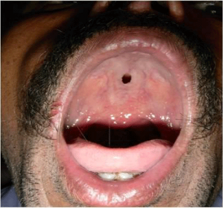
A 58 years old male had reported to outpatient department of Oral Medicine and Radiology at Sharad Pawar Dental College, Wardha with a complain of toothache in left posterior region of lower jaw. His past medical history and past dental history was not contributory. Clinical examination revealed food lodgment and initial proximal caries with 36. Soft tissue examination revealed presence of approximately 1X 1.5cm, oval palatal perforation suggestive of cleft palate and short bifid uvula (Figure 1). He had no local problems like speech difficulty or nasal regurgitation etc., and not ready for further evaluation regarding cleft palate and bifid uvula. Routine treatment protocol was advised for the carious tooth.
Case report 2
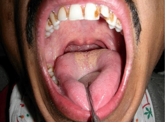
A 28 years old male who was an elder son of the patient described in first case report, had reported to outpatient department of Oral Medicine and Radiology at Sharad Pawar Dental College, Wardha with a complaint of decayed tooth in posterior region of right side of lower jaw. His past medical and dental history was not contributory. Clinical examination revealed deep caries with mandibular first molar. Examination of palate showed presence of approximately 1X 1cm, round palatal perforation suggestive of cleft palate and short bifid uvula as shown in (Figure 2). There was no history of speech difficulty, nasal twang or nasal regurgitation. He was also not ready for further evaluation regarding cleft palate and bifid uvula. The carious tooth was treated by conservative approach.
Case report 3
A 26 years old male who was a younger son of the patient described in first case report, had reported to same place with a complaint of discolored teeth due to deposits. Clinical examination revealed presence of extrinsic stains on the teeth. Examination of palate showed presence of younger son had bifid uvula and submucosal cleft, as shown in (Figure 3). There was no history of speech difficulty, nasal twang or nasal regurgitation. Systemic histosy was negative. With reference to evaluation of cleft palate and bifid uvula he had followed his father and elder brother (described in case report 1 and 2). Oral prophylaxis was advised as a routine protocol.
All of them were not willing for further evaluation or treatment of palate. Thus, it was not possible to explore any of the factors related to palate and uvula in them which can be considered as the limitation of this article. Even the etiological role of genetic factor may be suspected in the presented patients that is only on the basis of clinical presentation. This work was conducted after taking written consent from each of them and permission to utilize their photographs and information for publication.
Discussion
The patients reported in the present article had no symptoms related to bifid uvula as well as cleft palate. However, literature states that the patients having bifid uvula and cleft palate may suffer from a number of symptoms which may hamper the routine daily activities and thus they are usually diagnosed to have cleft palate and abnormality of morphology of uvula during infancy.
Various symptoms associated with bifid uvula and cleft palate may be inability to breastfeed, difficulty in bottle-feeding, nasal regurgitation and recurrent otitis media, delay in speech development etc. The speech had a characteristic hyper-nasal resonance with nasal air emission and an abnormal speech pattern with compensatory articulations [2].
Bifid uvula, although looks apparently benign, sometimes may be associated with anomalies leading to catastrophic complications. Cornelia de Lange syndrome is a rare congenital syndrome associated with bifid uvula and sub mucous cleft palate that causes problems in airway due anatomical distortion [3]. Bifid uvula may be associated with increased risk of schizophrenia, mild mental retardation, and chromosomal disorder, diagnosed by fluorescent in situ hybridization technique [4]. Loeys–Dietz syndrome (autosomal dominant) is a genetic syndrome with clinical features overlapped with Marfan syndrome, but etiology due to mutations in the genes encoding transforming growth factor beta receptor 1. Hypertelorism, cleft palate, or bifid uvula are the major findings. Arterial aneurysms/dissections, arterial tortuosity involving aortic and its branches, carotid, vertebral, extracranial artery, abdominal aorta and its branches, common iliac, and popliteal arteries are reported in this syndrome [5,6,7]. It is stated that bifid uvula may have been a warning sign of the syndrome with internal anatomical or functional changes without any external manifestation akin to the tip of an iceberg. Although cerebral aneurysm is very rare with bifid uvula, it may be a part of the above mentioned syndromes [8].
Thus, whenever anesthetists plans to conduct a case with bifid uvula (even though non-syndromic), they must ask for detailed family and genetical history, clinical examination relevant investigations, and specialty consultation [8]. Adequate preoperative preparation and, accordingly, intra-operative management can prevent unexpected complications in such patients.
Bifid uvula is often regarded as a marker for submucous cleft palate although this relationship has not been fully confirmed. The reason for the tacitly assumed connection between these two anomalies has, in part, been perpetuated by the generally accepted definition of submucous cleft palate as the triad of bifid uvula, notching of the hard palate, and muscular diastasis of the soft palate. Recently, investigations have provided evidence of more subtle manifestations of submucous cleft palate by the use of nasopharyngoscopic examination of the palate and pharynx [9].
SMCP is a rare cleft palate which is, despite the presence of a bifid uvula and symptoms of velopharyngeal insufficiency, often diagnosed late. In children with a bifid uvula and mild problems in speech, hearing and swallowing, it is important to be alert to SMCP because SMCP may account for these persistent mild complaints. Therefore, early detecting of SMCP can yield profits [10].
The possible treatments for bifid uvula depend on the severity of problem. In asymptomatic cases, as such no treatment required which was the situation in the patients described in the present article and thus they were not ready for any investigations or management. If the symptom includes speech difficulties, then a speech therapist could possibly help the patient learn how to talk well. Swallowing and feeding problems may also be addressed through appropriate therapy. Some patients may opt for the removal of the bifurcated uvula but others would opt for the surgical reconstruction of these abnormal tissues.
Treatment of SMCP is surgical repair which includes a V-Y palatal pushback and simultaneous transposition of a superiorly based pharyngeal flap. Similarly the interrelated problems of chronic otitis media and faulty speech production may be diminished by functional reorientation of the palatal muscles and simultaneous revision of the velopharyngeal portal [11].
- Neville BW, Damm DD, Allen CM, Bouquot JE (2009) Develpoemental defects of Oral and Maxillofacial region. In: Oral and Maxillofacial Pathology. 1st edition, Elsevier India Private ltd.
- Hasan A, Gardner A, Devlin M, Russell C (2014) Submucous Cleft Palate with Bifid Uvula. J of Pediatrics 165: 872. Link: https://goo.gl/oYV6zW
- Callea M, Montanari M, Radovich F, Clarich G, Yavuz I (2011) Bifid uvula and submucous cleft palate in cornelia de lange syndrome. J Int Dent Med Res 4: 74. Link: https://goo.gl/SV1n3c
- Vorstman JA, de Ranitz AG, Udink ten Cate FE, Beemer FA, Kahn RS (2002) A bifid uvula in a patient with schizophrenia as a sign of 22q11 deletion syndrome. Ned Tijdschr Geneeskd 146: 2033–2036. Link: https://goo.gl/5S8D5H
- Loeys BL, Schwarze U, Holm T, Callewaert BL, Thomas GH et al. (2006) Aneurysm syndromes caused by mutations in the TGF-beta receptor. N Engl J Med 355: 788–798. Link: https://goo.gl/eHFifg
- Johnson PT, Chen JK, Loeys BL, Dietzd HC, Fishman EK (2007) Loeys-Dietz Syndrome: MDCT Angiography Findings. AJR Am J Roentgenol 189: 29–35. Link: https://goo.gl/By8N2X
- Inoue Y1, Minatoya K2, Oda T1, Itonaga T1, Seike Y1 et al. (2016) Total Aortic Replacement for a 9-Year-Old Boy With Loeys-Dietz Syndrome. Ann Thorac Surg 101:1185-1188. Link: https://goo.gl/UXo9u3
- Samanta S, Samanta S (2013) Bifid uvula: Anesthetist don’t take it lightly! Saudi J Anaesth 7: 482–484. Link: https://goo.gl/T4zA9L
- Shprintzen RJ, Schwartz RH, Daniller A, Hoch L (1985) Morphologic significance of bifid uvula. Pediatrics 75: 553-561. Link: https://goo.gl/sp7X8E
- ten Dam E, van der Heijden P, Korsten-Meijer AG, Goorhuis-Brouwer SM, van der Laan BF (2013) Age of diagnosis and evaluation of consequences of submucous cleft palate. Int. J of Pediatrc Otorhinolaryngology 77: 1019-1024. Link: https://goo.gl/VLzX65
- Kinnebrew MC, McTigue DJ (1984) Submucousal cleft palate: review and two clinical reports. Pediatric Dentistry. Clinical reports 696: 252- 258. Link: https://goo.gl/wmS8gY
Article Alerts
Subscribe to our articles alerts and stay tuned.
 This work is licensed under a Creative Commons Attribution 4.0 International License.


 Save to Mendeley
