International Journal of Oral and Craniofacial Science
Idiopathic Thrombocytopenic Purpura (ITP) Topic Review and Case Report
Mohamed Ahmed1* and Alan Y Martinez2
2Director, division of Oral and Maxillofacial Surgery. Department of Oral Health, the MetroHealth System, Cleveland, Ohio, USA
Cite this as
Ahmed M, Martinez AY (2017) Idiopathic Thrombocytopenic Purpura (ITP) Topic Review and Case Report. Int J Oral Craniofac Sci 3(1): 008-011. DOI: 10.17352/2455-4634.000024Idiopathic thrombocytopenic purpura (ITP) is an abnormal decrease of platelets of unknown etiologic causes. Clinical manifestations include muco-cutaneous bleeding (petechia, purpura, ecchymosis), epistaxis and/or GI bleeding. Diagnosis is made by exclusion of other causes e.g. lab error, drug or medication interaction or an infection. Prevention and control of bleeding is the most important action to do before treating the condition. This is done by transfusion of platelets. The treatment consists of steroid therapy, Anti-D immunoglobulin [1,2,3], Thrombopoietin receptor agonists or even splenectomy in some severe cases. We are presenting a case of severe ITP that was identified in our dental clinic for an initial screening, our patient was admitted to the hospital and treated successfully with steroids, he presented with multiple oral and cutaneous lesions.
Introduction
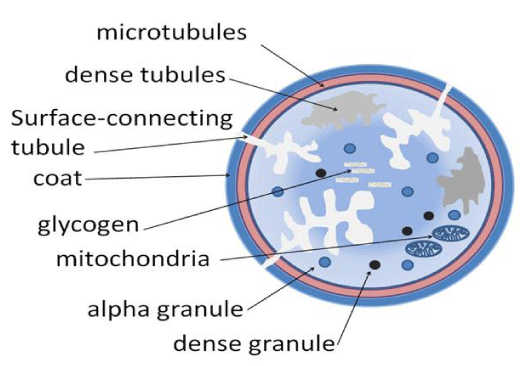
ITP is an acquired disorder that mainly affect the platelet count in the blood. Platelets are small colorless disk-shaped cell without a nucleus, it can found in great quantities in blood and are involved in clotting (Figure 1). ITP can be classified as acute, chronic or recurrent [5]. The acute form is a temporary condition, which persists for less than 3 months and affects mainly children while the chronic stage might last more than 12 months and affects adults more, the recurrent form is multiple incidents of thrombocytopenia in the period of over 3 months. Corticosteroids or I.V Immunoglobulin can treat idiopathic thrombocytopenia. ITP results from an auto-immune mechanism or from impaired thrombopoiesis. The literature shows that some ITP patients have platelet-associated autoantibodies in their blood [6]. Also, other patients have B-cells that produce antibodies attacking normal platelet antigens due to signal from CD4 T cells [7]. Another study demonstrates an increase in NK cells in patients with ITP. On the other hand, in vitro study showed suppression of megakaryopoies by autoantibodies in other ITP patients [8]. Also, the thrombopoietin, which regulates platelet production, is suggested to have an influence on the level of platelet count in blood.
Diagnosis of such cases relays mainly on the history, clinical and physical examination of the patient and Complete blood count. There might be different factors could lead to ITP. Patients at risk of HIV infection should be tested for antibodies [9].
To achieve a precise diagnosis, we should differentiate between ITP and TTP (thrombotic thrombocytopenic purpura [10] and disseminated intravascular coagulation (DIC) [11].
Case Presentation
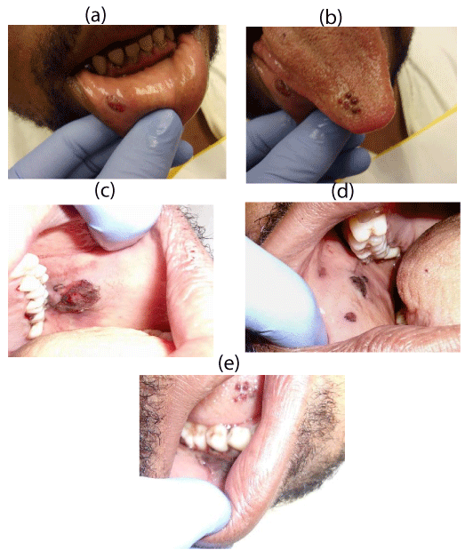
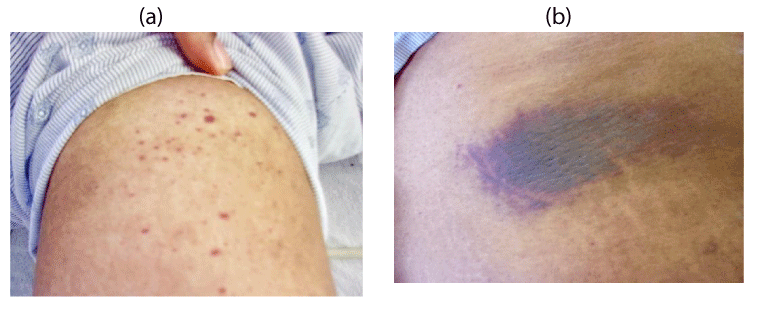
33 years old male presented in the Oral Health Department at MetroHealth Medical Center. His Chief Complain was “I can not eat and my whole mouth is hurting me’’. Patient denied past medical problems, hospitalizations and the use of any medications and no history of recent trauma. His pain started three days earlier. A comprehensive Intraoral examination was done and revealed that its related to hemorrhagic lesions in his oral cavity related to the buccal mucosa on both sides, the tip and lateral borders of the tongue, the lower lip and the attached mucosa around his teeth (Figure 2a-e). There were spots of clotted blood and other areas of active oozing blood from the gingiva. Extra-oral evaluation was done and revealed multiple petechia and ecchymosis all around his body (Figure 3a,b), which indicates a systemic cause rather than a local etiology.
The pain was severe with rating 9 out of 10 and unable to eat. Full clinical examination of these lesions revealed that they tend to bleed easily upon touch with granulation tissue all over them. They have a recent onset about a week ago which was confusing in the diagnosis. A consultation with the oral and maxillofacial surgery division was requested. Lab tests were ordered on the spot involving complete blood count, Prothrombin time and Partial prothrombin time. Tests revealed a massive drop in the platelets count, which was 2000 per microliter (normal values range from 150,000 to 400,000). The lab requested another sample to confirm the diagnosis and it was 1,000 platelet per microliter [Table 1]. The patient was admitted to the hospital to stabilize the condition. More blood samples were obtained and immediate platelet transfusion was started.
The pain was severe with rating 9 out of 10 and unable to eat. Full clinical examination of these lesions revealed that they tend to bleed easily upon touch with granulation tissue all over them. They have a recent onset about a week ago which was confusing in the diagnosis. A consultation with the oral and maxillofacial surgery division was requested. Lab tests were ordered on the spot involving complete blood count, Prothrombin time and Partial prothrombin time. Tests revealed a massive drop in the platelets count, which was 2000 per microliter (normal values range from 150,000 to 400,000). The lab requested another sample to confirm the diagnosis and it was 1,000 platelet per microliter [Table 2]. The patient was admitted to the hospital to stabilize the condition. More blood samples were obtained and immediate platelet transfusion was started.
Subsequently, the patient was diagnosed with Idiopathic thrombocytopenia purpura and type II diabetes.
The initial plan from the ED on 8/12/2016 was as follows:
Started dexamethasone HD for presumed ITP
40mg dexamethasone daily for total of 4 days
Proton pump inhibitors (PPI) and glucose control while on HD steroids. HbA1c elevated
IVIG if not responding to steroids or bleeding
Platelet transfusion only if evidence of bleeding
Continue with supportive care
Then he was admitted to the oncology medicine department for Possible ITP on 8/12/2016 and was given the following report:
ITP - unclear etiology
- Chest Xray and CT of abdomen and pelvis within normal limits.
- Hep B, Hep C and HIV serologies negative
- Steroid therapy: Dexamethasone 40 mg x 4 days
- If platelet count does not improve after 4 days or worsening bleeding; start on IVIG 400 mg/kg/day for 2-5 day
DM-II - newly diagnosed
- HbA1c: 12.2
- consult nutrition
- Start on Lantus 5 U qhs and SSI ACHS
Morbid Obesity
- Encourage lifestyle and diet modifications
- Follow up with nutrition and DM Education
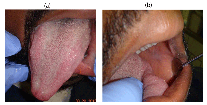
Patient returned back to the dental department after treatment on 8/29/16. Intraoral and extra oral evaluation was done. (Figure 4a,b) show complete healing of all lesions. The patient was pain free and in good spirits.
Diagnosis of such cases is made my exclusion of other causes such as Lab error, medications or drug interaction, infections like HCV or HIV, autoimmune disease or bone marrow disease like leukemia [12,13].
Management of similar cases will differ based on emergency vs chronic treatment approach. In emergencies the main goal is the prevention of bleeding and not cure [14]. This can be accomplished by raising the platelet count to a safe level. This safe level is from 30,000 to 50,000 platelet [15]. In such situations when platelet count needs to be increased rapidly, high dose methylprednisolone treatment can be used [16]. Alternative option would be blood transfusion to the patient that can help to improve the platelet count. Stabilization of the condition can be maintained with prednisone 1mg/kg/day and can be tapered off after platelet response. The duration of treatment is still controversial. Other lines of treatment can be IV immune globin 1mg/kg/day x 2days [17].
Conclusion
ITP is a common condition that can have serious complications if not treated promptly. Its diagnosis can be accomplished by the combination of clinical assessment and lab tests, which includes complete blood count, PT and PTT. It’s very important subject to dentists; they should be able to identify ITP to prevent complications that could occur. Referral of these cases to a center where a multidisciplinary team can manage this condition is also required.
- Boughton BJ, Chakraverty R, Baglin TP, Simpson A, Galvint G et al. (1988) The treatment of chronic idiopathic thrombocytopenia with anti-D (Rho) immunoglobulin: its effectiveness, safety and mechanism of action. Clin Lab Haematol 10: 275–84. Link: https://goo.gl/4qKdUj
- Shibuya A, Danya N, Shinozawa T (1994) Successful anti-D immunoglobulin therapy in refractory chronic idiopathic thrombocytopenic purpura showing reduction in thrombocytes even after splenectomy. Acta Paediatr Jpn 36: 297–300. Link: https://goo.gl/NVPs1u
- Longhurst, H. J., O’Grady, C., Evans, G., De Lord, C., Hughes, et al. (2002) Anti-d immunoglobulin treatment for thrombocytopenia associated with primary antibody deficiency. J Clin Pathol 64: 356-363. Link: https://goo.gl/G1gdSD
- Beards, G (2012) Diagram of the internal structure of a human blood platelet. Link:
- Stevens W, Koene H, Zwaginga JJ, Vreugdenhil G (2006) Chronic idiopathic thrombocytopenic purpura: present Strategy, Guidelines and new insights. Neth J Med 64: 356–363. Link: https://goo.gl/j9P8VD
- Cines DB, Bussel JB, Liebman HA, Luning Prak ET (2009) The ITP syndrome: pathogenic and clinical diversity. Blood 113: 6511-6521. Link: https://goo.gl/gFDGy7
- Audia, S., Rossato, M., Trad, M., Samson, M., Santegoets, et al. (2016) B cell depleting therapy regulates splenic and circulating T follicular helper cells in immune thrombocytopenia. Journal of Autoimmunity 11.002. Link: https://goo.gl/BbV1xJ
- Lu, J. (2013). Elevated NKT cell levels in adults with severe chronic immune thrombocytopenia. Experimental and Therapeutic Medicine. doi:10.3892/etm.2013.1386. Link: https://goo.gl/txTmSC
- Consolini, R., Legitimo, A., & Caparello, M. C. (2016). The centenary of immune Thrombocytopenia – part 1: Revising nomenclature and pathogenesis. Front Pediatr 4: 102. Link: https://goo.gl/UBF5cH
- Saifan C, Nasr R, Mehta S, Acharya PS, El-Sayegh S (2012) Thrombotic Thrombocytopenic Purpura. J Blood Disord Transfus S3:001. Link: https://goo.gl/Zl1Vo2
- Wada, H., Matsumoto, T., & Yamashita, Y (2014) Diagnosis and treatment of disseminated intravascular coagulation (DIC) according to four DIC guidelines. Journal of Intensive Care 2: 15. Link: https://goo.gl/gSJ90K
- Rodeghiero, F. (2016, September 07). Standardization of terminology, definitions and outcome criteria in immune thrombocytopenic purpura of adults and children: report from an international working group. The blood Journal 113: 2386-2393. Link: https://goo.gl/t3JZbC
- Douglas B, Cines, M.D, Victor S. Blanchette M.B, B.Chir (2002) IMMUNE THROMBOCYTOPENIC PURPURA. The New England Journal of Medicine 346: 995–1008. Link: https://goo.gl/J3fFLN
- George J N, Woolf SH, Gary ER,Jeffrey SW, Louis MA, et al. (1996). Idiopathic Thrombocytopenic Purpura: A Practice Guideline Developed by Explicit Methods for The American Society of Hematology. Blood Journal 88: 3–40. Link: https://goo.gl/oZ77kN
- Neunert C, Noroozi N, Norman G, Buchanan GR, Goy J, et al. (2015). Severe bleeding events in adults and children with primary immune thrombocytopenia: a systematic review. J Thromb Haemost, 13: 457–464. Link: https://goo.gl/AngnkF
- Akin M, Turgut S, Ayada C, Polat, Y., Balci, YL, et al. (2011) Relation between 3435C>T multidrug resistance 1 gene polymorphism with high dose methylprednisolone treatment of childhood acute idiopathic thrombocytopenic purpura. Gene 487: 80–83. doi:10.1016/j.gene.2011.06.019 Link: https://goo.gl/hpFeiM
- Fasano MB, Sullivan KE, Sarpong SB, Wood RA, Jones SM, et al. (1996) Sarcoidosis and common variable immunodeficiency: report of 8 cases and review of the literature. Medicine 75: 251–261. Link: https://goo.gl/SGTsGY
Article Alerts
Subscribe to our articles alerts and stay tuned.
 This work is licensed under a Creative Commons Attribution 4.0 International License.

 Save to Mendeley
