International Journal of Immunotherapy and Cancer Research
Titanium Mesh Reconstruction after Solitary Sternal Plasmacytoma Surgery-A Case report
Krdzalic G1*, Mujagic H2, Musanovic N1 and Krdzalic A3
2Former Scholar and Professor, Massachusetts General Hospital Cancer Center, 55 Fruit St.02114 Boston, USA
3Clinic for Cardiovascular Diseases, University Clinical Centre Tuzla, Trnovac bb, 75000 Tuzla, Bosnia and Herzegovina
Cite this as
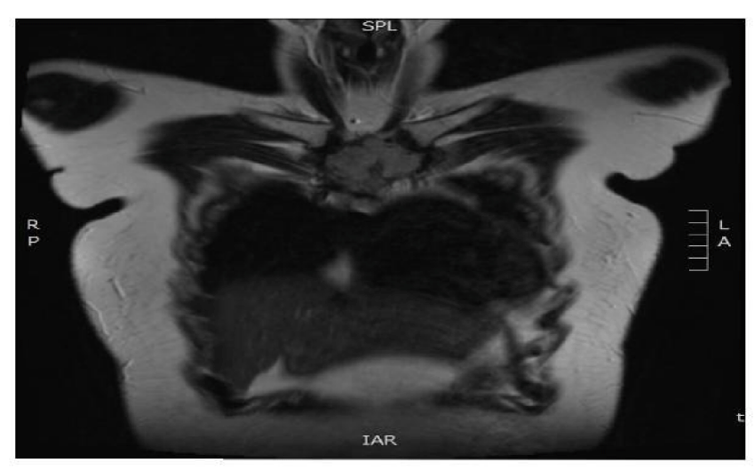
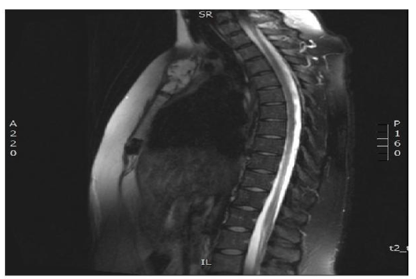
Krdzalic G, Mujagic H, Musanovic N, Krdzalic A (2017) Titanium mesh reconstruction after solitary sternal plasmacytoma surgery-A case report. Int J Immunother Cancer Res 3(1): 041-043. DOI: 10.17352/2455-8591.000018We present chest wall reconstruction with titanium mesh in a patient who underwent sternal resection due to solitary plasmacytoma (SP). A 35 year old female was admitted to The Thoracic Surgery Department of University Clinical Center Tuzla with pain and tender upper-sternal swelling. Thoracic magnetic resonance imaging (MRI) revealed hypo dense wll-shaped rounded mass involving manubrium streni which was 40mmx40mm in size measured by two right angle perpendicular diameters. Affected part was resected together with removal of sternoclavicular and costochondral junctions and reconstruction with titanium mesh was performed
Introduction
Primary sternal tumors are very rare and account for only ~ 1% of primary bone neoplasms [1]. The most common lesion is chondrosarcoma (33%) followed by mayeloma and plasmacytoma (30%), than (21%) lymphoma and occasional lesions like osteosarcoma, fobrosarcoma and Ewing sarcoma [2]. Surgical treatment requires adequate, wide margin resections and reconstruction of the anterior chest wall [3]. Resonstruction is essential for maintenance of respiratory function and for protection of mediastinal organs [4]. Here we report reconstruction of anterior chest wall with titanium mesh inserted between two polypropylene mesh sheets.
Case report
A 35-year-old female, with no significant past medical history, was admitted to our department with progressive anterior chest wall pain for the past 3 months. Physical examination revealed a palpable fixed mass involving sternal manubrium. Laboratory tests were unremarkable. MRI showed a solitary, well-shaped, hypo dense and round mass which involved manubrium sterni (40mm x 40mm in diameter) with no signs of subcutaneous invasion. A whole body computed tomography detected no metastatic deposits. The case was presented before thoracic team and it was decided to do radical resection.
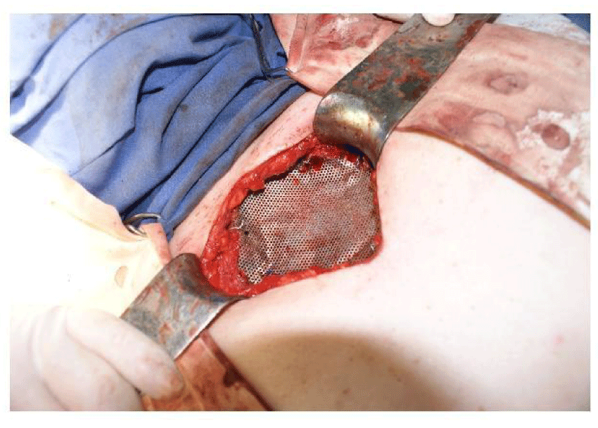
We performed wide manubrial resection including right and left sternocalvicular joints and upper sternal part. Complete specimen was sent to surgical pathology for rapid frozen section diagnosis and pathologist recommended to wait until final analysis. However, considering that all primary sternal tumors are malignant [5] we didn´t wait for final diagnosis but proceeded with resection of both clavicle sternal end and the first and the second rib cartilages. To reconstruct and stabilize anterior chest wall a titanium micro mesh (KLS Martin Germany - Ti100mm x 100mm/0.2mm thickness) was used. It was placed over polypropylene mesh to protect major vessels and pericard from direct contact with rigid titanium and fixed to the remaining edges of sternum, clavicles and ribs. Another polypropylene mesh was placed over titanium to avoid contact with subcutaneous tissue and to secure stability of thoracic wall. It was then covered with subcutaneous tissue without use of any myocutaneous flaps [Figures 1-3].
Two hours before surgery patient received Cefazolin 1g and 500mg every 8hours for 24 hours postopratively in five days. Intravenous tramadol and metamizol provided satisfactory analgesia after surgery. On 4th postoperative day, pleural drainage tube was removed, the patient recovered well and was discharged on postoperative day 7. The pathological examination revealed neoplastic plasma cell proliferation with excentric nuclei and eosinophilic cytoplasm. Immunohistochemical analysis was positive for CD138 and kappa-light chains and negative for lambda-light chains, S 100 protein, CKAE1 AE3, CK7 and EMA. The final pathological diagnosis was plasmacytoma. One year after surgery patient is doing well under hematological and surgical follow up.
Discussion
Primary tumors of the sternum are generally malignant and account for only 0.5-1% of all primary bone neoplasms. Sternal solitary plasmacytoma, a type of malignant tumor, is characterized by localized monoclonal plasma cell infiltration without evidence of multiple myeloma and represents less than 10% of all plasma cell neoplasms [6].
In an analysis of 11087 cases of bone tumors done at Mayo Clinic, only 66 (0.6%) were primary sternal. Of these 66 tumors, 22 (33%) were chondrosarcomas; 20 (30%) were myelomas including plasmacytomas; 14 (21%) lymphomas, 8 (12%) osteosarcomas, 1 (1.5%) fibrosarcoma and 1 (1.5%) Ewing tumors [2].
The most common site of SP is vertebral column. The involvement of ribs and sternum accounts for 10-15% of cases. Males are two times more frequently affected than females which makes sternal plasmacytoma being very rare. [5]. Radiological examination of sternal solitary plasmacytoma usually presents as an osteolytic expansive lesion or a typical ‘punched-out’ lytic lesion. Plasmacytoma destroys bone cortex in several places and invades soft tissues. On MR imaging, these tumors exhibit a low signal intensity on T1 and a high signal intensity on T2-weighted images [7].
Although radiotherapy is recommended for sternal SP, extensive wide resection and reconstruction of anterior chest wall is still treatment of choice [5,8,9]. The goal of wide resection is to ensure that all malignant tissue has been excised resulting in local control.
Reconstruction of strenal defect is essential to maintain original respiratory function and to protect mediastinal organs. The choice of reconstruction technique depends on the extent and localization of the defect using various prosthetic and homologous materials, including synthetic and metallic grafts, pedicled skin and muscle flaps, free skin grafts, fascia lata and autologous bone transplants [10].
Recently, titanium mesh has emerged as promising material for sternal reconstruction in cases of sternal defect larger than 5 cm without using additional autologous tissue. Due to its biocompability and flexibility titanium mesh has been acknowledged as versatile and easy to implement [11]. Polypropilene mesh is used more frequently for the reconstruction of sternal defects but its lack of rigidity may result in paradoxical chest wall motion [10]. Titanium mesh is may be complicated by infection or fragmentation of the graft, but it is more rigid and osteconductive than polypropilene mesh and mold to shape of the defect. It is easy to handle, minimally elastic, less visible on MRI than stainless steel and adapts well to surrounding soft tissues [10-12]. Our patient was extubated in the early postoperative period, and paradoxical respiration was not observed during 12 months postoperatively. In the present case the titanium mesh, embedded between two polypropylene meshes, was used for anterior chest reconstruction without use of musculocutaneous flap. The benefits for patients are simplifying the procedure, ideal rigidity and biocompability. Further advantages include minimal trauma, no infection and no change in pulmonary function [13].
In conclusion, we presented a wide manubrial resection and reconstruction with titanium mesh between two polypropylene meshes for a solitary plasmacytoma. This resection and reconstruction can be performed successfully and effectively in patients with large sternal tumors. We suggest that titanium mesh may be highly beneficial material for sternal reconstruction.
- He B, Huang Y, Li P, Xiaohe YE, Fuqiang lin, et al (2014) A rare case of primary chondrosarcoma arising from the sternum: A case report. Oncology Letters 8: 2233-2236. Link: https://goo.gl/dxSQhc
- Lee JH, Lee WS, Kim YH, Kim JD (2013) Solitary plasmacytoma of the sternum. Korean J ThoracCardiovasc Surg 46: 482-485. Link: https://goo.gl/L3sXtT
- McAfee MK, Pairolero PC, Bergstralh EJ, Piehler JM, Unni KK, et al. (1985) Chondrosarcoma of the chest wall. Factors affecting survival Ann Thorac Surg 40: 535-541. Link: https://goo.gl/ikpY92
- Beatttie EJ, Bloom ND, Harvey JC (1992) Thoracic Surgical Oncology. Churchill Livingstone. New York. Link: https://goo.gl/JrdSbW
- Dimarakis I, Thorpe J, Papagiannopoulos K (2004) Solitary Sternal Plasmacytoma: A Case Report And Current Review. The Internet Journal of Thoracic and Cardiovascular Surgery. Link: https://goo.gl/Zw9aF2
- Marcio T, Carolina F, Renata C, Sergio C, Henrique C, et al. (2016) Solitary Plasmocytoma: 19-Year Retrospective Study and Review of the Literature. Int J Hematol Therap 1: 1-5. Link: https://goo.gl/iWBTFv
- Zhao J, Li Y, Wu W, Zhang Z, Ding Y (2015) Solitary plasmacytoma of the sternum with a spiculated periosteal reaction: A case report. Oncol Lett 9: 191-194. Link: https://goo.gl/FpkdZU
- Pezzella AT, Fall SM, Pauling FW, Sadler TR (1989) Solitary plasmacytoma of the sternum: surgical resection with long-term follow-up. Ann Thorac Surg 48: 859–862. Link: https://goo.gl/AZyuLZ
- García Franco CE, Jiménez Hiscock L, Zapatero Gaviria J (2004) Solitary plasmacytoma in a rib.Arch Bronconeumol. 40: 100. Link: https://goo.gl/oSR2Fs
- Koto K, Sakabe T, Horie N, Ryu K, Murata H, et al. (2012) Chondrosarcoma from the sternum: reconstruction with titanium mesh and a transverse rectus abdominis myocutaneous flap after subtotal sternal excision. Med Sci Monit 18: 77-81. Link: https://goo.gl/JPxYUW
- Rong G, Kang H (2013) Local recurrence involving the sternum and ribs following mastectomy and titanium mesh implants for chest wall reconstruction: A case report. Onco lett 5: 1649-1652. Link: https://goo.gl/Vi9XwQ
- Hirai S, Nobuto H, Yokota K, Matsuura Y, Uegami S, et al. (2008) Surgical resection and reconstruction for primary malignant sternal tumor.Ann Thorac Surg 85: 1447-1448. Link: https://goo.gl/3i9m9S
- Ersöz E, Evman S, Alpay L, Akyıl M, Vayvada M, et al. (2014) Chondrosarcoma of the anterior chest wall: surgical resection and reconstruction with titanium mesh. J Thorac Dis 6: E230-E233. Link: https://goo.gl/R8amDE
Article Alerts
Subscribe to our articles alerts and stay tuned.
 This work is licensed under a Creative Commons Attribution 4.0 International License.


 Save to Mendeley
