International Journal of Dermatology and Clinical Research
Isolated bilateral facial palsy due to chicken pox- An unique presentation
Nandini Chatterjee1 and Chandan Chatterjee2*
2Associate Professor, Department of Pharmacology, ESIC Medical College, Joka, Diamond Harbor Road, Joka, Kolkata, India
Cite this as
Chatterjee N, Chatterjee C (2019) Isolated bilateral facial palsy due to chicken pox- An unique presentation. Int J Dermatol Clin Res 5(1): 001-001. DOI: 10.17352/2455-8605.000029A nineteen year old girl presented with lesions of chicken pox and inability to close both eyes properly for two days. She had difficulty in eating and smiling. Vesicular non pruritic rashes were visible in various stages of healing over face and body except palm and soles. She complained of bilateral headaches but no vomiting, or weakness in her limbs or alteration of her taste sensation. She was alert and cooperative with normal speech and memory. B/L lower motor neuron type of facial nerve palsy was found. Routine blood work up, serology for HIV and Lyme disease, chest x ray, CSF study and MRI brain were normal.
She was prescribed 60 mg of prednisolone and 4000 mg of acyclovir every day for 14days. She recovered completely from the neurodeficit within 14 days.
Case Report
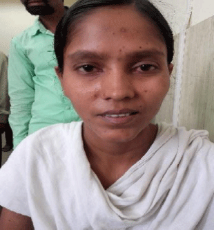
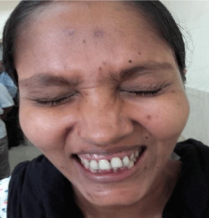
A nineteen year old girl presented with lesions of chicken pox and inability to close both eyes properly for two days. She had difficulty in eating and smiling. On examination, there was no anemia, cyanosis, jaundice, edema or lymphadenopathy. Vesicular non pruritic lesions were visible in various stages of healing over face and body except palm and soles. She complained of bilateral headaches but no vomiting, or weakness in her limbs and no alteration of her taste sensation. She was not on any medications and had no prior illness apart from the onset of chicken pox 7 days ago. Her vital signs were normal.
The head, ears, nose, and throat were all normal. The neck was supple, with a midline trachea. She was alert and cooperative with normal speech and memory.
B/L lower motor neuron type of facial nerve palsy examination of the other systems was normal. Routine blood work up, serology for HIV and Lyme disease, chest x ray, CSF study and MRI brain were performed which were normal.
She was prescribed 60 mg of prednisone and 4000 mg of acyclovir every day for 7 days. However, she was asked to present in next outdoor visit and all medications were continued for 7 more days. She recovered completely from the neuro deficit within 14 days (Figures 1-4).
Discussion
Isolated bilateral facial nerve is a rare neurological complication associated with varicella infection. Unilateral involvement reported only in 0.01-0.03% of the infections. Bilateral facial nerve palsy is a rare condition and hence presents a diagnostic challenge. Unlike the unilateral presentation, it is seldom secondary to Bell’s palsy. The majority of patients with bilateral facial palsy have Guillain-Barre Syndrome (GBS), multiple idiopathic cranial neuropathies, Lyme disease, sarcoidosis, meningitis (neoplastic or infectious), brain stem encephalitis, benign intracranial hypertension, leukemia, Melkersson-Rosenthal syndrome (a rare neurological disorder characterized by facial palsy, granulomatous cheilitis, and fissured tongue), diabetes mellitus, human immunodeficiency virus (HIV) infection, syphilis, infectious mononucleosis, malformations as Mobius Syndrome, vasculitis, or bilateral neurofibromas. The possibility of intrapontine and prepontine tumor should also be considered [1-3]. Thus, it should be carefully investigated before establishing the diagnosis of Bell idiopathic palsy [4].
The most common infectious cause of bilateral FNP is Lyme disease, caused by spirochete Borrelia burgdorferi, whose carrier is a common tick [5]. Bilateral FNP can be seen in about 30–35% of patients with Lyme disease. Diagnosis is serologic, and IgM antibodies increase in the second week and tend to decrease with treatment, while IgG antibodies appear late with reaching its peak in the second or third month, and it can indefinitely remain positive [6,7]. Common ticks are found in northern Himalayan regions of India. Therefore tick borne lyme disease is not very uncommon supported by two articles published in two reputed journals of Dermatology [8,9].
Bilateral palsies usually reflect an underlying systemic pathology whereas unilateral peripheral facial palsies are usually idiopathic (presumably virally related). This one is a rare exception of this hypothesis where a patient with viral infection (chicken pox) presented with bilateral facial palsy in the absence of GBS or meningitis.
- Stahl N, Ferit T (1989) Recurrent bilateral peripheral facial palsy. J Laryngol Otol. 103: 117-119. Link: https://goo.gl/xt2ALq
- McIntosh WE, Brenner JF, Aschenbrenner JE (1987) Bilateral facial paralysis as the sole presenting feature of sarcoidosis: report of a case. J Am Osteopath Assoc. 87: 245-247. Link: https://goo.gl/si9YkW
- Haydar A, Hujairi NM, Tawil A (2003) Bilateral facial paralysis: what's the cause? Med J Am 179: 553. Link: https://goo.gl/W6CfhN
- Hoitsma E, Faber CG, Drent M (2004) Neurosarcoidosis: a clinical dilemma. Lancet Neurol 3: 397-407. Link: https://goo.gl/DnGpcf
- Zajicek JP, Scolding NJ, Foster O (1999) Central nervous system sarcoidosis—diagnosis and management. Q J Med 92: 103-117. Link: https://goo.gl/nRkYSz
- Steenerson RL (1986) Bilateral facial paralysis. Am J Otol. 7: 99-103. Link: https://goo.gl/B5cnkN
- Gevers G, Lemkens P (2003) Bilateral simultaneous facial paralysis—differential diagnosis and treatment options. Acta Otorhinolaryngologica Belg. 57: 139-146. Link: https://goo.gl/sMDQud
- Jairath V, Sehrawat M, Jindal N, Jain VK, Aggarwal P (2014) Lyme disease in Haryana, India.IJDVL 80: 320-323. Link: https://goo.gl/44HhTx
- Sharma A, Guleria S, Sharma R, Sharma A (2017). Lyme disease: A case report with typical and atypical lesions. IDOJ 8: 124-127. Link: https://goo.gl/wWR7na
Article Alerts
Subscribe to our articles alerts and stay tuned.
 This work is licensed under a Creative Commons Attribution 4.0 International License.


 Save to Mendeley
