Imaging Journal of Clinical and Medical Sciences
Myocarditis in arrhythmogenic right ventricular dysplasia. A casual finding or a part of the disease pathophysiology?
Ana Margarida Barrigó Fernandes Bernardo1*, Ana Rita Santos2, Bruno Piçarra2, João Pais2, Mafalda Carrington2 and José Aguiar2
2Department of Cardiology, Hospital Espírito Santo de Évora, Portugal
Cite this as
Fernandes Bernardo AMB, Santos AR, Piçarra B, Pais J, Carrington M, et al. (2020) Myocarditis in arrhythmogenic right ventricular dysplasia. A casual finding or a part of the disease pathophysiology? Imaging J Clin Medical Sci 7(1): 001-002. DOI: 10.17352/2455-8702.000130Myocarditis in Arrhythmogenic Right Ventricular Dysplasia (ARVD) is characterized massive inflammatory cell infiltrates founded after acute myocardial necrosis in the early stages of the disease. This inflammatory response didn’t mean necessarily an infective process and could lead to changes in phenotype and to an abrupt progression of the disease.
Arrhythmogenic dysplasia of the right ventricle is the most frequent in young individuals and may have different clinical presentations: From asymptomatic condition to sudden death. Its diagnosis is based on well-defined clinical and imaging criteria in 2010 “Diagnosis of arrythmogenic right ventricular cardiomyopathy dysplasia: Proposed modification of the Task Force criteria”.
The major Cardiac Magnetic Resonance (CMR) criteria for ARVD are:
Regional right ventricle akinesia on dyskinesia or dyssynchronous RV contraction and one of the following:
- Ratio of RV end-diastolic volume to BSA ≥110mL/m2 (male) or ≥100mL/m2 (female); OR
- RV ejection fraction ≤40%.
The authors present the images of two patients with simultaneously myocarditis and major CMR criteria for ARVD.
Case 1
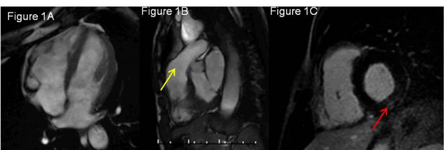
52 y/o male patient without cardiovascular risk factors that was admitted in the Emergency department (ER) with palpitations and syncope. The Electrocardiogram (EKG) showed a wide-QRS tachycardia with 220 bpm. In the etiologic evaluation, he performed a CMR that revealed a dilated Right Ventricular (RV) high with an end-diastolic volume of 152mL/m2 (Figure 1A), RV dysfunction with Ejection Fraction (RVEF) of 27%; regional wall motion abnormalities with dyskinesia of RV infundibulum (Figure 1B) and subepicardial late enhancement in the basal inferolateral segment of the left ventricular (Figure 1C) suggestive of myocarditis.
A genetic test for right ventricular dysplasia was performed, which was negative.
Case 2
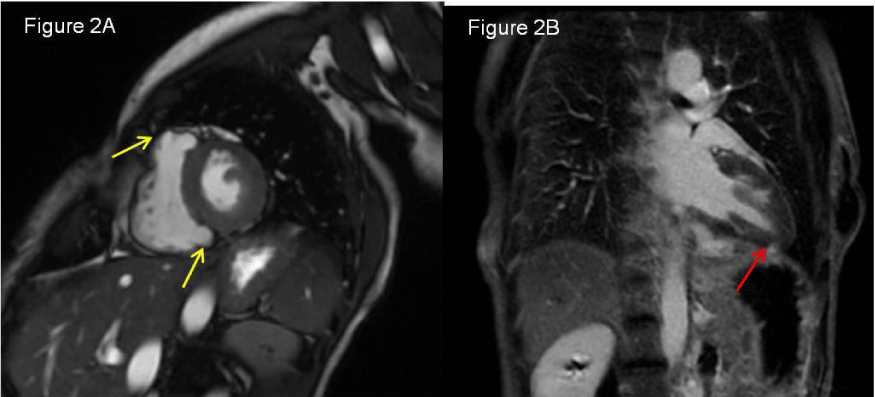
39 y/o female patient with dyslipidemia admitted with chest pain and dyspnea. The electrocardiogram showed sinus rhythm and negative T waves in leads V1-V4. The coronary angiography showed normal coronaries. A CMR was performed and revealed a high normal RV volume with an end-diastolic volume of 93mL/m2, RV dysfunction with Ejection Fraction (RVEF) of 30%; small aneurysms in the RV outflow tract (Figure 2A) and subepicardial late enhancement in the apical inferior segment of the left ventricular (Figure 2B) suggestive of myocarditis.
A genetic test for right ventricular dysplasia was performed, which was negative.
Article Alerts
Subscribe to our articles alerts and stay tuned.
 This work is licensed under a Creative Commons Attribution 4.0 International License.


 Save to Mendeley
