Imaging Journal of Clinical and Medical Sciences
Invasive yeast infection in patient with Acute pancreatitis
Jana Simonova1, Lukas Cuchrac1, Jozef Firment1, Viktoria Takacova2, Ladislav Vasko3 and Janka Vaskova3*
2Department of Medical and Clinical Microbiology, Faculty of Medicine, Pavol Jozef Šafárik University in Košice, Tr SNP 1, 040 66 Košice, Slovak Republic
3Department of Medical and Clinical Biochemistry, Faculty of Medicine, Pavol Jozef Šafárik University in Košice, Tr SNP 1, 040 66 Košice, Slovak Republic
Cite this as
Simonova J, Cuchrac L, Firment J, Takacova V, Vasko L, et al. (2017) Invasive yeast infection in patient with Acute pancreatitis. Imaging J Clin Medical Sci 4(1): 006-010. DOI: 10.17352/2455-8702.000034The incidence of invasive yeast infections is rising in patients hospitalised in intensive care. Their early diagnosis is problematic, although predictive models (Pitt’s colonization index, Leon score and others) can be helpful when deciding to initiate treatment. The incidence of candidiasis significantly increases the mortality of patients, although the timely commencement of antimycotic therapy is a key factor in increasing their survival. In the present patient, antibiotic treatment with carbapenem was initiated after CT verification of necrosis with formation of abscesses and the onset of inflammatory parameters. After surgical review, septic shock developed with multi-organ failure. Microbiological examinations of biological materials also revealed non-C. albicans yeast was present in haemocultures, so echinocandin and anidulafungin were included in the treatment. C. parapsilosis persisted in haemocultures, while negative haemoculture was obtained 14 days after the inclusion of liposomal amphotericin B.
Abbrevations
ECIL: The European Conference on Infections in Leukaemia; ERCP: Endoscopic Retrograde Cholangiopancreatogram; ICU: Intensive Care Unit; MPR: Multiplanar Reconstruction; IDSA: The Infectious Diseases Society of America; MIC: Minimal Inhibitory Concentration; CVC: Central Venous Catheter
Introduction
Invasive yeast infections are serious systemic infections associated with high mortality. In large faculty hospitals, candidiasis is the fourth most common infection in the blood stream. The incidence of invasive yeast infections in hematology departments is decreasing, and candidiasis is currently a problem of intensive medicine where its incidence is either remaining stable or even rising. Data from Slovakia confirm an increase in the incidence of candidiasis, with the current incidence at 2.16/100,000 habitants per year. Compared to the last published national epidemiological study, the ratio of C. albicans and non- C. albicans (non-C) candidiasis has also changed in favour of non-C. albicans strains of yeast. The greatest increase was recorded for C. parapsilosis and C. glabrata [1].
Acute necrotising pancreatitis is a life-threatening illness and proper timing of antimicrobial treatment is one of the key therapeutic approaches used to treat it. Antimycotic treatment is often part of this treatment, as these patients are at high risk of developing an invasive yeast infection. According to ECIL-6 recommendations, echinocandins and liposomal amphotericin B are the first-line drugs in the initial and first-line treatment of candidiasis in both neutropenic and non-neutropenic patients.
Case Report
A 31-year-old female patient without significant history of illness was admitted to the Intensive Care Unit (ICU) of 1st Surgical Clinic at the L. Pasteur University Hospital in Košice in July due to her deteriorating clinical condition in CT-verified biliar acute pancreatitis. She rejected the proposed cholecystectomy. After conservative treatment and improvement of clinical conditions, she was transferred to the department in Bardejov. Abdominal CTs (immediately after admission and 2 month later) were provided. 6 days after the second CT, the patient was re-admitted to the ICU of 1st Surgical Clinic due to the deterioration of clinical conditions and elevation of inflammatory parameters. She was conscious, orientated, cardio-pulmonally compensated, the belly was raised, more difficult to palpate, palpably sensitive in the epigastrium, no symptoms of peritoneal irritation, and peristalsis was audible. Increased inflammatory parameters were found - CRP 155.8 mg/l, PCT 0.10 μg/l. The serum amylase value was 44.30 ukat/l, and serum lipase was 34.10 ukat/l. A CT scan of the pancreas with MPR reconstructions showed entire pancreas destruction by multiple large necrotising colitic pathological sites. These changes exhibit as manifestations of extensive necrotising pancreatitis with the formation of abscess lesions. There were bulky fluid deposits localised peripancreatically and a progression of fluid deposits throughout the abdominal cavity. Liquid deposits in both pleural spaces were presented, although the patient rejected the proposed surgical revision.
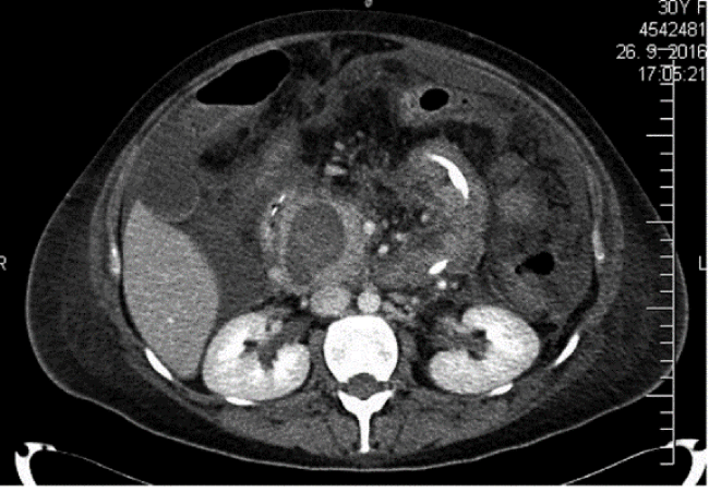
The conservative treatment continued with administration of antibiotics 6 g/24 h, parenteral nutrition, endoscopically introduced three-lumen probe for enteral nutrition into the jejunum, vitamin therapy, and analgesic spasmolytic infusions. The patient’s clinical condition deteriorated, and inflammatory parameters increased (CRP 313.23 mg/l, PCT 0.36 μg/l, leukocytes 12.11 x 10⁹/l). Control CT of abdomen verified a necrotising, abscessing form of pancreatitis with further increase in free fluid in the abdominal cavity, and abscesses in the peripancreatic and left subphrenic space (Figure 1).
Immediately after receiving patient approval on the 19th day, surgical intervention was carried out - laparotomy, evacuation of 4000 ml ascites, cholecystectomy, necrectomy, evacuation of parapancreatic abscesses, and adhesiolysis. Six insulating abdominal drains and a drainage tube were introduced into the bursa omentalis. The patient was significantly haemodynamically unstable during surgery, and was admitted to the 1st Clinic of Anesthesiology and Intensive Medicine, L. Pasteur University Hospital after surgery. The patient was continuously administrated analgesics, and kept on artificial-lung ventilation support. Circulation was supported by a high dose of noradrenaline. Blood was collected for culture examination in BD BactecTM haemocultic media (Plus Aerobic/F, Plus Anaerobic/F, Mycosis IS/F). The 6-day duration of meropenem treatment resulted in the initial elevation of inflammatory parameters. For this reason, initial antibiotic treatment was adjusted to take into account the sensitivity of Pseudomonas aeruginosa isolated from the endotracheal canal to treatment with piperacillin/tazobactam 4.5 g/6 hrs administered in prolonged 3 hour infusions, linezolid 600 mg/12 hrs administered via continuous i.v. infusion, and metronidazol 500 mg/8 hrs administered intravenously. The antimycotic, fluconazole, was then added to the treatment at 800 mg/day. In order to promote cellular and humoral immunity, Polyoxidonium 6 mg s.c. and Flebogamma 5 g were added for 3 consecutive days. Due to the documented fluidothorax on the left side, a chest drain was introduced. The abdominal cavity was flushed with saline and iodine disinfectant. Repeated haemosubstitutive treatment was administered by transfusions of erythrocytes and frozen human plasma.
On the second postoperative day, a epidural catheter was placed in the chest area for continuous epidural analgesia with Breivik mixture. After haemodynamic stabilisation of the patient and reduction in the dose of continuously administered noradrenaline, we continued enteral nutrition with a nasojejunal probe in combination with supplementary parenteral nutrition. The patient was extubated, but continued on non-invasive ventilation with a facial mask applying higher positive end-expiratory pressure values. The patient no longer required catecholamine support, remained consciousness, breathed spontaneously, did not report pain, and continued antibiotic therapy, with combined enteral and parenteral nutrition. High values of inflammatory parameters persisted, we repeatedly registered shivers with a body temperature above 38.5 °C, and leucopenia developed - leukocytes were under 4x10⁹/l for several days. The first results of the cultivation (Table 1), were obtained. Due to the fact that the patient was at high risk for an invasive yeast infection, standard dosing of echinocandin-anidulafungin was included in the treatment. All invasive inputs were replaced to prevent the possible presence of biofilms. An ocular examination was performed with negative results. Two days later, a definitive yeast identification was obtained using the MALDI Bio Typer system with a conclusion of C. parapsilosis from the blood and from the fixed abdominal drain No. 3. C. albicans 105/1 ml was identified from urine. The results of MIC tests for the tested antimycotics are summarized in table 2.
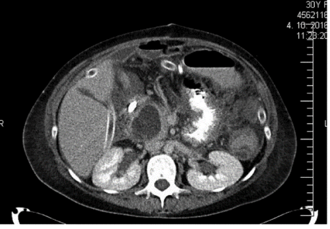
The patient’s clinical condition improved after treatment with anidulafungin, with inflammatory parameters showing a decreasing tendency. On the 26th day, a CT scan of the abdomen verified the development of abscess formation and peripancreatically localised pseudocysts (Figure 2). On the same day, laparotomy, evacuation of the pseudocysts and abscesses located subphrenally to the right and left, and evacuation of intercostal abscesses was performed. In the culture examinations, multi-resistant gram-negative enterobacteria prevailed, along with gram-positive bacteria, especially Enterococcus faecium. Antibiotic treatment was adjusted accordingly. The patient was conscious, spontaneously breathing, and the left chest drain with 955 ml of serosanguinolent content was taken out. Despite treatment with echinocandin, C. parapsilosis was repeatedly detected in both tracheal and haemocultures. Although the patient was effectively treated with anidulafungin (repetaed MIC value 0.380 μg/ml), haemodynamically stabilised, with adjusted leukopenia, the high inflammatory parameters persisted. Therefore, on 34th day, according to the IDSA guidelines, a lipid form of amphotericin B was included in the treatment. After its inclusion at a dose of 400 mg i.v. daily, together with targeted antibacterial therapy, a gradual decrease in inflammatory parameters was registered. Lipid treatment with amphotericin B lasted for a total of 29 days.
On the 41st day, leukocyte values were 9.26x10⁹/l, CRP 24 mg/l, PCT 0.121 μg/l and IL-6 20.6 ng/l. Renal parameters and mineralogy were monitored daily. The serum values of urea, creatinine, and potassium are presented in table 3. The patient was transferred to the Intensive Care Unit of 1st Surgical Clinic. The first negative haemoculture from blood collection was obtained on the 48th day. The patient was enterally nourished, intensively rehabilitated, and all invasive inputs were cancelled.
The patient’s temperature, and an increase in serum amylase values, were measured again 47 days after detection of a negative haemoculture. CT of the abdomen was performed verifying the paracolic abscess on the right. The following day, the patient was re-examined and a drain was introduced into the abscess. Abscess cavity material was positive for Pseudomonas aeruginosa, without the presence of yeasts. Gradually, there was a decrease in pus, but the safety drain was left, draining into a stomic bag. The surgical wound was healed secondary, and local wet therapy was applied. The patient received a pancreatic diet per os, and prescribed home enteral oral nutrition along with pancreatic enzymes. The patient was released home 10 days after re-examination. The dehiscent surgical wound was treated in a surgical clinic at regular intervals; the drain was discontinued. One month later, the patient was re-admitted to the 1st Surgical Clinic with continued resection over the middle portion of the surgical wound forming a pancreatic-cutaneous fistula with extensive phlegmous skin. Upon second hospitalisation, values of leukocytes 8.49x10⁹/l, CRP 28.76 mg/l, albumin 42.3 g/l, serum amylase 1.35 ukat/l were detected. A high amylase content (2916.9 ukat/l) was confirmed in secretion. An ERCP examination was carried out, which indicated a leak in the ductus pancreaticus major. It was not possible to introduce the stent into the leaking area. After endoscopic surgery, the necessity for pancreaticocutaneous fistula surgery was not indicated. After 10 days of hospitalisation, the patient was released home. Currently, the patient is at home, regularly monitored by the surgeon. During the check-up after 10 days, the belly was freely throbbing, the surgical wound was dehiscent, locally treated, with persistent section of pancreatic juice in the area of operative wound, and stomic sack adhesion.
Five months later, the patient was re-admitted to the 1st Surgical Clinic for dyspeptic disorders caused by pancreatic pseudocysts, as verified by CT. The pseudocystogastroanastomosis was performed according to Jurasza. After surgery, the patient was fully loaded with food, and the presence of the yeast was not confirmed in the extracted biological materials from the abdominal cavity. The patient was released home 14 days after admission.
Discussion
Acute necrotising pancreatitis is a life-threatening disease often complicated by the development of septic shock with multi-organ failure. Patients are often colonised by multi-resistant bacteria, but also primary sterile necrosis is infected. Preventive administration of antibiotics to prevent pancreatic necrosis infection is not recommended [2]. In the present patient, antibiotic treatment with carbapenem was initiated after a CT verification of necrosis with formation of abscesses in the onset of inflammatory parameters. After a surgical review, septic shock with multi-organ failure (circulating, GIT, kidney, breathing, coagulation) developed. All available biological materials were collected for microbiological examinations during the surgical procedure, including blood collection into BD BactecTM haemocultic media. We continued with broad-spectrum antibiotic therapy, artificial lung ventilation, complex treatment of septic shock, and support for vital functions.
The decision to include antimycotics in the treatment was based on the outcome of various scoring systems. According to [3], a patient at high-risk of invasive yeast infection is considered to be one undergoing broad-spectrum antibiotic therapy, a central venous catheter (CVC), parenteral nutrition, a large surgical procedure linked to UPV, haemodialysis, a stay longer than 3 days in ICU, treatment with corticosteroids or immunosuppressive therapy, and pancreatitis. León’s so-called Candida scores include a score based on 4 factors. If a patient reaches > 2.5 points, it is recommended to start antimycotic treatment. Our patient fulfilled many of these criteria - she was hospitalised for 3 weeks at ICU, underwent extensive surgery for severe necrotising pancreatitis, had a CVC, was in septic shock, and had undergone a procedure linked to UPV. Studies investigating the effect of pre-emptive antimycotic treatment strategy in surgical patients for intra-abdominal infection in both community and hospital settings have also been published. However, they have not confirmed the clear efficacy of pre-emptive echinocandins, but concluded that patients with nosocomial intra-abdominal infections were closest to a significant difference in favour of the pre-treated group of micafungin [4].
Candidiasis is a manifestation of invasive yeast infection. The presence of yeast in the blood is never considered a possible contamination of the sample, there is always a need to look for the source from which the yeast was washed into the blood. Clinical signs of invasive yeast infection are non-specific and do not differ from symptoms of bacterial infections. In the described patient, we recorded the temperature outputs with adequate antibiotic treatment covering the spectrum of diagnosed bacteria. The relationship between hospital mortality and the delay in antimycotic therapy intervention has been demonstrated by several authors [5,6]. Although, C. albicans is the most common yeast isolate presented in haemocultures, the non-C ratio has increased in recent years, especially C. glabrata and C. parapsilosis [7]. In Luis Pasteur University hospital, among the 101 investigated samples of biological material taken from primary sterile sites between January 2013 and June 2015, C. albicans were identified in most patients from ICU (48 cases, 48.6%). C. parapsilosis was identified in 8 samples (7.9%) and was the third most commonly identified non-C albicans yeast after C. glabrata and C. krusei [8]. Upon notification by microbiologists that non-C. albicans yeast was present in haemocultures, echinocandin was included in the treatment. Anidulafungin was chosen for empirical treatment due to its broad spectrum fungicidal effect on isolates of the non-C. albicans yeast, resulting in minimal pharmacological interactions, linear pharmacokinetics, high volume of distribution, minimal toxicity, degradation and biofilm transfer. Echinocandins (AII) are indicated for the treatment of candidiasis in non-neutropenic patients according to ESCMID Guidelines 2012 and ECIL-6 recommendations [9]. The IDSA guidelines from 2016 equally recommend echinocandins or liposomal amphotericin B [6]. CVC extraction is an equally important factor for reducing the mortality of non-neutropenic patients with candidiasis [6,10]. The recommended duration of antimycotic treatment for a documented candidate is 14 days after the first negative haemoculture, in case of possible metastatic spread of the candida infection. Haemocultures should be taken every day or at least every other day. Minimum inhibitory concentrations (MIC) for C. parapsilosis are higher for echinocandins than for other yeasts [7,11].
Several authors report the high sensitivity of C. parapsilosis to fluconazole (91-96%) and voriconazole (95-98%) [12-14]. The anidulafungin MIC value for C. parapsilosis was 0.380 μg/ml in the female patient, fluconazole MIC 2 μg / mL. Despite the administration of anidulafungin at recommended doses and the improvement in the patient’s clinical status, C. parapsilosis was repeatedly present in haemocultures even after 14 days of treatment. For this reason, liposomal amphotericin B was introduced into the treatment regimen at a dose of 5 mg/kg/day. After the inclusion of liposomal amphotericin B, the first negative haemoculture was obtained after 14 days of use. As there was no confirmed metastatic spread of the infection by the available examinations, the treatment was deemed to be successful and ended. Treatment with the lipid form of amphotericin B is considered to be effective and safe while adhering to recommendations for its application. Through sufficient hydration, adequate perfusion blood pressure and excluding other nephrotoxic substances, the risk of nephrotoxicity may be significantly reduced.
Conclusion
This case report points to the need for a comprehensive and multidisciplinary approach in the treatment of a patient with invasive intra-abdominal infection. Surgical treatment of the source of infection and subsequent intensive support of vital functions is a key condition for successful treatment. A vital aspect of treatment is also the rapid identification of the pathogen and the early initiation of effective antimicrobial therapy. Antimycotic treatment is often a part of this therapy. Further study is required to assess the unique role of antimycotics in preventive and early-term treatment strategies in high-risk non-neutropenic patients hospitalized on ICU.
- Drgona (2013) Infectious complications of oncological patients - selected chapters (in Slovak). Commenius University in Bratislava, Slovakia, 24.
- Tenner S, Baillie J, DeWitt J, Vege SS, American College of Gastroenterology (2013) American College of Gastroenterology guideline: management of acute pancreatitis. Am J Gastroenterol 108: 1400-1415. Link: https://goo.gl/tjfnyb
- Ostrosky-Zeichner L, Shohan S, Vazquez J, Reboli A, Betts R, et al. (2014) MSG-01: A randomized, double-blind, placebo-controlled trial of caspofungin prophylaxis followed by preemptive therapy for invasive candidiasis in high-risk adults in the critical care setting. Clin Infect Dis 58: 1219-1226. Link: https://goo.gl/ipQEbN
- Knitsch W, Vincent JL, Utzolino S, Francois B, Dinva T, et al. (2015) A randomized, placebo-controlled trial of preemptive antifungal therapy for the prevention of invasive candidiasis following gastrointestinl surgery for intra-abdominal infections. Clin Infect Dis 61: 1671-1678. Link: https://goo.gl/fYBxb7
- Rajendran R, Sherry L, Deshpande A, Johnson EM, Hanson MF, et al. (2016) A prospective surveillance study of candidaemia: epidemiology, risk factors, antifungal treatment and outcome in hospitalized patients. Front Microbiol 7: 915. Link: https://goo.gl/yT14Ua
- Pappas PG, Kauffman CA, Andes DR, Clancy CJ, Marr KA, et al. (2016) Clinical practice guideline for the management of candidiasis: 2016 update by the Infectious Diseases Society of America. Clin Infect Dis 62: e1-15. Link: https://goo.gl/D1RNhx
- Kauffman CA (2016) Treatment of candidaemia and invasive candidiasis in adults. UpToDate®, Edited by Marr KA, Wolters Cluver. Link: https://goo.gl/kMwvtn
- Hrabovský V, Taká?ová V, Schréterová E, Pastvová L, Hrabovská Z, et al. (2017) Distribution and antifungal susceptibility of yeasts isolates from intensive care unit patients. Folia Microbiol (Praha). Link: https://goo.gl/y4FqvB
- Tissot F, Agrawal S, Pagano L, Petrikkos G, Groll AH, et al. (2016) ECIL-6 guidelines for the treatment of invasive candidiasis, aspergillosis and mucormycosis in leukemia and hematopoietic stem cell transplant patients. Haematologica 102: 433-444. Link: https://goo.gl/ejCrrU
- Andes DR, Safdar N, Baddley JW, Playford G, Reboli AC, et al. (2012) Impact of treatment strategy on outcomes in patients with candidemia and other forms of invasive candidiasis: a patient-level quantitative review of randomized trials. Clin Infect Dis 54: 1110-1122. Link: https://goo.gl/ZhKh9k
- Reboli AC (2014) Editorial commentary: Is the debate about treatment of Candida parapsilosis complex infections with echinocandins much ado about nothing? Clin Infect Dis 58: 1422-1423. Link: https://goo.gl/Z87nEb
- Cleveland AA, Farley MM, Harrison LH, Stein B, Hollick R, et al. (2012) Changes in incidence and antifungal drug resistance in candidemia: results from population-based laboratory surveillance in Atlanta and Baltimore, 2008-2011. Clin Infect Dis 55: 1352-1361. Link: https://goo.gl/dZ8nSP
- Pfaller MA, Moet GJ, Messer SA, Jones RN, Castanheira M (2011) Candida bloodstream infections: comparison of species distributions and antifungal resistance patterns in community-onset and nosocomial isolates in the SENTRY Antimicrobial Surveillance Program, 2008-2009. Antimicrob Agents Chemother 55: 561-566. Link: https://goo.gl/4VKqdw
- Posteraro B, Spanu T, Fiori B, De Maio F,De Carolis E, et al. (2015) Antifungaal susceptibility profiles of bloodstream yeast isolates by Sensititre YeastOne over nine years at a large Italian teaching hospital. Antimicrob Agents Chemother 59: 3944-3955. Link: https://goo.gl/dqkJw7
Article Alerts
Subscribe to our articles alerts and stay tuned.
 This work is licensed under a Creative Commons Attribution 4.0 International License.


 Save to Mendeley
