Imaging Journal of Clinical and Medical Sciences
Neuroimage of the Subventricular Zone Associated with Intrauterine Ischemic Infarction
Ramos-Zuñiga R*, Rochin-Mozqueda JA and García-Mercado CJ
Cite this as
Ramos-Zuñiga R, Rochin-Mozqueda JA, García-Mercado CJ (2016) Neuroimage of the Subventricular Zone Associated with Intrauterine Ischemic Infarction. Imaging J Clin Med Sciences 3(1): 012-013. DOI: 10.17352/2455-8702.000028Introduction: Recent studies that characterize the subventricular zone (SVZ) have identified the role that it represents for neurogenesis, neuromigration processes and the formation of cortical neural networks. There are two neurogenic niches in the adult human brain, the SVZ and the dentate gyrus of the hippocampus, where neural stem cells have a significant potential of differentiation to become glial cells and neurons. Alterations resulting from structural lesions affecting the human SVZ may represent neurodevelopment disorders.
Methods: We present a case about a 5 year-old male child that had an intrauterine stroke, presenting hypoactivity and secondarily generalized partial seizures at birth. The nuclear magnetic resonance imaging demonstrates a delimitated area between the encephalomalacia area and the ex-vacuo ventricular dilatation that corresponds to the ventricular wall, which highlights an important transitional component of the subventricular zone.
Result: The present case contributes to the evidence of the failure in cortical formation when the precise migrating mechanism from the SVZ is not correctly developed.
Conclusion: New technologies will allow to set up anatomo-functional correlations of the SVZ and to establish potential links with neurodevelopmental pathologies associated with a primary lesion
Abbrevations
SVZ: Subventricular Zone
Clinical image
The patient is a 5 year-old male child, product of the second gestation, in a 36 year old mother without comorbidities. During pregnancy, risk of abortion was presented since the third month of gestation and was managed with obstetrical care and rest. The product was obtained by vaginal birth at 40 weeks of gestation, presenting hypoactivity and secondarily generalized partial seizures. It was decided to initiate treatment with phenytoin. During his neurological evolution, he presented backwardness in the psychomotor development, right hemiparesia that progressed to spasticity (spastic hemiparesia), language and communication limitation, subsequently he recurred with convulsive crisis. Currently the patient is treated with levetiracetam and clonazepam, presenting appropriate control.
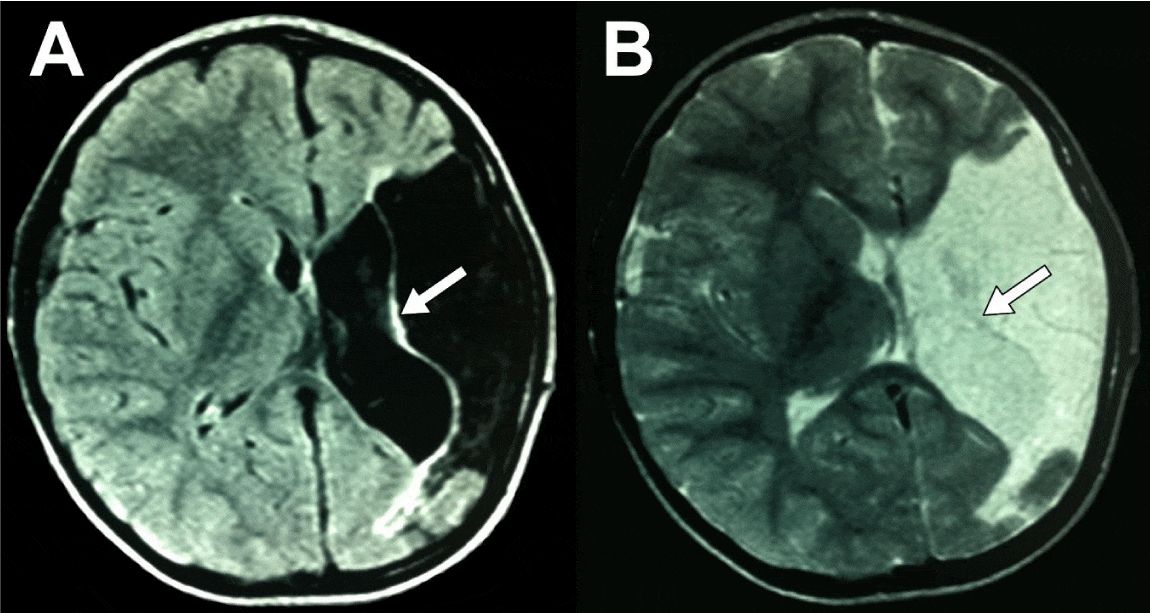
The nuclear magnetic resonance imaging (NMRI) findings demonstrated hypointensity in T1 (Figure 1A) and T2 (Figure 1B) by encephalomalacia secondary to infarction in the middle cerebral artery area. Delimiting between the infarction area and the ex-vacuo ventricular dilatation, the ventricular wall (white arrows), which highlights an important transitional component of the subventricular zone (SVZ). This zone is hard to identify clearly in the normal magnetic resonance imaging.
Recent studies that characterize the SVZ have identified the role that it represents for neurogenesis, neuromigration processes and the formation of cortical neural networks [1]. Today, we know that there are two neurogenic niches in the adult human brain: the SVZ and the dentate gyrus of the hippocampus. In these places, neural stem cells have a significant potential of differentiation to become glial cells and neurons [1,2]. The proliferative source in late pregnancy seems to be the external SVZ [3]. The SVZ should form a “protomap” that is differenciated towards a cortical columnar structure, via a process of radial organized migration [1].
This example of neuroimaging associated to the presence of an ischemic intrauterine infarction allows to evidence a clear delimitation of the SVZ with greater detail than when it is evaluated in the NMRI with a normal brain structure. Alterations resulting from structural lesions affecting the human SVZ may represent neurodevelopment disorders, epilepsy, heterotopias, phakomatosis, so it requires a more specific focus in the neuroimaging of the SVZ [1].
This is particularly relevant for understanding the tangential and radial patterns of neural migration and corticogenesis. NMRI is a tool that offers a unique view of the anatomical structure, and each time more on the underlying microstructures of the human brain. It has been identified a volume increasing per week of 15% in supratentorial volume, 18% in the cortical plate while the ventricular cavity only augments 9% [1,3,4].
The present case contributes to the evidence of failure in cortical formation when the precise migrating mechanism from the SVZ is not correctly developed. New technologies will allow to set up anatomo-functional correlations of the SVZ and to establish potential links with neurodevelopmental pathologies associated with a primary lesion.
- Kolasinski J, Takahashi E, Stevens AA, Benner T, Fischl B, et al. (2013) Radial and tangential neuronal migration pathways in the human fetal brain: anatomically distinct patterns of diffusion MRI coherence. Neuroimage 79: 412–422.
- Ramos-Zuñiga R, González-Perez OR, Macias-Ornelas A, Capilla-González V, Quiñones-Hinojosa A (2012) Ethical Implications in the Use of Embryonic and Adult Neural Stem Cells. Stem Cells Int 2012: 470949
- Scott JA, Habas PA, Kim K, Rajagopalan V, Hamzelou KS, Corbett-Detig JM, et al. (2011) Growth trajectories of the human fetal brain tissues estimated from 3D reconstructed in utero MRI. Int J Dev Neurosci 29: 529–536.
- Serrano nimble CV, Hernández Herrera RJ, Flores Santos R, Garcia Quintanilla F (2011) Spontaneous cerebral infarction in a newborn's term. BOL Med Hosp Infant Mex 68: 374-379.
Article Alerts
Subscribe to our articles alerts and stay tuned.
 This work is licensed under a Creative Commons Attribution 4.0 International License.


 Save to Mendeley
