International Journal of Clinical Endocrinology and Metabolism
Parathyroid adenoma-An incidental diagnosis
Nandini Chatterjee1 and Chandan Chatterjee2*
2Associate Professor, Department of Pharmacology. ESI Medical College and Hospital, Joka, Kolkata 700104, India
Cite this as
Chatterjee N, Chatterjee C (2018) Parathyroid adenoma-An incidental diagnosis. Int J Clin Endocrinol Metab 4(1): 012-013. DOI: 10.17352/ijcem.000032A 50 year old man was referred to us with inadequate control of blood pressure for last few years. He was being treated as a case of essential hypertension on a four drug regime. Subsequent work up led to the diagnosis of primary hyper parathyroidism due to left inferior parathyroid adenoma.
Introduction
Parathyroid related disorders have a whole gamut of clinical presentations.
56-80% of cases have increased blood pressure as well as target organ damage. However, primary hyperparathyroidism is an uncommon endocrine cause of hypertension, prevalence being less than 0.01% [1].
Case Report
A 50 year old man presented with hypertension for the last ten years. The patient was on medication but had inadequate control of blood pressure .He was otherwise asymptomatic except some vague complaints of body ache and occasional abdominal pain that was largely overlooked so far. On examination, he had average build, weight- 55kg there was absence of pallor, jaundice, edema, cyanosis or clubbing. B P was 150/90 mm Hg. Examination of respiratory, neurological, and gastrointestinal systems were essentially normal.
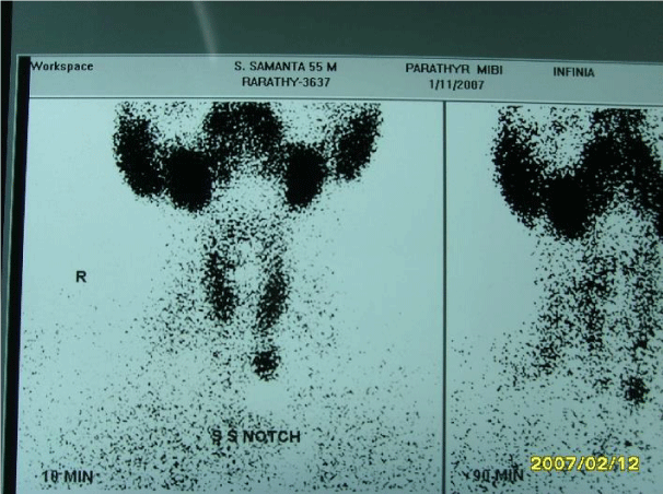
Investigations revealed a normal complete hemogram, blood urea 34 mg/dl creatinine 1.5 mg/dl, fasting blood sugar 87 mg/dl, serum sodium 139 meq/l, potassium 3.8 meq/l. USG KUB showed calcifications in the pyramids of both kidneys. Kidney size and cortical echo pattern as well as thickness were normal. The detection of renal calcifications prompted us to perform serum calcium, phosphate and magnesium. The results were 11 mg/dl, 3.3 mg/dl and 3.8 mg/dl respectively.24 hour urinary calcium was 480 mg/day (normal < 300mg/day ) Immediately serum parathyroid level was sent for and it came out to be 234.2 pg/ml (15-65 pg/ml). Tc 99m sestamibi subtraction scan showed focal uptake of radiotracer just distal to the inferior pole of the left lobe of thyroid gland consistent with a left inferior parathyroid adenoma. He underwent surgical excision after which he is doing well on one drug.
Discussion
The patient presented at the fifth decade- thus secondary hypertension was not thought of at the beginning. For years his antihypertensive doses were escalated without much result. The detection of renal calcifications were a pointer to diagnosis. Metastatic calcifications are due to deposition of calcium salts in normal tissues. Common sites involved are walls of blood vessels, peri-articular areas, skin, soft tissues and viscera notably lung, heart, kidney, intestine and brain.
Essentially a disbalance of calcium and phosphate homeostasis plays the key role in pathogenesis of this state [2]. The increased calcium-phosphate product has been proposed as the determining factor. In primary hyperparathyroidism though phosphate is initially low, progressive renal impairment may later contribute to raise calcium phosphate product metabolism [2,3].
It has been documented that serum phosphate > 8-9mg/dl and/or calcium phosphate product >70 mg/dl, local tissue injury, increased parathormone level-all contribute to heterotopic calcification [2,4].
The mechanism of high BP in hyperparathyroidism is controversial. Plasma cortisol and plasma renin activity were found to be higher inhypercalcemic states but plasma cortisol and catecholamines were found to be normal [1]. Endothelial dysfunction with decreased NO, stiffness of great arteries, and increased responsiveness to vasoconstrictors and TGF B1 have been put forward as hypotheses [5-7].
However it has been documented that simple surgery of adenoma leads to control of BP which was also demonstrated in our case.
- Rossi GP (2011) Hyperparathyroidism, arterial hypertension and aortic stiffness: A possible bidirectional link between the adrenal cortex and the parathyroid glands that causes vascular damage? Hypertension Research 34: 286–288. Link: https://goo.gl/6c5LUs
- Chakrabarti N, Chattopadhyay C (2010) Dysparathyroidism: A clinical window. Ann Saudi Med 30: 497-498. Link: https://goo.gl/Co83Jc
- Valdivielso P, Lopez J, Garrido A, Sanchez Carrillo JJ (2006) Metastatic calcification and severe hypercalcemia in a case of parathyroid carcinoma .J Endocrinol Invest 29: 641-644.
- Martin KJ, Gonzalez EA, Slatopdsky E (2004) Renal Osteodystrophy, Ed Brennert B M.7th edition, Saunders 2255-2304.
- Martina V, Bruno GA, Brancaleoni V, Zumpano E, Tagliabue M, et al. (1998) Calcium blood level modulates endogenous nitric oxide action: effects of para thyroidectomy in patients with hyperparathyroidism. J Endocrinol 156: 231–235. Link: https://goo.gl/Y2qM8g
- Tordjman KM, Yaron M, Izkhakov E, Osher E, Shenkerman G, et al. (2010) Cardiovascular risk factors and arterial rigidity are similar in asymptomatic normocalcemic and hypercalcemic primary hyperparathyroidism. Eur J Endocrinol 162: 925–933. Link: https://goo.gl/wt1x26
- Chhokar VS, Sun Y, Bhattacharya SK, Ahokas RA, Myers LK (2005) Hyperparathyroidism and the calcium paradox of aldosteronism. Circulation 111: 871–878. Link: https://goo.gl/Rj3Wt8
Article Alerts
Subscribe to our articles alerts and stay tuned.
 This work is licensed under a Creative Commons Attribution 4.0 International License.


 Save to Mendeley
