International Journal of Clinical Endocrinology and Metabolism
Two cases of traumatic isolated ACTH deficiency
Tatsuo Ishizuka1*, Motochika Asano2, Kei Fujioka1, Ichiro Mori1, Kazuo Kajita2 and Hiroyuki Morita2
2Department of General Internal Medicine, Gifu University Graduate School of Medicine, 1-1 Yanagido, Gifu 501-1194, Japan
Cite this as
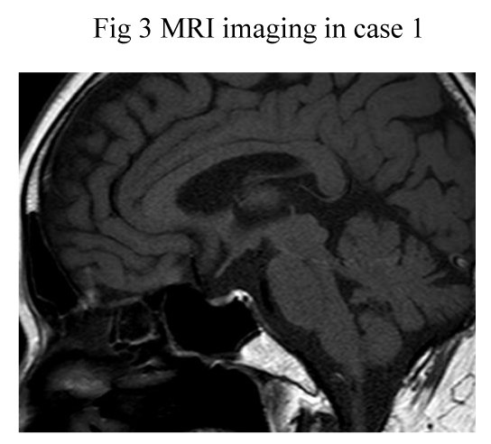
Ishizuka T, Asano M, Fujioka K, Mori I, Kajita K, et al. (2018) Two cases of traumatic isolated ACTH deficiency. Int J Clin Endocrinol Metab 4(1): 004-007. DOI: 10.17352/ijcem.000030Case 1: A 65- year-old man was accidentally injured by wooden hammer on his top of head on 34 years before. He was suffered from vomiting, diarrhea and hypotension, and the laboratory examination revealed increased CRP level, hyponatremia and decreased plasma cortisol and ACTH levels, suggesting isolated ACTH deficiency and Crohn disease diagnosed by colonoscopic biopsy, and finally transferred to University Hospital. LH-RH, TRH, CRH and GHRP stimulation tests showed normal response of plasma pituitary hormones except for no response of plasma ACTH and cortisol levels by CRH stimulation. ACTH stimulation test showed no response of plasma cortisol levels although hydrocotisone replacement therapy had already been started. MRI imaging showed bottom of anterior lobe was crushed and pituitary gland was atrophied, which suggested brain might be injured by any strong trauma.
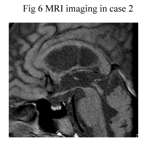
Case 2: An 83-years old man was injured on brain contusion by staff’s violence in nursing home, and introduce to our hospital to remove brain hematoma on 6 months before. He presented transient loss of consciousness because of hypoglycemia. Laboratory examinations revealed hyponatremia, and low levels of plasma ACTH and cortisol. Endocrinological examination showed normal LH-RH and TRH stimulations tests, basal GH and IGF-1 levels, and no response of plasma ACTH and cortisol levels by CRH stimulation, showing traumatic isolated ACTH deficiency. MRI imaging showed atrophic pituitary gland. These results suggest that traumatic isolated ACTH deficiency may be able to appear for short and long period after brain injury.
Introduction
Isolated ACTH deficiency is a rare disease characterized by secondary adrenal insufficiency with low or absent cortisol production and normal secretion of pituitary hormones other than ACTH [1]. Isolated ACTH deficiency has been caused by traumatic injury [2], lymphocytic hypophysitis due to autoimmune etiology [3,4], genetic origin in neonatal or childhood [1], and unknown origin. Previous reports have demonstrated that traumatic brain injury—mediated hypopituitarism could be more frequently occured [5-7]. High prevalence of neuroendoctine dysfunction in patients with traumatic brain injury has been reported [8].
In this study we have shown two cases of traumatic isolated ACTH deficiency.
Case Presentation
Case 1
A 65- year-old man was accidentally injured by wooden hammer on his top of head on 34 years before. He was suffered from vomiting, diarrhea and hypotension, and the laboratory examination revealed increased CRP (4.0 mg/dl) level, hyponatremia and decreased plasma cortisol (less than 1 μg/dl) and ACTH (less than 2.0 pg/ml) levels, suggesting isolated ACTH deficiency and Crohn disease diagnosed by colonoscopic biopsy, and finally transferred to University Hospital.
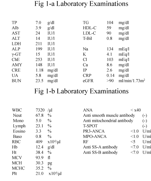
Laboratory examinations showed normal values exept for slightly hyponatremia and leukocytosis during hydrocortisone replacement therapy (Figures 1-a,b). Thyroid function was normal. Basal ACTH and cortisol levels were suppressed by hydrocortisone replacement therapy. LH-RH, TRH and CRH and GHRP stimulations tests showed normal response of plasma pituitary hormones except for no response of plasma ACTH and cortisol levels by CRH stimulation and hyperresponse of PRL by TRH stimulation with high level of PRL (Figures 2-a,b). ACTH stimulation test showed no response of plasma cortisol levels although hydrocotisone replacement therapy had already been started (Figure 2-b). MRI (T1 weighted image) imaging showed bottom of cerebral anterior lobe was crushed and anterior pituitary gland was atrophied, which suggested brain might be injured by any strong trauma (Figure 3).
Case 2
An 83-years old man was injured on brain contusion by staff’s violence in nursing home, and introduce to Gifu Municipal hospital to remove brain hematoma on 6 months before. He presented transient loss of consciousness because of hypoglycemia (37 mg/dl), and transferred to our hospital.
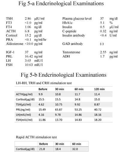
Laboratory examinations revealed hyponatremia (126 mEq/l), normal HbA1c (5.1 %) level (Figures 4-a,b, Figures 5-a), and low levels of plasma ACTH (6.8 pg/ml), cortisol (15.2 μg/dl) and suppression of PRA (less than 0.1 ng/ml/hr) and aldosterone (less than 10.0 pg/ml) levels during saline infusion (Figure 4-c, Figure 5-a).
Endocrinological examination showed normal LH-RH and TRH stimulation test, normal plasma basal GH (3.73 ng/ml) and IGF-1 (37 ng/ml) levels and no response of plasma ACTH and cortisol levels by CRH stimulation, showing traumatic isolated ACTH deficiency (Figures 5-a,b). ACTH stimulation test showed delayed low response of plasma cortisol levels (Figure 5-b). MRI imaging showed anterior lobe of atrophic pituitary gland (Figure 6).
Discussion
It has been reported that the percentage of probability in the appearance of endocrinological abnormality was 15-68%, especially in hypopituitarism of anterior lobe was 27.5% and that hypofunction in hypophysico–pituitary axis occurs within half year after brain trauma. Gonadal hypofunction and insufficiency of growth hormone secretion occurred in highest rate, and in the next rate hypoadrenocortism and hypothyroidism occurred. Schneider et al. [2] also reported that grade of brain damage evaluated in Glasgow Coma Scale (GCS) was correlated with occurrence rate in hypothalamo-pituitary hypofuction as follows: Severe damage (GCS 9-12 points), moderate damage (9-12 points) and slight damage ( 13-15 poins) was 35.3%, 10.9% and 16.8%, respectively. However, even if in the case of slight brain damage, occurrences of endocrinological abnormality should be taken care. They conclude that hypopituitarism is a common complication of both traumatic brain injury and aneurysmal subarachnoid hemorrhage. This systematic review showed ACTH deficiency was occurred in 0-19.2% after traumatic brain injury. Tanriverdi et al. [9], also reported that some 5.8% of the traumatic brain injury patients had TSH deficiency, 41.6% had gonadotropin deficiency, 9.8% had ACTH deficiency, and 20.4% had GH deficiency, and that pituitary function may improve or worsen in a considerable number of patients over 12 months. A patient presented in case 1 occurred clinical symptoms of hypopituitarism for 34 years after severe traumatic injury. Recent report indicated 3cases of isolated ACTH or TSH deficiency following mild traumatic brain injury with long-term follow (10 days- 20 years) [10], which was similar to our case 1. Sixty-five years-old man occurred both isolated ACTH deficiency and Crhon’s disease at the same time in case 1. Kalambokis et al. [11], had been reported that isolated ACTH deficiency associated with Croh’s disease without traumatic brain injury which might be associated with immune reactions. Therefore, etiology of our case 1 might be a little relevance for complication of Crhon’s disease. Recently, old men and women received violent brain traumas in old people’s home have been happened. Clinical symptoms and results of laboratory examinations such as nausea vomiting, hyponatremia and increased CRP levels should be paid attention.
Conclusion
Two cases of these disorders were treated with 15-20 mg of hydrocortisone and continued to live in good health. These results suggest that traumatic isolated ACTH deficiency may be able to appear for short and long period after brain injury.
Disclosure
None of the authors have any potential conflict of interest associated with this research.
The ethical committee in the Gifu Municipal Hospital have been approved in this study.
- Andrioli M, Pecori Giraldi F, Cavagnini F (2006) Isolated corticotrophin deficiency. Pituitary 9: 289-295. Link: https://goo.gl/tQmZ1d
- Schneider HJ, Kreitschmann-Andermahr I, Ghigo E, Karl Stalla G, Agha A, et al. (2007) Hypothalamo-pituitary dysfunction following traumatic brain injury and aneurysmal subarachnoid hemorrhage. A systematic review JAMA 298: 1429-1438. Link: https://goo.gl/Xfkyhz
- Escobar-Morale H, Serrano-Gotarredona Jm Varela C (1994) Isolated adrenocorticotropic hormone deficiency due to probable lymphocytic hypophsitis in a man. J Endocrinol Invest17: 127-131. Link: https://goo.gl/gZdfGt
- Fasten HK, Nadia C, Mouna FM, Mahdi K, Faouzi A, et al. (2013) Isolated adrenocorticotropic hormone deficiency due to probable lymphocytic hypophysitis in a woman.Indian J Endocrinol Metab17: S107-S110. Link: https://goo.gl/JCCsoX
- Benbenga S, Campenni A, Ruggeri RM, Trimarchi F (2000) Clinical review 113: hypopituitarism secondary to head trauma. J Clin Endocrinol Metab85: 1353-1361. Link: https://goo.gl/UD831x
- Kelly DF, Gonzalo IT, Cohan P, Berman N, Swerdlo HR, et al. (2000) Hypopituitarism following traumatic brain injury and aneurysmal subarachnoid hemorrhage: a prelim report. J Neurosurg 93: 743-752. Link: https://goo.gl/4ryx1Z
- Edwards OM, Clarl JD (1986) Post-traumatic hypopituitarism. Six cases and review of the literature. Medicine 65: 281-290. Link: https://goo.gl/xBZ9vS
- Liberman SA, Oberoi AL Gilkison CR, Masel BE, Urban RJ (2001) Prevalence of neuroendocrine dysfunction in patients recovering from traumatic brain injury. J Clin Endocrinol Metab86: 2752-2756. Link: https://goo.gl/Ucw9P3
- Tanriverdi F, Senyurek H, Unluhizarci K Selcuklu A, Casanueva FF, et al. (2006) High risk of hypopituitarism after traumatic brain injury: A prospective investing of anterior pituitary function in the acute phase and 12 months after trauma. J Clin Endocrinol Metab91: 2105-2111. Link: https://goo.gl/Xopcvc
- Baek CO, Kim Y, Jm Kim JH, Park JH (2015) Isolated adrenocorticotropic hormone or thyrotropin deficiency following long-term follow up. Korean J Neurotrauma11: 139-143. Link: https://goo.gl/zXLLHs
- Kalambokis G, Vassiliou V, Vergos T, Christou I, Tsatsoulis A, et al. (2004) Isolated ACTH deficiency associated with Crohn’s disease. J Endocrinolo Invest 27: 961-964. Link: https://goo.gl/7pkuvd
Article Alerts
Subscribe to our articles alerts and stay tuned.
 This work is licensed under a Creative Commons Attribution 4.0 International License.


 Save to Mendeley
