Global Journal of Rare Diseases
Aarskog-Scott Syndrome: Clinico-Radiological Illustration of a Rare Case
Akanksha Gupta, Ravi Prakash Sasankoti Mohan*, Sumit Goel and Swati Gupta
Cite this as
Gupta A, Ravi Prakash SM, Sumit G, Gupta S (2016) Aarskog-Scott Syndrome: Clinico-Radiological Illustration of a Rare Case. Glob J Rare Dis 1(1): 010-013. DOI: 10.17352/2640-7876.000004Aarskog-Scott Syndrome is a rare syndrome and is estimated to occur in 1 in 1 million individuals in general population. It is transmitted in an X-linked recessive manner and occurs due to FGD1 gene mutation. It has facial, genital and digital hands symptoms, musculoskeletal anomalies and affected growth. Here we present a case of Aarskog-Scott syndrome in an 18-year-old male patient with the chief complaint of irregularly placed teeth. In addition to all the classical features, presence of talon cusp was an interesting intraoral finding which has never been reported previously in dental literature.
Introduction
Aarskog-Scott Syndrome, also known as Aarskog syndrome, Faciogenital syndrome, Faciodigitogenital syndrome, Faciogenital dysplasia, Shawl scrotum syndrome, Aarskog disease, FGDY [1,2]. Aarskog Scott syndrome was first described by Norwegian paediatrician named Dagfinn Aarskog in 1970. The American paediatrician Charles I Junior Scott also described the syndrome in 1971. Aarskog described a syndrome of short stature with facial and genital anomalies in seven males from two generations in a family [1,2]. Many reports over two decades have shown that males show full expression and that in females expression, if present, is limited to minor signs only; therefore X-linked recessive, X-linked semi-dominant or sex influenced autosomal dominant inheritance have been suggested [1,2].
Case Report
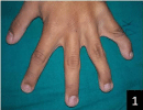
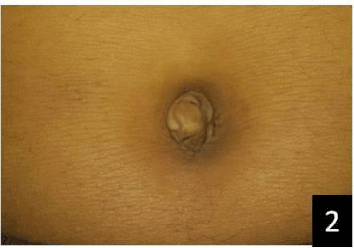
An 18-year-old male patient came to our department with the chief complaint of irregularly placed front teeth in upper and lower jaw since 4-5 years. Patient had not undergone any previous dental treatment. There was history of undescended testis which he got operated at the age of five and is now normal. Patient got Growth hormone stimulation test done at the age of 4 yrs. Peak Growth Hormone Concentration in Blood was 5 ng/mL indicating growth hormone deficiency. Patient had normal intelligence. General physical examination revealed short strature, large palms and feet, long neck, sloping shoulders and proportionate body with hyperelastic skin. There was clindodactyly (Figure 1) and everted umbilicus (Figure 2).
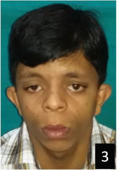
On extraoral examination, triangular-elongated face, widow’s peak, puffy enlarged lips, ocular hypertelorism, ptosis, slight antimongoloid obliquity of palpebral fissure, small and short nose, long philtrum, broad nasal bridge, slight crease below lower lip and fleshy earlobes were observed (Figure 3). Patient had bimaxillary protrusion.
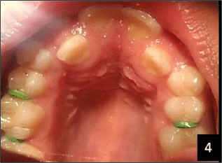
Intraoral examination revealed that there was Angle’s Class I molar relationship bilaterally. He had palatally placed upper lateral incisors, labially placed canines and crowding in maxillary and mandibular anterior region. There was high arched palate (Figure 4).
Patient was subjected to radiographic examination.
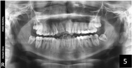
Orthopantomogram revealed talon cusp in 11 and 21 with crowding in maxillary and mandibular anterior region. The roots of mandibular premolars were short and blunt but the root formation was complete. There was presence of unerupted maxillary and mandibular third molars (Figure 5).
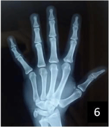
In the hand-wrist radiograph, clindodactyly was present and fusion of epiphysis and diaphysis of radius could be appreciated (Figure 6).
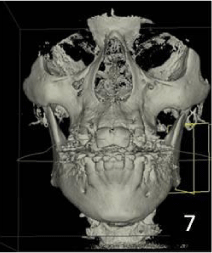
Sections of cone beam computed tomography did not reveal any other significant finding (Figure 7).
Patient was referred to the department of Orthodontics and was advised to consult the endocrinologist for growth hormone evaluation. Growth hormone levels came out to be normal.
Discussion
Aarskog-Scott syndrome is transmitted in an X-linked recessive manner. The sons of female carriers are at 50% risk of being carrier themselves. Females may have mild manifestations of the syndrome. The syndrome is caused by mutation in a gene called FGDY I in band p11.21 on the X chromosome [1,2].
FGD1 encodes a guanine nucleotide exchange factor (GEF) which activates Cdc42, a member of the Rho (Ras homology) family of the p21GTPases. By activating Cdc42, FGD1 protein stimulates fibroblasts to form filopodia, cytoskeletal elements involved in cellular signaling, adhesion and migration. Through Cdc42, FGD1 protein also activates the c-Jun N-terminal kinase (JNK) signaling cascade, a pathway that regulates cell growth, apoptosis and cellular differentiation. It appears likely that the primary defect in Aarskog-Scott syndrome is an abnormality of FGD1/Cdc42 signaling resulting in anomalous embryonic development and abnormal endochondral and intramembranous bone formation [1,2]. We could not carry out the mutation analysis in our case to identify the specific mutation in FGD1 gene due to lack of facility and cost restrain but the classical clinical features of the patient lead to the diagnosis of this syndrome.
There is a typical triad of the syndrome which includes altered facial appearance, digital anomalies and genital abnormalities [3].
Patient usually presents with round face, prominent metopic ridge, widow’s peak hair anomaly, ocular hypertelorism, ptosis, antimongoloid obliquity of palpebral fissure, small and short nose, anteverted nares, broad philtrum, broad nasal bridge, maxillary and mandibular hypoplasia, slight crease below lower lip, low set ears with fleshy earlobes and incomplete outfolding of upper helices [4,5]. In our case, there was ocular hypertelorism, ptosis, slight antimongoloid obliquity of palpabral fissure, small and short nose, broad nasal bridge, long philtrum, slight crease below lower lip, puffy enlarged lips and fleshy earlobes.
Digital anomalies are present in the form of short and broad hands, hypermobility of fingers, cutaneous syndactyly, clindodactyly with hypoplasia of middle phalanges and transverse palmer crease [4,5]. Out of these, clindodactyly could be appreciated in our patient clinically as well as on hand-wrist radiograph.
Genital abnormalities in this syndrome include shawl scrotum, cryptorchidism and phimosis [4,5]. Our patient gave the history of undescended testis which he got operated at the age of five.
There is delayed puberty and slight to moderate short stature in 71% of the patients who suffer from this syndrome [4,5]. Our patient had a short strature, large palms and feet and proportionate body.
Most individuals are of normal intelligence, as was seen in our case. Hyperactive and attention deficits are present in 61% of these individuals, which usually regress after 12-14 years of age. In individuals with subnormal intelligence, hyperactive and attention deficits are present in 84% cases [4-6].
Dental abnormalities in these patients include hypodontia, retarded dental eruption and broad central upper incisors [5]. Patient suffers from orthodontic problems which was the chief complaint of our patient as well. Our patient had palatally placed upper lateral incisors and labially placed canines. There was crowding in maxillary and mandibular anterior region. Patient had Angle’s Class I molar relationship bilaterally. Presence of talon cusp in maxillary central incisors was an interesting finding as it has not been reported in any case of Aarskog-Scott syndrome before. According to the classification by Hattab et al., the cusps on the maxillary central incisors in our case are type 3 or trace talon cusps [7].
Some ocular features are also observed in such patients such as strabismus, nystagmus, amblyopia, bilateral blepharoptosis, astigmatism, hyperopia, anisometropia, corneal enlargement, deficient ocular elevation, blue sclera, posterior embryotoxon, ophthalmoplegia and tortuosity of retinal vessels. Musculoskeletal anomalies such as cervical vertebral anomalies, spina bifida occulta, mild pectus excavatum, genu recurvatum, joint restriction and prominent hernias may be present. Some other rare signs have been reported in few patients such as gastrointestinal obstruction and volvulus. There is rare perinatal occurrence of severe cerebrovascular accidents. Congenital heart defects have also been reported [4,8].
Dental and skeletal development should be assessed in patient suffering from this syndrome. Orthopantomogram of our patient revealed that although the roots of mandibular premolars were short and blunt but the root apex of all the erupted teeth was complete which is the 10th stage of tooth calcification as classified by Nolla [9]. Hand-wrist radiographs showed that there was fusion of epiphysis and diaphysis of radius which is the 4th stage of bone maturation as classified by Leonard S Fishman [10]. These findings indicate the normal dental and skeletal development of the patient.
Prenatal diagnosis by ultrasonography is possible if there is positive family history of a previously affected child by demonstrating ocular hypertelorism, short long bones, vertebral defects and digital anomalies in a male fetus [11].
There are numerous treatments that exist and increase the quality of life. It is important to establish early contact with the dental services as some teeth may be missing. The dental services should be familiar with the problems children with abnormal growth patterns can experience as bone growth and maturity may be delayed. Bite abnormalities may require orthodontic treatment as was required by our patient. Testicles that do not descend into the scrotum are treated surgically [12]. Our patient had gone through this surgical procedure when he was five years old. Inguinal hernia is also treated surgically during childhood [13].
Growth failure is one of the main features of this syndrome and may start prenatally. Growth rate is low during the first year of life and becomes distinct between 1 and 3 years of age. During childhood, growth rate is slow and height usually remains below the 3rd centile [14]. Growth hormone deficiency has been reported in this syndrome in the early years of patient’s life [15]. Our patient also suffered from growth hormone deficiency in his early years of life and had short strature at the time of presentation in our department. Response to growth hormone treatment was evaluated in 21 patients with this syndrome enrolled in the KIGS database. After 3 years of treatment with a standard dose (0.22 mg/kg/week), a beneficial effect on growth increment was shown [14]. Our patient, however, did not undergo any such therapy.
Conclusion
Aarskog-Scott syndrome is a rare syndrome which is characterized by a typical triad which includes altered facial appearance, digital anomalies and genital abnormalities. Growth is affected and puberty is delayed in these patients. There are numerous dental anomalies and patient requires orthodontic treatment. Presence of talon cusp on maxillary central incisors is an interesting intraoral finding as it has never been reported in this syndrome before. More case reports are required to consider talon cusp as a consistent finding. Radiography, ophthalmic examination and mutation analysis helps to establish the diagnosis. Genetic counseling is important for these patients. There are numerous treatments that exist and increase the quality of life.
- Aarskog D (1970) A familial syndrome of short stature associated with facial dysplasia and genital anomalies. J Pediatr 77: 856–861. Link: https://goo.gl/N2trfj
- Scott CI (1971) Unusual facies, joint hypermobility, genital anomaly and short stature: a new dysmorphic syndrome. Birth Defects Orig Artic Ser 7: 240–246. Link: https://goo.gl/lZf79u
- Closs LQ, Tovo M, Dias C, Corradi DP, Vargas IA (2015) Aarskog-Scott Syndrome: A Review and Case Report. Int J Clin Pediatr Dent 5: 209-212. Link: https://goo.gl/FcpS76
- Porteous MEM, Goudie DR (1991) Aarskog syndrome. J Med Genet 28: 44-47. Link: https://goo.gl/K0jwfT
- Bozorgmehr B (2006) Aarskog-Scott syndrome: Report of 7 cases and review of literature. Genet 3rd millennium 4: 954-956. Link: https://goo.gl/u7fTd4
- Teebi AS, Naguib KK, Al-awadi SA, Al-saleh QA (1988) New autosomal recessive faciodigitogenital syndrome. J Med Genet 25: 400-106. Link: https://goo.gl/OGHfC0
- Ozcelik B, Atila B (2001) Bilateral Palatal Talon Cusps on Permanent Maxillary Lateral Incisors: A Case Report. Eur J Dent 5: 113-116. Link: https://goo.gl/0RfjK9
- Fernandez I, Tsukahara M, Mito H, Yoshii H, Uchida M, et al. (1994) Congenital heart defects in Aarskog syndrome. Am J Med Genet 50: 318-322. Link: https://goo.gl/XnNq6X
- Panchbhai AS (2011) Dental radiographic indicators, a key to age estimation. Dentomaxillofacial Radiol 40: 199–212. Link: https://goo.gl/0IlInx
- Fishman LS (1982) Radiographic Evaluation of Skeletal Maturation. Angle Orthod 52: 88-112. Link: https://goo.gl/gMKYli
- Sepulveda W, Dezerega V, Horvath E, Aracena M (1999) Prenatal Sonographic Diagnosis of Aarskog Syndrome. J Ultrasound Med 18: 707-710. Link: https://goo.gl/QGrAfp
- Waaler PE (2008) Clinical and cytogenetic studies in undescended testes. Acta Paediatr 65: 553-558. Link: https://goo.gl/GYFJfA
- Andrassy RJ, Murthy S, Woolley MM (1979) Aarskog syndrome: Significance for the surgeon. J Pediatr Surg 14: 462-464. Link: https://goo.gl/5Nt3Vj
- Şıklar Z, Berberoğlu M (2014) Syndromic Disorders with Short Stature. J Clin Res Pediatr Endocrinol 6: 1–8. Link: https://goo.gl/I6YoWf
- Kodama M, Fujimoto S, Namikawa T, Matsuda I (1981) Aarskog syndrome with isolated growth hormone deficiency. Eur J Pediatr 135: 273–276. Link: https://goo.gl/tyxCPU
Article Alerts
Subscribe to our articles alerts and stay tuned.
 This work is licensed under a Creative Commons Attribution 4.0 International License.
 Save to Mendeley
