Global Journal of Medical and Clinical Case Reports
Antenatal ultrasonography should be for all. Phocomelia: An extremely rare congenital anomaly- A case report
Pradyumna Pan*
Cite this as
Pan P (2019) Antenatal ultrasonography should be for all. Phocomelia: An extremely rare congenital anomaly- A case report . Glob J Medical Clin Case Rep 6(1): 001-002. DOI: 10.17352/2455-5282.000066While rarely seen in the present-day, Phocomelia syndrome is a sporadic congenital malformation disrupting the normal growth and progressing of the musculoskeletal system.
Introduction
The term phocomelia comes from the Greek: fóke-“seal,” plus melos-“limb. It is a very rare congenital birth defect affecting the limbs [1]. In recent times it is very unusual to see phocomelia babies at term. This case concerns a 3-months-old boy with multiple skeletal anomalies, delivered at term and is reported owing to its rarity and term live birth.
Case Report
We report a case of a 3months baby who presented to our outpatient department with multiple defects of hand and legs since birth. He was born to a 37-year-old G5P4 with a history of non-consanguineous marriage. She was an un-booked case from a tribal community with no previous antenatal visits. She delivered vaginally a term live 2.5 kg male baby with multiple anomalies. There was no history of chronic illnesses, drug intake, exposure to known teratogenic agents, radiation exposure, and family history of congenital anomalies. He is the fourth living child of the family born from the five pregnancy of the mother. The mother’s living children are healthy, but a second baby was delivered stillborn with lower limb agenesis.
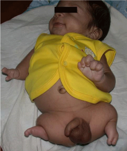
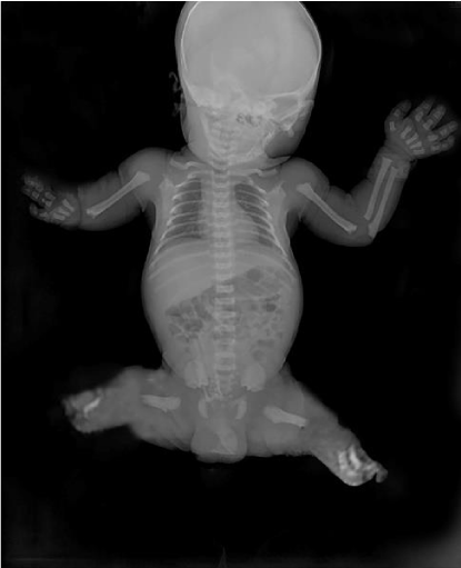
Physical examination showed right arm to be well formed up to elbow with absent forearm and malformed hand with normal thumb and syndactyly of two fingers. The left upper limb was normal. Both the lower limbs are malformed with short thigh, absent leg and one digit in right foot and 2 digits in the left foot (Figure 1). Systemic examination revealed no abnormality. The testicles are descended and there were no associated skull and pelvic anomalies. Radiograph of the baby revealed right short humerus while both the lower limbs showed very scanty attempted formation of the femur with an absence of tibia-fibula on either side (Figure 2). Ultrasonography of the abdomen was normal. The child lost to follow up subsequently.
Discussion
The term Phocomelia was first used by Etienne Geoffroy Saint in 1836 and its prevalence is 0.62 in 1,00,000 births [2]. This malformation was seen with thalidomide embryopathy. In light of the different related clinical patterns of defects, Phocomelia was further classified in to Al-Awadi/Raas-Rothschild Syndrome syndrome (AARR syndrome), Roberts/SC phocomelia, Schinzel phocomelia and Zimmer phocomelia [3].
After fertilization, development of limb bud begins from lateral (paraxial and somatic) plate mesoderm at 26th day and stretches out until day 56 [4]. The exact mechanism underlying the development of phocomelia remains unclear. Nevertheless, it could be hypothesized that during the defined critical period, between days 24 and 33, defects in angiogenesis could produce a loss of mesenchyme and distal fate of limbs [5]. Limb formation begins within the morphogenetic limb field. The mesenchymal cells from the lateral plate mesoderm proliferate causing the ectoderm above to project out thus creating a limb bud. The developing limb can be delineated into three fundamental zones; the proximal stylopod, the middle zeugopod and the distal autopod. This intricate patterning process is governed by a three dimensional signaling system that defines a proximodistal axis controlled of fibroblast growth factors (FGFs) from the apical ectodermal ridge (AER). An anteroposterior axis which is under the control of sonic hedgehog (SHH) from the posterior mesenchyme (zone of polarizing activity), and lastly dorsoventral axis regulated by bone morphogenetic proteins (BMPs), engrailed (EN1) from the ventral ectoderm and by Wnt7a from the dorsal ectoderm. Wnt7a specify the dorsoventral axis by prompting the expression of the transcription factor Lmx1 in the dorsal mesenchyme of the developing limb. Wnt7a belongs to a large family of secreted signaling proteins that consists of two chief functional groups: the Wnt1 and the Wnt5a. The binding of Wnt to the Frizzled, Lrp, and alternative receptors causes a stabilization of intracellular b-catenin, transported into the nucleus and interacts with the transcription factors Lef1 and Tcf to control gene expression. This pathway is regulated by the cytosolic component of the Wnt signal, Dishevelled (Dsh) phosphoprotein. Thus errors in the gene expression results in limb malformations.
Fuhrmann syndrome is a type of skeletal dysplasia caused by mutations to the WNT7A gene and is inherited in an autosomal recessive manner very similar to Al-Awadi-Raas-Rothschild syndrome. The genetic mutations in Fuhrmann syndrome results in partial loss of the function of the gene whereas in Al-Awadi-Raas-Rothschild syndrome result in complete loss of function of the gene. The syndrome is allelic to Schinzel phocomelia, which results from null mutations in the same gene.
FATCO syndrome (Fibular aplasia, tibial campomelia, and oligosyndactyly) is a syndrome of undetermined genetic basis and inheritance having close alikeness to Furhmann syndrome and Al-Awadi syndrome.
Phocomelia Syndrome may occur as a result to a mutation in the gene or can be the outcome of genetic transmission between family members. Thus, there is significantly increased risk for phocomelia when parents have consanguinity. Phocomelia Syndrome is usually diagnosed on a prenatal ultrasound of the fetus.
Treatments for Phocomelia Syndrome are designed while the child is an infant and emphases upon the severity of symptoms of the affected individual. Most of the treatment is supportive, allowing the infant to live a more normal life. Genetic counselling for the child and the family is needed.
Conclusion
Prenatal diagnosis should be offered to all and when detected before viability, termination of pregnancy should be offered.
- Newman CGH (1986) The thalidomide syndrome: risk of exposure and spectrum of malformations. Clin Perinatol 139: 555-573. Link: https://tinyurl.com/y5ck6vrc
- Rao RR, Rao BN (2012) Tetra phocomelia-a case report. Indian Journal of Medical Case Reports 1: 20-22. Link: https://tinyurl.com/y25gcqos
- Esma A, Hayrullah A, Mehmet EA, Ozgur P (2010) Al-Awadi/Raas-Rothschild Syndrome in a Newborn with Additional Anomalies. J Clin Res Ped Endo 2: 49-51. Link: https://tinyurl.com/y5lez5ms
- Eva Bermejo S, Lourdes C, Emmanuelle A, SebastianoB, Fabrizio B, et al. (2011) Phocomelia: a worldwide descriptive epidemiologic study in a large series of cases from the international clearinghouse for birth defects surveillance and research, and overview of the literature. Am J Med Genet C Semin Med Genet 157C: 305-320. Link: https://tinyurl.com/y5zfddyg
- Galloway JL, Delgado I, Ros MA, Tabin CJ (2009) A reevaluation of X-irradiation-induced phocomelia and proximodistal limb patterning. Nature 460: 400-404. Link: https://tinyurl.com/y24ksqt4

Article Alerts
Subscribe to our articles alerts and stay tuned.
 This work is licensed under a Creative Commons Attribution 4.0 International License.


 Save to Mendeley
