Global Journal of Medical and Clinical Clinical Image
Iatrogenic Venous Air Embolism (VAE) in a patient secondary to contrast injection; a rare case
Ramsha Fatima Qureshi*, Raisa Altaf, Faiza Hassan, Nimra Ali, Hina Naseer and Syeda Baneen Fatima
Cite this as
Qureshi RF, Altaf R, Hassan F, Ali N, Naseer H, et al. (2023) Iatrogenic Venous Air Embolism (VAE) in a patient secondary to contrast injection; a rare case. Glob J Medical Clin Case Rep 10(2): 013-015. DOI: 10.17352/2455-5282.000170Copyright License
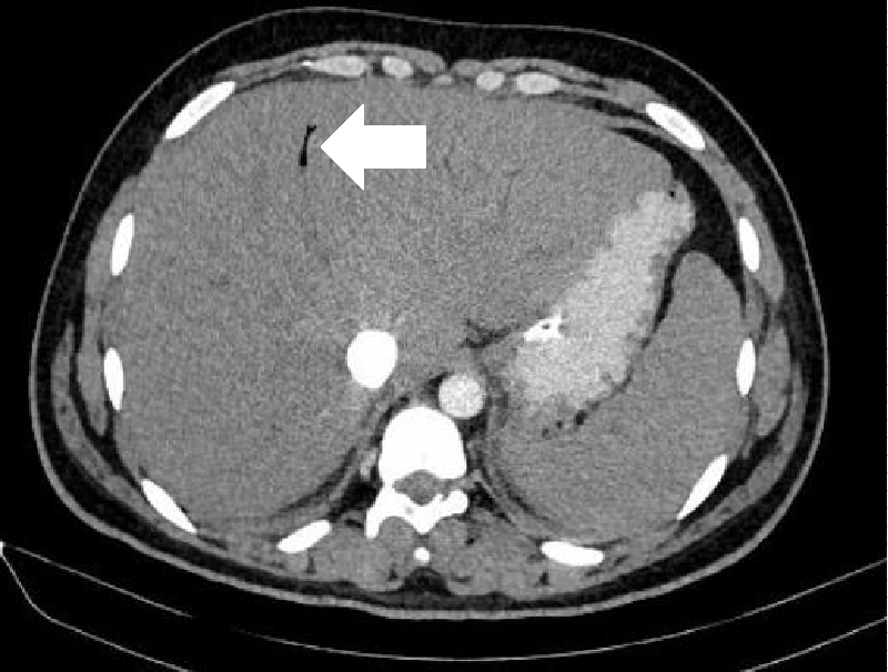
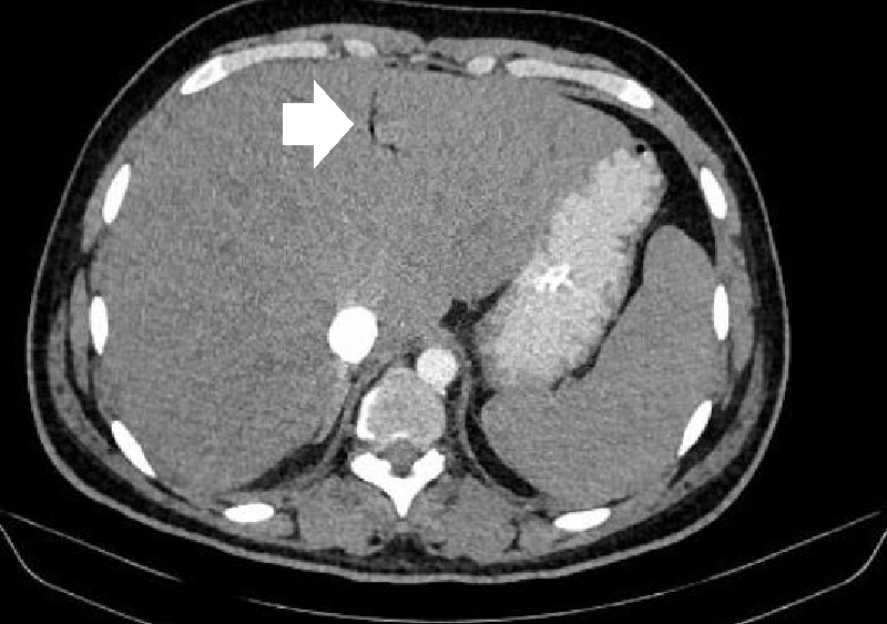
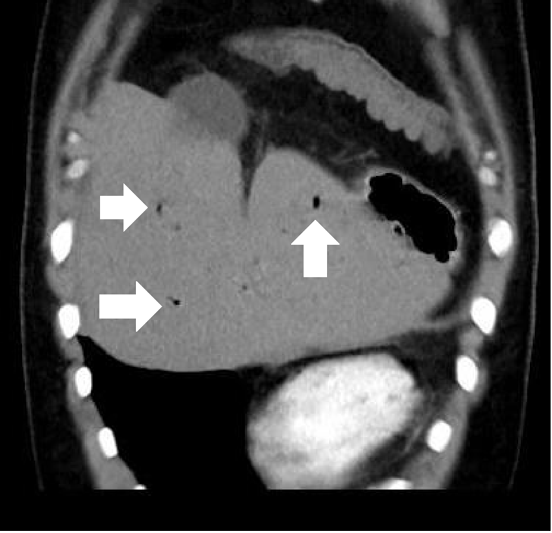
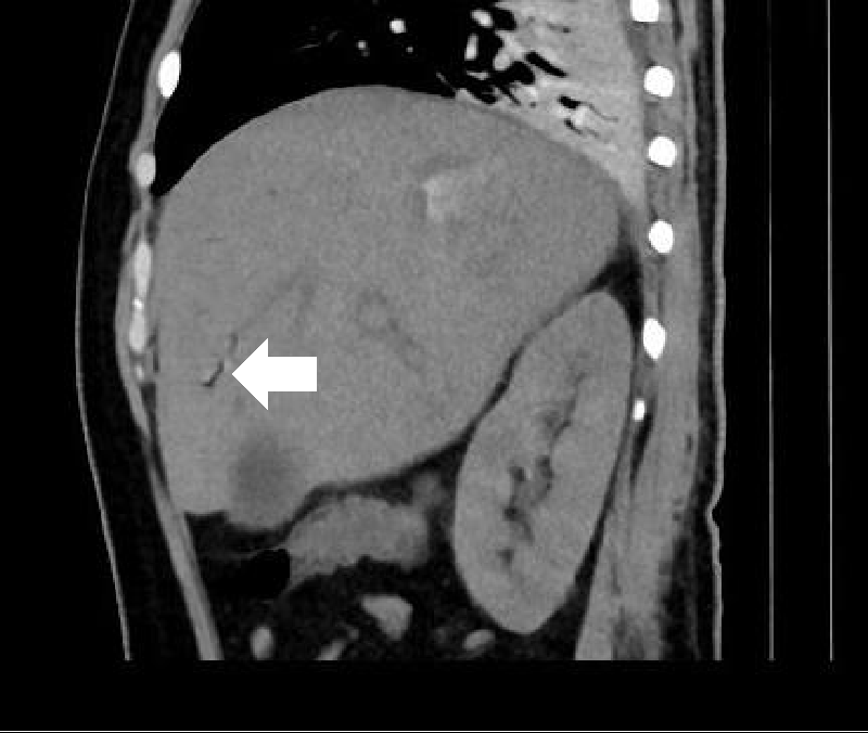
© 2023 Qureshi RF, et al. This is an open-access article distributed under the terms of the Creative Commons Attribution License, which permits unrestricted use, distribution, and reproduction in any medium, provided the original author and source are credited.Venous Air Embolism (VAE) can occur in patients while injecting contrast material, we have reported a case of a 26 yrs old patient who came with the complaint of abdominal pain, vomiting, and fever with no known Co-morbid. CT whole abdomen with contrast was performed and the patient was injected with contrast, which resulted in an air embolism. On administration of contrast, air is seen in the hepatic vein which was not seen on plain and delayed images. 93 ml of contrast material was intravenously injected by hand and followed by a drip infusion of 100 ml of contrast material.
Introduction
Venous Air Embolism (VAE) during or after intravenous contrast media injection is being increasingly recognized with the increased use of CT as a diagnostic modality in modern medicine. In most cases, an air embolus within the venous system is detected incidentally. Studies have shown that small volumes of air within the venous system are usually not detected clinically, but rather radiographically, as most patients are usually asymptomatic. Larger air emboli, however, when present within the venous system may have catastrophic physiological effects on circulatory hemodynamics and may potentially lead to mortality.
We hereby present a case of an incidental finding of iatrogenic air embolism that was seen on the CT abdomen in the hepatic vein while the administration of contrast. After a few hours under examination, the patient developed tachypnea and became unstable.
Hence contrast material should be administered vigilantly to avoid the risk of clinically significant air embolism.
Case report
Our patient was a 26-year-old male without any significant medical history who came with a complaint of abdominal pain and jaundice. Ultrasound was unremarkable so a CT scan with contrast was advised, where on evaluation he was diagnosed with colitis. Iohexol (iodinated) contrast was used. The purpose of contrast-enhanced CT is to find pathology by enhancing the contrast between a lesion and normal surrounding structures. Further workup was done and the patient had enteric fever. The scan was done with 193 ml of contrast medium injected automatically through a power injector into the right femoral vein through the CVP line.
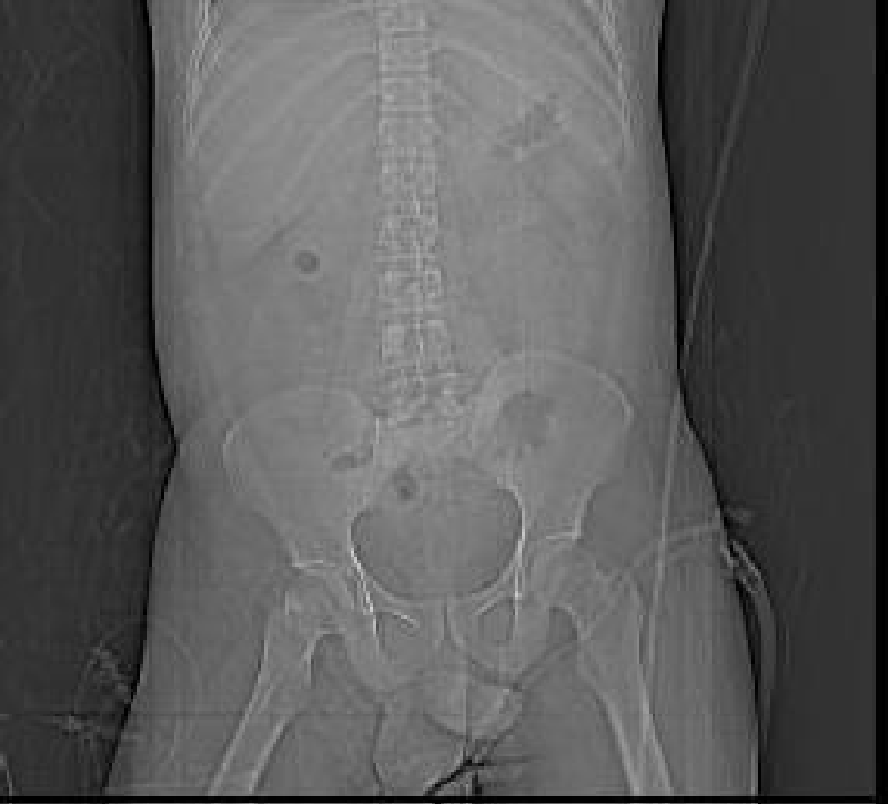
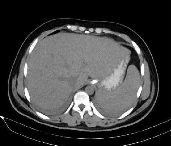
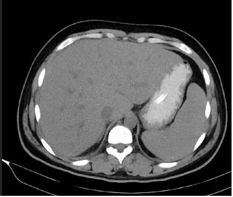
While reviewing the scan, the air was seen in the hepatic vein on arterial and Porto venous phase but no air was seen in Porto venous system or biliary track, neither on scout (Figure 1) nor on plain (Figure 2) nor on delayed images (Figures 3-7) which was likely due to the iatrogenic air embolism.
After a few hours of CT scan, he developed tachypnea and he became vitally unstable and got intubated. He took 2 days to stabilize and got extubated. Later on, an echocardiogram was done which was unremarkable.
Discussion
Venous air embolism after intravenous administration of contrast before CT scanning has been reported to occur in 11% - 23% of patients [1]. Accidental injection of 100 cc of air has been reported as fatal, however, multiple factors such as body position, injection speed, total amount of air injected, and general health status play a part in fatal cases of venous air embolism [1,4]. So it is possible to detect cases of subclinical venous air embolism on CT by the technologists and radiologists as immediately after the contrast injection, the head, or thorax can be scanned and analyzed for smaller volumes of venous air emboli, which may not be detected clinically [4-5]. In humans, it has been estimated that 300 mL to 500 mL of air injected at a rate of about 100mL\sec can result in cardiac arrest [6]. As little as 0.05 mL of air can result in end-organ ischemia and infarction [2].
Air embolism can occur by either a passive or active process. Passive entry of air into the venous circulation may occur during the insertion or removal of central venous catheters. Venous air embolism can also occur as a result of gas being actively forced into the venous circulation such as can transpire during pressure infusions of intravenous contrast medium. Because pressure differences increase as the height above the heart increases, venous air embolism is more likely to occur during procedures on the head and neck [7-11]. It is an unusual but potentially dangerous complication in left heart catheterization. With the use of two-dimensional echocardiography, microbubbles can be found in the ventricles [12]. To look for embolisms from cardiac sources, Transthoracic echocardiography is used. In a structurally normal heart, the next step is to look for a patent foramen ovale, which could indicate a paradoxical embolus. The right-to-left shunt can be diagnosed via a transthoracic contrast examination. Transesophageal echocardiography is required to describe the anatomy of the atrial septum, determine its suitability for device closure, and, ideally, confirm that the shunt is caused by a patent foramen ovale rather than a pulmonary shunt or other atrial septal defects.
Air in the hepatic and portal vein is most likely a manifestation of penetrating Crohn’s disease. It is described as linear radiolucencies that extend to the liver’s periphery within 2 cm. Two hypotheses have been proposed regarding how the air in the portal and hepatic veins is formed: first, that gas embolisation may be caused by elevated intraluminal pressure and mucosal disruption, and second, that gas-producing organisms may be present inside the portal venous system as a result of sepsis [12-16].
The clinical symptoms of air embolism are nonspecific. Symptoms include loss of consciousness, acute onset of shortness of breath, focal paralysis, loss of sensation in an extremity, seizure, vertigo, nausea, chest pain, vomiting, and a sense of impending death. The physical examination can reveal cyanosis, crepitus, tachypnea and hypotension [2].
The immediate treatment in cases of venous air embolism is to make use of the Durant position, that is, make the patient lie in left lateral decubitus with head down. The possible hypothesis is that this position helps to keep the air bubble toward the apex of the right ventricle so that the possibility of the bubble obstructing the outflow tract or gaining access to the pulmonary arterial system would be minimized [3].
Careful handling of intravenous injections of contrast material, including proper insertion of cannula, and checking for any visible free air in the tubing or in the infusion bottle before loading the contrast, (inserting central lines with patients in the Trendelenburg position could be helpful) which should go a long way in minimizing the incidence of venous air embolism after CT examination.
Conclusion
To conclude, radiologists, and technologists (healthcare professionals) should be aware of vascular air embolism, which is a common iatrogenic benign finding detected on CT scans. Asymptomatic venous air emboli do occur frequently and should suggest the diagnosis in the proper given clinical background to preclude a search for other sources of air.
- Sodhi KS, Das PJ, Malhotra P, Khandelwal N. Venous air embolism after intravenous contrast administration for computed tomography. J Emerg Med. 2012 Apr;42(4):450-1. doi: 10.1016/j.jemermed.2009.11.025. Epub 2010 Jan 25. PMID: 20097505.
- Ie SR, Rozans MH, Szerlip HM. Air embolism after intravenous injection of contrast material. South Med J. 1999 Sep;92(9):930-3. doi: 10.1097/00007611-199909000-00019. PMID: 10498176.
- Ie SR, Rozans MH, Szerlip HM. Air embolism after intravenous injection of contrast material. South Med J. 1999 Sep;92(9):930-3. doi: 10.1097/00007611-199909000-00019. PMID: 10498176.
- Woodring JH, Fried AM. Nonfatal venous air embolism after contrast-enhanced CT. Radiology. 1988 May;167(2):405-7. doi: 10.1148/radiology.167.2.3357948. PMID: 3357948.
- Rubinstein D, Symonds D. Gas in the cavernous sinus. AJNR Am J Neuroradiol. 1994 Mar;15(3):561-6. PMID: 8197958; PMCID: PMC8334314.
- Orebaugh SL. Venous air embolism: clinical and experimental considerations. Crit Care Med. 1992 Aug;20(8):1169-77. doi: 10.1097/00003246-199208000-00017. PMID: 1643897.
- O'Quin RJ, Lakshminarayan S. Venous air embolism. Arch Intern Med. 1982 Nov;142(12):2173-6. PMID: 7138162.
- Goodwin J, Gnanapandithan K. Air embolism after peripheral IV contrast injection. Cleve Clin J Med. 2020 Nov 23;87(12):718-720. doi: 10.3949/ccjm.87a.20001. PMID: 33229386; PMCID: PMC8048767.
- Rana BS, Thomas MR, Calvert PA, Monaghan MJ, Hildick-Smith D. Echocardiographic evaluation of patent foramen ovale prior to device closure. JACC Cardiovasc Imaging. 2010 Jul;3(7):749-60. doi: 10.1016/j.jcmg.2010.01.007. PMID: 20633854.
- Schawkat K, Litmanovich D, Appel E, Ghorishi A, Selim M, Manning WJ, Nakhaei M, Biglione B, Camacho A, Brook OR. Embolic Events After Computed Tomography Contrast Injection in Patients With Interatrial Shunts: A Cohort Study. J Thorac Imaging. 2022 Sep 1;37(5):331-335. doi: 10.1097/RTI.0000000000000663. Epub 2022 Jun 23. PMID: 35797552.
- Moser J, Sheard S, Patel J, Sayer C, Madden B, Vlahos I. Iatrogenic peripheral pulmonary air embolism following intravenous contrast administration for CT pulmonary angiography: proposal of the "double bronchus sign". BJR Case Rep. 2017 Feb 13;3(2):20160097. doi: 10.1259/bjrcr.20160097. PMID: 30363281; PMCID: PMC6159241.
- Davies A, Mathur N, Lau T, Crossett M, Lau KK. Venous air embolism in CT coronary angiography. J Med Imaging Radiat Oncol. 2022 Apr;66(3):351-356. doi: 10.1111/1754-9485.13316. Epub 2021 Aug 20. PMID: 34415110.
- Kearney KR, Smith MD, Xie GY, Gurley JC. Massive air embolus to the left ventricle: diagnosis and monitoring by serial echocardiography. J Am Soc Echocardiogr. 1997 Nov-Dec;10(9):982-7. doi: 10.1016/s0894-7317(97)80017-x. PMID: 9440078.
- Jia X, Li X, Li J, Jin C, Chen J, Huang X, Wang Y, Guo J, Yang J. Reducing the incidence of venous air embolism in contrast-enhanced CT angiography using preflushing of the power injector. Clin Radiol. 2020 Jun;75(6):479.e1-479.e7. doi: 10.1016/j.crad.2019.12.025. Epub 2020 Feb 5. PMID: 32035624.
- Czibur A, Ferchaw M, Vazquez D, Mikell S, Schwenke C, Endrikat J. Comparison of 2 CT Contrast Media Injection Systems: Visual Air Identification and Injector Face Cleaning. Radiol Technol. 2020 Jan;91(3):214-222. PMID: 32060078.
- Cunningham G, Cameron G, De Cruz P. Hepatic portal venous gas in Crohn's disease. BMJ Case Rep. 2014 Sep 26;2014:bcr2014206244. doi: 10.1136/bcr-2014-206244. PMID: 25260428; PMCID: PMC4180553.

Article Alerts
Subscribe to our articles alerts and stay tuned.
 This work is licensed under a Creative Commons Attribution 4.0 International License.

 Save to Mendeley
