Global Journal of Ecology
Physical principles of the mossbauer effect for solving inner-nuclear phenomena in solids
AK Shokanov1, MF Vereshchak2, IA Manakova3, YA Smikhan4* and YA Ospanbekov5
2Institute of Nuclear Physics, Almaty, Kazakhstan
3Institute of Nuclear Physics, Almaty, Kazakhstan
4Kazakh National Pedagogical University named after Abay, Almaty, Kazakhstan
5Kazakh National Pedagogical University named after Abay, Almaty, Kazakhstan
Cite this as
Shokanov AK, Vereshchak MF, Manakova IA, Smikhan YA, Ospanbekov YA (2021) Physical principles of the mossbauer effect for solving inner-nuclear phenomena in solids. Glob J Ecol 6(1): 003-007. DOI: 10.17352/gje.000037In this work, using a number of examples, it is shown that one of the promising methods for studying the physicochemical state of matter is Mossbauer spectroscopy with all the variety of its methodological approaches. The processes occurring in the shell of atoms have a negligible effect on intranuclear phenomena, and often turn out to be inaccessible for detection by other methods. Mossbauer spectroscopy reveals these influences.
Four methodological approaches have been implemented and an experimental base has been created for observing the resonant absorption of quanta by 57Fe nuclei. Techniques for absorption (MS), Emission Mossbauer Spectroscopy (EMS), Characteristic X-ray Radiation (CXR), Conversion Electron Mossbauer Spectroscopy (CEMS) allow, practically without restrictions on the elemental composition and geometric dimensions of samples (from bulk to nanometer), to carry out studies.
More than 60 years have passed since the discovery of the Mossbauer effect. But even now, Mossbauer spectroscopy continues to develop intensively and is widely used in various fields of physics and chemistry, Geology and Mineralogy, biology and medicine, materials science and industry. Thus, Mossbauer spectroscopy is a powerful tool for studying the Mineralogy of iron-containing minerals and has been widely used for laboratory analysis, samples of lunar soil and meteorites. However, until 2004, no interplanetary expedition used the Mossbauer spectrometer. This situation changed with the landing of two American Rovers on Mars. The «Spirit» and «Opportunity» devices contained miniaturized Mossbauer spectrometers, which successfully worked for 90 Martian days and made a significant contribution to the determination of the mineralogical features of the Martian soil. In particular, the mineral hematite was discovered, indicating the existence of water on the surface of the planet in the distant past.
Mossbauer spectroscopy allows us to determine the valence state of resonant atoms, the degree of symmetry of the crystal around them, the presence of a magnetic field or electric field gradient, etc. From these data, we can judge the phase composition or structure of the crystal lattice of samples.
One of the most urgent problems of modern solid state physics is related to the study of the structural state of near-surface layers of materials. The problem is that the main physical and chemical properties of materials are determined by the state of the atoms in these layers. For example, during oxidation and corrosion, laser treatment of material surfaces, ion implantation, plastic deformation, surface modification by applying thin coatings to their surface using the ion-plasma method followed by heat treatment, etc.
A large number of monographs and original articles have been devoted to the study of the physical and chemical state of matter using Mossbauer spectroscopy with all its variety of methodological approaches [1-5]. It is known that the decay of a resonantly excited nucleus can occur through two channels: 1) re-emission of the resonant γ - quantum; 2) internal conversion process in which, along with conversion and Auger electrons, characteristic x-ray radiation is also emitted.
In the case of the Mossbauer effect on 57Fe nuclei, 10 Mossbauer quanta with an energy of 14.4 Kev are formed per 100 Mossbauer quanta captured by the nuclei without loss of energy for recoil due to the internal conversion of the Mossbauer transition. At the same time, 90 internal conversion electrons with an energy of 7.3 kev will be illuminated, the emission of which is accompanied by the illumination of 27 x-ray quanta of characteristic radiation with an energy of 6.4 Kev and 63 Auger electrons with energies close to 5.6 Kev. Therefore, it is possible to implement four methods for observing the resonant absorption of γ- quanta by 57Fe nuclei. All these methodological approaches are described in detail in the literature have their advantages and disadvantages and are used based on specific tasks.
In this case, the attenuation of the primary beam of γ-quanta is measured due to their interaction with the absorber containing the Mossbauer isotope (for example, 57Fe). It should be noted that Nuclear Gamma Resonance (NGR) spectra obtained in the absorption geometry carry information averaged over the thickness of the absorber about the state of the atoms of the resonant isotope. At the same time, the absorber, for known reasons, must be of optimal thickness, which limits the applicability of the technique as a non-destructive method for monitoring the state of the sample.
The method of transmission in the methodological plan is quite simple, so it is widely used in various fields of science and technology. It is not surprising that most of the work using the Mossbauer effect is performed in the absorption geometry. However, to solve a number of problems, the most preferable method may be resonant scattering without recoil and secondary radiation accompanying this process. This primarily applies to the study of near-surface layers of massive samples and thin films.
The pass-through EMC is used in three cases:
1) the object of the study is an absorber containing the Mossbauer isotope 57Fe, and 57Co is used as a source of gamma radiation in a paramagnetic matrix with cubic syngony;
2) a sample that does not contain the Mossbauer isotope is examined. In this case, the source of γ - quanta is a radioactive preparation introduced by a known technology into the sample under study. A material with a paramagnetic cubic structure is used as an absorber. Thus, the EMC was successfully used in the study of the interaction of point defects in molybdenum and niobium by the method of nuclear gamma resonance [6].
3) the method of selective registration of Primary dislodged Atoms (PVA) and their final States was implemented in the INP Kazakhstan using the u-150 isochronous cyclotron [7,8]. The method allows selectively probing small local areas of the sample, significantly increasing the sensitivity of the JAG to radiation effects, and studying the structure of the material in the PVA inhibition zone. The method consists in registering the emission JAGR spectrum of the Mossbauer radionuclide 57Co formed in the nuclear reaction of an irradiated sample. This method allows you to extend the application of JAG to objects that did not contain a Mossbauer element in the original state. A detailed description of the technique and unique results of austenitic-martensitic transformations in the PVA region of stainless steel irradiated with protons is contained in [7].
Since all of these processes are associated with the resonant absorption of γ-quanta, registering any of the secondary radiation can observe the effect of nuclear γ-resonance.
The internal conversion process is most likely to occur on K-electrons. The ratio of the number of emitted electrons to the number of emitted γ- quanta is called the internal conversion coefficient, and for 57Fe it is quite large and equal to 9.0.
In the case of a Fe absorber, the electron energy is equal to 7.3 Kev, which corresponds to an effective layer thickness of about 100 nm.
Therefore, obtaining resonant spectra with registration of electron radiation, it is possible to study relatively thin layers of solids, i.e., significantly expand the range of scientific and practical problems to be solved in comparison with the absorption method [9,11]. In addition, it is possible to obtain values of the resonant effect greater than in the absorption geometry, since the internal conversion coefficients of the Mossbauer transitions are relatively large.
In the middle of 70 years of the last century, these features of electronic mössbauer spectroscopy attracted the attention of researchers, and by the end of the decade, the number of published works on this technique has increased dramatically and continues to grow [12-15].
To date, several types of devices have been created for obtaining JAGR spectra with registration of conversion and associated Auger electrons. They can be divided into two groups: devices based on the selection of different energy groups from the total flow of electrons escaping from the absorber, as well as devices that register the integral flow of electrons. Each of these types of devices has its own characteristics.
For the first time, Mossbauer spectra with registration of various energy groups of conversion electrons were obtained for 119Sn in [16], and for 57Fe in [17]. A detailed description of the spectrometer for observing nuclear gamma resonance by conversion electrons is given in [18].
The literature describes several types of mössbauer spectrometers that differ in the ways of registering conversion electrons:
a) devices based on secondary electronic multipliers [19];
b) gas-discharge proportional electron detectors [20];
C) gas-discharge avalanche electron detector [21].
These methods of registering conversion and Auger electrons were implemented in the Laboratory of nuclear gamma-resonance spectroscopy of the Institute of Nuclear Physics (INP) of the Republic of Kazakhstan. Two types of detectors were designed and manufactured. The design, operating principle, and specific features of each of them are listed below.
The main element of the detector is a cassette (Figure 1) containing the test sample – cathode and a tungsten filament stretched parallel to the sample – anode. In the gap (working volume) between the cathode and the anode (3 mm), a potential difference of 1.0 Kev is created. The cassette is placed in a vacuum chamber, the front part of which is closed by a flange with an aluminum foil window for the passage of γ-quanta to the sample and filtration of x-rays 57Co. To reduce the background of photoelectrons formed by γ and by their characteristic radiation, the inner surface of the camera is covered with plexiglass. The vacuum chamber is connected to a pre-vacuum pump and a gas cylinder with a he-8% CH4 mixture. When preparing the detector for operation, the chamber is vacuumed and filled with a helium-methane mixture to atmospheric pressure.
When resonant absorption of - quanta from the sample surface, a conversion electron flies out, which loses energy for ionization and excitation of the gas mixture molecules, resulting in the creation of an amount of electron-ion pairs proportional to the energy of the electron with which it began to move in the gas. In the electric field of the detector’s working volume, positive ions drift to the cathode, and electrons drift to the anode. Near the anode, conditions are created for the development of avalanche formation, due to which gas amplification is carried out. The electric charge collected at the anode is proportional to the number of primary ion pairs. More detailed information about the method of electron registration by a gas-discharge proportional detector can be found in [16,18].
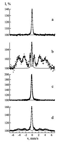
For rice. 2 shows the Mossbauer spectra obtained using Emission Mossbauer Spectroscopy (EMS) on 57Co (57Fe) cores and Conversion Emission Mossbauer Spectroscopy (CEMS) on 57Fe cores of X18N10T stainless steel subjected to heat treatment and plastic deformation.
The sample for research by emission Mossbauer spectroscopy was prepared by applying radioactive 57Co to a stainless steel foil X18N10T by electrolytic method followed by thermal diffusion of the isotope into the sample matrix at a temperature of 700°C in high vacuum.
A radioactive preparation of non-carrier cobalt chloride obtained on the u-150 isochronous cyclotron of the NNP NNC RK by the 58Ni(p,2n)57Co nuclear reaction was isolated from a Nickel target by radiochemical method after it was irradiated with protons.
Part of the foil was used for KEMS. Heat treatment and plastic deformation of the samples were performed under the same conditions. It is known [4] that the most effective resonant γ-quanta come from a depth of 1-2 microns. Therefore, in the case of EMS, information about the state of the mössbauer atoms is obtained from the volume of the sample up to 2 microns thick, and in the case of CEMS – from the near-surface layer up to 0.1 microns thick.
From Figure 2 it is seen that the spectra of annealed samples show monolines characteristic of austenitic stainless steel structure. The spectra of rolled samples (the degree of plastic deformation of 30%) against the background of the austenitic phase show the presence of martensite. Moreover, the presence of martensite in the near-surface layer is much greater than in the volume. Consequently, the austenitic-martensitic transformation during rolling on rollers is more intensively affected by the surface of steel.
The design of a gas-discharge avalanche detector is not much different from the design of a gas-discharge proportional electron detector. The proportional detector uses tungsten as the anode material, which has a large cross-section of the photoelectric effect, which leads to the formation of non-resonant photoelectrons and, as a result, to a decrease in the resonant effect. The difference between an avalanche detector and a proportional one is due to the design of the anode and the gas filling. In the avalanche detector, instead of a tungsten filament, a beryllium plate transparent for γ-quanta is used as the anode. Its surface, facing the cathode, is polished. The gap (3 mm) between the cathode (test sample) and the anode is the sensitive volume of the detector. When preparing the detector for operation, the chamber was pumped out with a fore-vacuum pump and filled with acetone or alcohol vapors to a pressure of 80 kgf / cm2.
The flow of γ-quanta from the Mossbauer source passes through an aluminum foil filter window, then through a beryllium plate and a sensitive volume, and falls on the sample surface. Given that the detector is filled with gas at a relatively low pressure, the registration of γ and x-ray quanta can be ignored. A low-energy electron released from the sample surface causes gas ionization, which creates an electron-ion avalanche in a strong electric field of a sensitive volume, and in the power supply chain, the detector is a source of a voltage pulse corresponding to the electron registration act. When the voltage at the anode increases, the counting rate increases monotonously until an independent gas discharge begins in the detector. Stable operation of the detector is observed when the voltage at the anode is 10% lower than the limit. A detailed analysis of the operation of the gas-discharge avalanche electron detector can be found in [2]. Note only some advantages over other methods of detecting electrons.
In an avalanche detector, smaller electron energies correspond to larger pulse amplitudes, which causes a reduced efficiency to register non-resonant medium-energy electrons. Since the detector is ineffective against other types of radiation, it is possible to lower the background level, i.e. increase the resonant effect and conduct studies of radioactive samples.
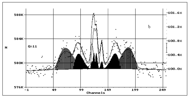
Figure 3 shows the spectrum of a sample cut from the spent fuel rod cover of the BN-350 power reactor with subsequent thinning to a thickness of 0.34 mm by grinding and electropolishing. Despite the high activity of the sample, it was possible to obtain a satisfactory Mossbauer spectrum via an electronic channel using a gas-discharge avalanche detector.
A methodological basis has been created for observing the resonant absorption of γ - quanta by 57Fe nuclei. It is shown that the methods of absorption, reflection, emission and conversion Mossbauer spectroscopy allow almost without restrictions on the element composition and geometric dimensions, to study metals, alloys and chemical compounds of metallurgical, geological, mineralogical, biological and other fields of science and technology [22-24]. The created CEMS technique allows us to study iron-containing samples with a natural content of 57Fe. It is shown that the use of CEMS significantly expands the range of scientific and practical problems that can be solved using nuclear gamma-resonance spectroscopy.
- Goldansky VI (1970) Chemical applications of mössbauer spectroscopy. Mir 502.
- Spinel VS (1969) Resonance of x - rays in crystals. Moscow: Nauka.
- Wertheim G (1966) The Messbauer Effect. Principles and application. Moscow: Mir.
- Irkaev SM, Kuzmin RN, Opalenko AA (1970) Nuclear gamma resonance (equipment and methods). Moscow: Iz-vo MSU 207.
- Kalashnikova VI, Kozodaev MS (1966) Detectors of elementary particles. Moscow: Nauka 150.
- Shokanov AK (1985) Investigation of interaction of point defects in molybdenum and niobium by the method of nuclear gamma resonance: dissertation of the candidate of physical and mathematical Sciences.
- Ibragimov ShSh, Vereshchak MF, Zhantikin TM (1986) Study of plastic deformation and proton irradiation influence on phase transitions in C18N9T stainless steel. Hyperfine Interactions 29: 1293-1296. Link: https://bit.ly/3i3ggOr
- Zhetbaev AK, Vereshchak MF, Zhantikin TM (1986) Investigation of radiation demage in amorphous Fe70Ni10P13C7. Hyperfine Interactions 29: 1297-1300. Link: https://bit.ly/2XqEetw
- Manakova IA, Vereshchak MF, Sergeeva LS, Shokanov AK, Antonyuk VI, et al. (2010) Regularities of thermally induced phase formation in - Fe with titanium coating. FMM 109: 483-496.
- Sergeeva LS, Vereshchak MF, Manakova IA, Antonyuk VI, Rusakov VS, et al. (2006) Investigation of the kinetics of phase formation on the surface of a Fe-Ti alloy with a titanium coating. Bulletin of NSC RK 4: 28-36.
- Manakova IA, Vereshchak MF, Sergeeva LS, Yaskevich VI, Antonyuk VI, et al. (2011) Diffusion and phase formation in a layered Fe(10 µm)- Ti(2 µm) system during isothermal annealing. Bulletin of the NAC 110-116.
- Vereshchak MF, Kadyrzhanov KK, Plaksin DA, Rusakov VS, Suslov EE, et al. (2003) Messbauer studies of the iron-aluminum layered system. Habercisi, Physical Series 94: 100.
- Ozernoy AN, Vereshchak MF, Manakova IA, Tleubergenov ZhK, Bedelbekova KA (2017) Nuclear gamma-resonance spectroscopy in the study of nanoscale composites. Nuclear Physics and Engineering 81: 492-496.
- Ozernoy AN, Vereshchak MF, Bedelbekova KA, Manakova IA, Sergeeva LS, et al. (2010) Investigation of nanoscale metal coatings by emission Messbauer spectroscopy. Proceedings of the 7th International scientific conference" Chaos and structures in nonlinear systems. Theory and experiment. Karaganda 358-361.
- Shokanov AK, Vereshchak MF, Manakova IA, Ozernoy AN, Tleubergenov ZhK, et al. (2020) Messbauer studies of iron carbides formed by thermal vacuum deposition. Crystallography 65: 363-366. Link: https://bit.ly/2Lk7504
- Bonchev ZW, Jordanov A, Minkova A (1969) Nucl. Method of analysis of thin surface layers by the Mössbauer effect. Nucl Instr and Meth 70: 36-40.
- Gruzin PL, Petrikin YuV, Rodin a.m. (1971) Observation of the Messbauer effect on 57Fe nuclei by conversion electrons. XII Meeting on nuclear spectroscopy and nuclear theory. Dubna 196.
- Gruzin PL, Petrikin YuV, Stukan RA (1975) Installation for observing nuclear -resonance by conversion electrons. LTE 48.
- Gonzer W (1983) Mössbauer spectroscopy. Unusual applications of the method. Mir 244c.
- Swanson KR, Spijkerman JJ (1970) Analysis of Thin Surface Layers by Fe‐57 Mössbauer Backscattering Spectrometry. J Appl Phys 41: 3155. Link: https://bit.ly/35qPhaA
- Petrikin YuV, Alekseev VN, Bychkov VA (1983) Gas discharge detector for electronic JAGR spectroscopy. Plant laboratory 46-48.
- Vereshchak MF, Manakova IA, Shokanov AK (2019) Messbauer research of minerals contained in the coals of Kazakhstan. Bulletin of the NAC 13-16.
- Vereshchak MF, Zhetbayev AK, Kaipov DK, Satpayev KK (1972) The Messbauer Effect on impurity atoms of 57Fe in quartz single crystals. Solid State Physics 14: 3082 3083.
- Ni LP, Vereshchak MF, Zhetbaev AK, Solenko TV, Goldman MM (1980) Application of Messbauer spectroscopy for the study of sodium aluminoferrites. Izvestiya Vuzov, non-Ferrous metallurgy 136-138.
Article Alerts
Subscribe to our articles alerts and stay tuned.
 This work is licensed under a Creative Commons Attribution 4.0 International License.


 Save to Mendeley
