Global Journal of Cancer Therapy
Atypical Fibroadenoma Presentation of a Case
Juana Teresa Santiago Pérez1*, José Miguel González Barcena2, Yuranis Gárciga Martin3 and Yahumara Suarez Morales3
21st grade specialist and Assistant Professor of General Surgery, University of Medical Sciences of Havana, Cuba 31st degree specialist in General Surgery, Cuba
Cite this as
Santiago Pérez JT, González Barcena JM, Martin YG, Morales YS (2020) Atypical Fibroadenoma Presentation of a Case. Glob J Cancer Ther 6(1): 042-044. DOI: 10.17352/2581-5407.000035Fibroadenoma is the most frequent benign tumor of the breast, although its classification as a true benign tumor or as a mastopathy related to circulating estrogens is discussed today.
They usually occur below 30 years and constitute 76% of breast masses between 10 and 20 years. Bilateralism has been reported between 12 and 16% of fibroadenomas. The type called Juvenile or Giant only reaches 2% of all Fibroadenomas and 7% of all lesions of the breast in children under 20 years.
The most common form of presentation is like a solid, mobile and painless tumor that can acquire a large size.
The diagnosis of these masses is made with physical examination and imaging techniques, mainly ultrasound.
The diagnostic confirmation is given by the histological study.
The surgical treatment must be individualized in each case, taking into account its particularities.
We present the case of a 72-year-old woman who goes to an outpatient clinic for a giant tumor in the breast, imaging and histological studies are possible, and by joint decision in the multidisciplinary team the surgical treatment is decided and surprisingly the histological result , results in a giant fibroadenoma of the breast.
Introduction
Fibroadenoma is the most common benign breast lesion in women below 30 years. They are usually lesions associated with early ages of life [1] Its most common form of presentation is a hard, painless and mobile mass that can have a large size. The diagnosis is made regularly with imaging tests, with ultrasound being the method of choice in young patients, although mammography and nuclear magnetic resonance imaging are diagnostic tools, in women over 35 years. From the histological point of view it is a mixed tumor in which there is proliferation of the intralobular stromal connective tissue, and a cluster multiplication of the ducts [2,3].
Objective
Show a 72-year-old patient with a giant fibroadenoma, whose clinical presentation was unusual.
Method
The corresponding complementary studies were performed: Imaging (ultrasound) mammography is not performed due to local breast conditions; BAAF is performed that is hemorrhagic, so the surgical intervention is decided, after discussion in the multidisciplinary team.
Presentation of the case
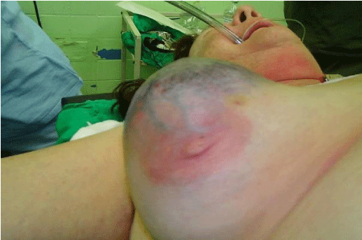
Patient AMP, a 72-year-old female, with a history of hypertension, who has been systematically treated with Captopril and remains under control, reports that since February 2018, she has observed a non-painful increase in the volume of the right breast, with redness of the area and which has grown markedly only in three months. In the rest of the interrogation, there are no data that point to the risk factors for breast cancer.
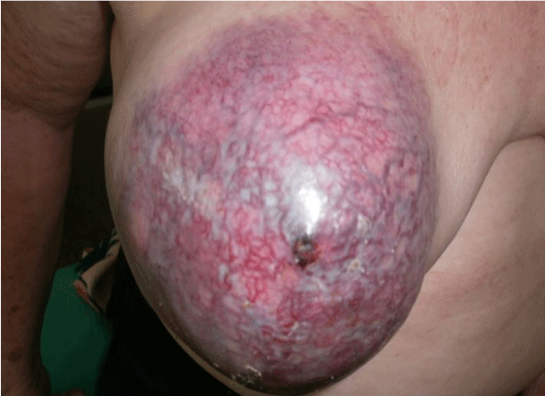
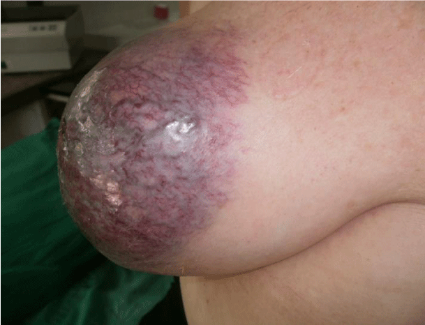
The physical examination shows skin with an increase in the important vascularization, which occupies the central region of the right breast, in a radiated way towards the four quadrants, a tumor mass of firm consistency (not stony) is not felt painfully defined but not defined edges irregular.
The axillary region is examined and ipsilateral lymph nodes, not supraclavicular nodes, are confirmed Figure 1a,b.
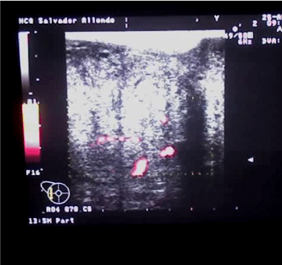
Imaging studies (ultrasound) are performed; Mammography is not performed due to the characteristics of the physical examination of the breast, considering that it was not suitable for the compression of the organ required by this study.
Figure 2 shows the particularities found:
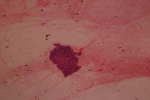
It also proceeds to perform fine needle aspiration biopsy, which does not provide guiding results, as the blades showed extended hemorrhagic, not useful for diagnosis Figure 3.
After discussing the case thoroughly by the multidisciplinary team, it is decided to perform the exeresis of the lesion and confirmation by transoperative biopsy.
This biopsy is negative for neoplastic cells and suggests waiting for paraffin cuts Figure 4a,b.
Pathological anatomy
The results of the paraffin study show the characteristics of a giant fibroadenoma, which in the macroscopic description measured 10 x 13 centimeters. In the histological sections the cellularity of the stroma was demonstrated, as well as the epithelial component Figure 5.
Discussion
Fibroadenoma is a very common benign breast lesion. They are composed of glandular and stromal tissue (connective). Within benign lesions, in terms of frequency, they are only preceded by the Fibrocystic Breast Condition (breast dysplasia) as it is also known [3-5].
Many times they appear during puberty, and in younger girls but they are more frequent in women of little more than twenty years. Other authors report their age of onset, between the ages of 20 and 35 years of age [5]. However, this type of injury is unlikely to appear in postmenopausal women [5,6].
Usually, the reason for consultation is the appearance of a lump in one of the breasts, not painful and fast growing. On physical examination, when they are palpable they can be well defined, they are movable, sometimes giving an elusive sensation, during the exploration. The size ranges from a few millimeters to several centimeters (most are 1 to 3 cm). In those very small lesions, the diagnosis is a fortuitous finding [6-9].
Mammography and ultrasound represent the two fundamental diagnostic pillars in terms of imaging tests. The definitive diagnosis is the histological study, which in large tumors is essential for the diagnosis of benignity, in addition, the histological study will allow the differential diagnosis to be completed with other entities: lipomas, hamartomas, fatty necrosis, giant abscesses, papillomas, etc [7-10].
The treatment should be individualized and whenever possible, especially in large tumors, the removal or excision of the tumor will be performed for the complete pathological study of the piece. In cases of giant tumors, a mastectomy may be necessary. Some authors in these cases advocate a mastectomy with posterior breast reconstruction [9,11,12].
When it becomes necessary to assume aggressive surgical treatment, it is important to take into account the aesthetic result as well as protect the breast, areola and nipple unit since in girls and adolescent patients, it is still in full development [3,8,9].
In the case at hand, we had to resort to mastectomy, as evidently the lesion invaded the entire breast and on the other hand, the result of the paraffin biopsy was really a surprise when reporting a benign lesion, taking into account the age of the patient and the preoperative studies that did not provide the necessary results due to the condition of the skin or the vascularization of the lesion.
This case was against all expected expectations, due to the age of the patient and the peculiarities of tumor presentation.
Conclusion
An unusual form of presentation of the Giant Fibroadenoma is presented, since this disease is usually shown in early ages of female life and it is not usual to appear in older people as it happened in this patient. In addition, it is not frequent to take the skin as it happened in this case, which led the multidisciplinary team to the surgical decision as the first treatment weapon despite the dimensions of the tumor, since the diagnostic means could not offer convincing results.
- Sklair-Levy M, Sella T, Alweiss T, Craciun I, Libson E, et al. (2008) Incidence and management of complex fibroadenomas. AJR Am J Roentgenol 190: 214-218. Link: https://bit.ly/3eE0AQ1
- Smith GEC, Burrows P (2008) Ultrasound diagnosis of fibroadenoma - is biopsy always necessary?. Clin Radiol 63: 511-515. Link: https://bit.ly/3p6NLCJ
- Michalina K, Brian Y, Suela S, Heidi M (2017) Giant juvenile fibroadenoma in a 9-year-old: A case presentation and review of the current literature. Breast Dis 37: 95-98. Link: https://bit.ly/3lcSReb
- Gao Y, Saksena MA, Brachtel EFl, terMeulen DC, Rafferty EA (2015) How to approach breast lesions in children and adolescents. Eur J Radiol 84: 1350-1364. Link: https://bit.ly/3keDoZL
- Sosin M, Pulcrano M, Feldman ED, Patel KM, Nahabedian MY, et al. (2015) Giant juvenile fibroadenoma: A systematic review with diagnostic and treatment recommendations. Gland Surgery 4: 312-321. Link: https://bit.ly/2IansuN
- Rafeek N, Rangasami R, Dhanraj K, Joseph S (2016) Multimodality approach in the diagnosis and management of bilateral giant juvenile breast fibroadenoma. BMJ Case Rep 2016. Link: https://bit.ly/3eHxC1F
- Duflos C, Plu-Bureau G, Thibaud E, Kuttenn F (2012) Breast diseases in adolescents. Pediatric and adolescent gynecology. Evidence-based clinical practice 2nd ed. 22: 208-221. Link: https://bit.ly/3n7KelK
- Van Osdol AD, Landercasper J, Andersen JJ, Ellis RL, Gensch EM, et al. (2014) Determining whether excision of all fibroepithelial lesions of the breast is needed to exclude phyllodes tumor: Upgrade rate of fibroepithelial lesions of the breast to phyllodes tumor. JAMA Surg 149: 1081-1085. Link: https://bit.ly/35cc8qE
- Matz D, Kerivan L, Reintgen M, Akman K, Lozicki A, et al.(2013) Breast preservation in women with giant juvenile fibroadenoma. Clin Breast Cancer 13: 219-222. Link: https://bit.ly/3kjwG4U
- Ross DS, Giri DD, Akram MM, Catalano JP, Olcese C, et al. (2017) Fibroepithelial lesions in the breast of adolescent females: A clinicopathological study of 54 cases. Breast J 23: 182-192. Link: https://bit.ly/3pdVcrs
- Cabrera G, Monduy R, Frías O, Cabrera CA (2017) Tumor phyllodes de la mama. Rev Ciencias Médicas Pinar del Río 15: 11. Link: https://bit.ly/2I96OM1
- Torres L (2012) Tumor phyllodes de la mama: estudio de 32 años. Finlay 2: 8. Link: https://bit.ly/3pbxOLq

Article Alerts
Subscribe to our articles alerts and stay tuned.
 This work is licensed under a Creative Commons Attribution 4.0 International License.


 Save to Mendeley
