Forensic Sci Today
Rigor Mortis and News obtained by the Body’s Scientific Reconstruction of the Turin Shroud Man
M Bevilacqua1, G Concheri2, S Concheri3, G Fanti4* and S Rodella5
2Department of Civil Environmental and Architectural Engineering, University of Padua, Italy
3Orthopedic Clinic, S. Antonio Hospital, Padua, Italy
4Department of Industrial Engineering, University of Padua, Italy
5Rodella’s study, Vigonovo (Venezia), Italy.
(authors are listed in alphabetic order)
Cite this as
Bevilacqua M, Concheri G, Concheri S, Fanti G, Rodella S (2018) Rigor Mortis and News obtained by the Body’s Scientific Reconstruction of the Turin Shroud Man. Forensic Sci Today 4(1): 001-008. DOI: 10.17352/pjfst.000010After various tentative to artistically build a tridimensional form of the Turin Shroud (TS) Man, the authors faced the problem of the scientific construction of a 3D model of this Man with an accuracy of the order of 1 cm.
It is well known that the TS wrapped a human body heavily tortured, but it is not easy to build a 3D model starting only from the 2D information shown on two body images visible on a linen sheet and on the hypothesis that these images really corresponds to a real man wrapped in it.
The study showed that, while the two TS body images (frontal and dorsal) seem to appear not coherent with the image of a human body because distorted in many parts, these two images are perfectly coherent with the distortions provoked by a man wrapped in this linen sheet.
This model confirmed the evident rigor mortis of the human body and evidenced the particular posture corresponding to the position on the cross that also showed a rotation never detected previously of the human body around his spine. The study of this 3D model partially confirmed previous results but also evidenced interesting news, like the position of the exit hole of the nail posed on the palm of the hand.
The Gospels place the Resurrection in the early hours of Sunday, at least 36 hours after death. The effect of the preservation of rigidity up to this moment may be not consistent with the thanato-chronological alterations expected in a man in the same pre-mortal clinical condition, but this problem has been solved by the “mixture of aloe and myrrh” that allowed the preservation of the corpse, thus prolonging the rigor mortis for tens of hours. In fact, this body seems not to have undergone any significant putrefactive phenomenon.
Introduction
The Turin Shroud (TS), is a mortuary linen sheet 4.4 m long and 1.1 m wide [1-3]. That wrapped the corpse of a tortured man, scourged, thorn-crowned, crucified and stabbed in the side [4-6]. A double - front and back - human image is impressed on the TS and it is not explainable by science nor reproduced [7].
Many believe that the TS is the burial cloth in which Jesus Christ was wrapped, but others still think that it is an artist’s work because the 1988 radiocarbon result [8]. Declared it Medieval. Nevertheless this result is debatable [9], for the presence of a systematic effect and more recent studies demonstrated that the TS’ age is compatible with that in which Jesus Christ lived in Palestine [10,11].
There are some clues indicating [12] that the TS was in Palestine in the first century A.D. and then taken to Edessa (current Sanliurfa in Turkey). The strict similarity between the TS face and that of Christ on Byzantine coins (especially the golden solidi and histamena), minted starting from the VII century A.D. up to the Sack of Constantinople in 1204 and later, demonstrates that the TS was known during the Byzantine empire [13]. After this period, the “Shroud of Christ” appeared in Europe in 1353 at Lirey in France and it was fire damaged in 1532, at Chambéry in France.
The TS shows many marks: those produced by blood, fire, and water, that, with the creases, partially obscure the body image, causing scientific analysis to be less easy, but it is clear that this funeral cloth really wrapped a tortured dead man [14,15] characterized by an evident rigor mortis [16].
The monochromatic dark-yellow color of the TS body image has a peculiar feature: the variable intensity of this color can be related to the hypothetical body-cloth distance. In this way the TS image contains 3D information [17,18]. For this reason various scholars tried to reconstruct a 3D Man of the TS trying to interpret the 3D information there contained.
For example L. Mattei [19], L.J. M. Miñarro and P. Soons [20-22] built a life-size sculpture while Fanti et al. [11, 32, 33] built a 3D numerical manikin of the TS Man. Nevertheless, these studies produced interesting results but not completely corresponding to the posture of the TS Man because of the intrinsic difficulty to obtain a sufficiently good result only starting from a 2D information reproduced on a linen sheet with some 3D data obtainable by the hypothesis of a particular draping configuration. In fact the above-mentioned color intensity versus body-cloth distance relation needs a hypothesis that quantifies this distance too. The following section will synthetically explain how the authors have been able to improve these results after building the new 3D model.
Based on the evident rigor mortis of the TS Man, well evidenced during the studies performed to build the new 3D model, this paper revises and improves the results previously published by the same authors [23-26]. They again confirm the sentence: “Only a man sustained by a faith in his mission, conscious of his martyrdom, though young and strong, could bear such a massacre with a deep and absolute peace of mind!” [25].
How the new 3D Model was obtained
In general, a sculptor produces his handwork either by copying a real subject, perhaps adding some subjective artistic interpretation, or by producing an artwork of his own, based on inspiration. This case is instead particular and someway opposite: the aim of the sculptor Sergio Rodella and the supporting scientific group was to rigorously produce a 3D model, starting only from both the information obtainable by a 2D linen sheet reproducing a double image of a man - the TS - and the information relative to the body morphology and size of a standard man. The 3D information related to the color intensity of the body image was instead used to identify the proximity areas between the linen and the body, and therefore the posture of the Man.
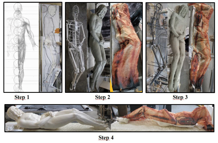
To build the 3D model, a wrapping of the linen around a standard body was iteratively supposed. Linear distances measurements between characteristic points were performed directly on the 2D body image and afterwards flexible physical replicas of such lengths were shaped according to the relevant body morphology, iterating the process in order to reach convergence between frontal and dorsal images. Once defined the 2D median section, a trial-and-error procedure has been followed starting from the construction of an iron skeleton of the TS Man covered by modeling clay, figure 1, and wrapping it with a TS copy in order to match the characteristic points previously recognized on the 2D body images.
Once evaluated the incongruences, the modeling clay was removed, the skeleton adjusted also on the basis of additional models of head, legs, hands and feet properly prepared for the purpose and the wrapping procedure was repeated up to the detection of a congruence of the order of one centimeter between the TS copy and the 3D model.
As a result, the compatibility of the 2D information of the TS image has been verified with the data relative to a standard man also accounting for the sheet distortions produced by the wrapping a 3D human body. In this case, the compatibility was reached selecting a man whose legs are about 3 cm longer than the standard. 2D human shapes, that appeared not real from a first sight on the TS image, resulted instead perfectly coherent with the distortions produced by the imprint of a 3D man on a wrapped sheet.
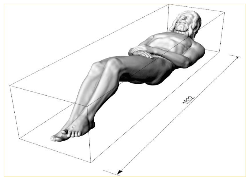
In addition, the more or less tight configuration of the TS wrapping the Man has been studied; in fact the 3D relation of the body image intensity with the cloth distance has been detected but not quantified. A memory about this problem has been presented [27]. In reference to the face wrapping and a relatively tight wrapping resulted: the maximum face-cloth distance resulted of 10 mm (Figure 2).
Thanato-cronology of the Shroud Man
According to the Gospel account, Jesus was deposed not before 5:00 pm, just after Pilate’s consensus to Joseph of Arimathea to depose the body of Jesus and after he had the time to purchase the sepulchral linens.
Jesus was violently scourged with numerous skin wounds, ragged-bruised; he was fatigued by the enormous workload endured during the transport of the cross to Calvary; he had been massacred by the falls to the ground when carrying the heavy cross, nailed and bleeding, exhausted on the cross for causalgia and the effort to breathe, thirsty and feverish in a state of traumatic, hypovolemic, orthostatic, hypoxic, metabolic shock, all conditions which have made an almost immediate onset of rigidity after death. However this death occurred in full consciousness and presupposes the onset of a very short duration post-mortal flaccidity.
The temporal evolution of the rigor mortis must have been much faster than the normal one observed in a subject dead of natural death, without nutritional affections, without having suffered violence and with muscular integrity, which develops an intense, late and lasting rigidity.
The thanato-chronology deals with the death timing and it both considers the destructive transformative and the consecutive thanatological phenomena of the cadaver, that are modifications immediately occurring after the post-mortal period [28].
Thereafter Jesus, at 5:00 pm, was in a state of complete and persistent rigidity not only until the burial despite the manual operations to which he was subjected but also until resurrection.
But here things get complicated. If the TS shows the body image of Jesus, why does it represent a man still in its evident rigor mortis if, in similar pre-mortal physical conditions, another man would have gone through advanced destructive and putrefactive processes? What delayed these processes? Perhaps He resurrected long before Sunday? Is perhaps the TS a fake?
For brevity and greater clarity, we have summarized in table 1 the probable thanato-chronology of the TS Man by comparing it with that of a standard man, according to the literature data [29], and at the same time with the events reported in the Gospels.
Results of the body reconstruction of the TS Man
The construction of the 3D model of the TS Man allowed to obtain the following results, some of them new and not previously expected. The resulting human body produced by the detailed study is of extraordinary beauty, well proportioned, fixed in his post-mortal collapse still hanging on the cross, apart from the recomposition of the head, arms and feet; He shows evident signs of intense muscular effort along the whole body, isometric type, with muscular prominence, and respiratory reliefs.
The 3D model was built in reference to the photo of the TS made by Giandurante in 2002 and then was referred to a man 180 cm tall; Figure 2 shows the resulting posture of the model that from the top of the head to the tiptoes measures 183 cm. Nevertheless it must be considered that the reference photo was taken in 2002 just after the heavy intervention during which the patches, put on the Relic in the XVI Century after the Chambéry fire, were removed and the TS was stretched up to 8 cm from one edge to the opposite one. In this occasion it results that the TS Man was stretched of about 3 cm and therefore the reference model was 177 cm tall before 2002.
The body, having the feet fixed on the cross and the arms stretched with the hands nailed, seems to have slipped along the stipes so that the trunk is only a little detached from the stipes at mid-dorsal level while the shoulders, the neck and the head inflected forward.
The rigor is not coherent with a supine human body, but with a man crucified in a vertical position with his head bent forward [11], caught between the shoulders. These last remained in a raised position even when his arms, in rigor, were moved down [25].
The head, tilted forward of 40°, is 16 cm distant from the stipes at the nape of the neck.
The face was a little different from the usual iconography derived from the TS because the distortions due to the sheets wrapping and wrinkles. It is a bit thin, despite the swelling from beatings, the eyes are sunken (the right eye is slightly more retracted), the eyelids are lowered (the right eye seems almost ajar), the nose is sharp, slightly deformed by the fracture of the nasal septum and the facial muscles are relaxed (it is known that the muscles of the head are the first to contract and the first to relax). Apart from the alterations due to traumas and dehydration, we begin to glimpse the real face of the TS Man. The face obtained expresses a majestic serenity, arousing an intense emotional participation.
Two columns of hair on the face sides probably hide the chin-band, applied during the deposition.
The head is bent forward, bent to the right of 10 degrees; the nape is high and tense.
The right shoulder is lowered by 2.5 cm while the vertebral column is in axis.
The right humerus is lowered by 3 cm, then dislocated or subluxated at the shoulder joint.
The thorax is hyper-expanded, in inspiration, and the maiores pectorales muscles are contracted and prominent.
The lance wound is placed in the sixth intercostal space between the anterior axillary line and the hemiclavear. Route hypothesis spear: the path of the lance with the soldier on the ground. Thorax in inspiration [26].
The shoulder blades are approached to the vertebral column, fixed in rigor mortis, proper to a person who had his arms nailed on the patibulum.
The dorso-lumbar musculature is tense; the lumbar lordosis is light, like a body that slid down the cross when it collapsed.
The right hand is extended, flat, with fingers that lap the outer edge of the left thigh, elongated for the humeral dislocation.
The left hand is superimposed on the right with flexed fingers that seem to “hold” the other hand: this supports our hypothesis: the “claw hand” due to proximal lesion of the ulnar nerve was probably recomposed by stretching the fingers that then contracted partially again preventing the separation of the hands.
The arms give the impression of a flaccidity with the elbows not tense but resting on the sides, sign of poor rigidity. This is compatible with the orthostatic hypoperfusion of the muscles of the upper body in a man hanging from the cross.
The left thumb is retracted; the right is covered by the overlying hand.
The exit hole of the nail on the back of the left hand is placed under the carpo-metacarpal joint just distal to the bases of the third and fourth metacarpal bones. It’s an unexpected datum because a more proximal nailing was hypothesized in Ref [25].
The epigastrium is hollowed, in the inspiration phase and in the position of a man with arms stretched out by nailing. The hypogastrium is prominent, probably due to the incipient putrefactive phenomena with intestinal gas formation, while not all the other parts of the human body evidence any sign of putrefaction. According to the authors, it is the proof that if the TS Man was Jesus, he was a true man up to the resurrection.
The buttocks are well contracted, fixed by the rigidity. The right, better imprinted, was interpreted with a greater flattening on the 3D model, leading to think to a greater pressure of this body’s side on the cross.
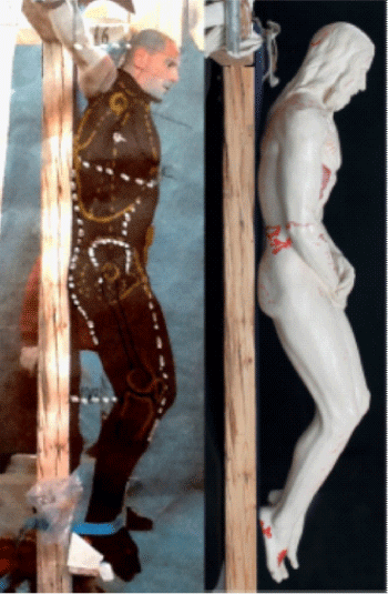
The knees are bent: the right one with an angle of 45°; the left of 50°. The left knee is a bit more raised than the right one. This flexion resulted similar than that found in our in vivo experiment, of a young man of the same age and height, tied to the cross (Figure 3).
The thighs and legs are closely juxtaposed.
The feet are plantar hyperflexion and parallel, the left higher. The right foot rests on the stipes only with the heel while the plant is slightly detached, with a foot-stipes angle of 16°. This suggests that under the foot, some subtle support was perhaps put, like a rudimentary suppedaneum, or, more likely, it was practiced a hollow with by means of a gouge on the stipes also to increase the suffering of the condemned. The left foot is also slightly turned inwards, resting on the right side of the malleolus. Probably the feet were moved from their superimposition but the rigor mortis fixed them in this position.
The entrance hole of the nail, in the right foot, has been identified between the third and fourth metatarsal bone, taking into account the denser part of the TS bloodstain on the back of the foot; the exit hole is easily recognizable at about 40% of the length of the foot starting from the heel. The path of the nail is compatible with the nailing of the left foot on the right, with our experiments on plaster casts figure 4. Evidently the nail went out from the soft parts of the sole of the foot.
The entrance hole in the left foot has been hypothetically identified in the first intermetatarsal space, a position that allows the crossing of the feet and their nailing.
Comments
The scientific reconstruction of the 3D model of the TS Man allows the following comments
1. The lengthening of the right upper limb and the lowering of the right shoulder are confirmed. These two data together with the posture of the hand, the extended fingers, and probably the retraction of the right eye confirm the author’s opinion that the TS Man suffered a severe trauma to the neck, chest and shoulder, probably by falling under the weight of the cross: He also had the dislocation, or at least subluxation, of the right shoulder and injury to the entire right brachial plexus with limb paralysis, ipsilateral Claude-Bernard-Horner syndrome (palpebral ptosis, enophthalmos, miosis) due to paralysis of the stellate ganglion located at the neck base.
2. The same posture of the left hand with flexed fingers seems compatible with a claw-like hand attenuated during the recomposing maneuvers of the corpse on the sepulchral stone. (In fact, the typical hand of a crucifix is the so-called “blessing hand” from injury to the median nerve). This validates the author’s hypothesis that the TS’ convict suffered a strong stretching of the left arm with ulnar nerve injury during the crucifixion.
3. Head position. The iconographic tradition depicts Jesus Christ on the cross with his head bent forward, but turned to the right and this likely originated from direct testimony. This posture of the head can be explained by the consequences of the fall of Jesus, which has led to a paralysis of the posterior muscles of the neck saving the sternocleidomastoideus and so with a posture similar to a person with wryneck. It’s logic to think that the head has been straightened during the positioning of the body in the sepulcher, before being placed in the TS. This operation was quite easy, despite the rigor mortis, since the right of the neck muscles were largely paralyzed.
4. A nail with an exit hole on the back of the hand in the third intermetacarpal space, near the bases of the third and fourth metacarpal bones, until now has never been suggested neither from us nor from other scholars because it appears unlikely. Indeed, a nailing at this level would be little performable and inadequate to support the applied forces because without ligaments. In this case, the hand would have torn and probably would not have supported the body if cords posed along the arms on the patibulum did not bind it.
We tried to imagine a Roman soldier piercing his wrist immediately proximal to the transverse ligament of the carpus (knowing that this ligament “holds” ...) and then tilting the nail trying to reach the hypothetic preformed hole on the wood of the patibulum, which was probably distant a little further. (Due to the low quality of the iron nail, the hole in the patibulum was probably preformed.)
As the nail progresses obliquely under the transverse ligament of the carpus (and in direct contact with the median nerve!), it would have travelled the soft tissues of the palm, thus reaching the bone plane at the beginning of the space between the third and fourth metacarpal bones.
At that point the soldier would have straightened the nail (evidently deforming and “pulling” the soft palm tissues, including ligamentum carpi transversus) and the nail would come out dorsally at the above mentioned level, and finally penetrating the hole in the wood. In other words the nail would then have been used as a “lever” that allowed the soldier to additionally pull the hand by using the hole in the wood. This could explain why the nail remained at the proximal end of the space between the third and fourth metacarpal bones,, without being dragged more distally.
If, in fact, the nail was introduced “transversely” and not obliquely as here hypothesized, or if even the palm of the entry hole was located in the most proximal part of the space between the third and fourth metacarpal bones,, we expect a different situation from that of the TS, and described by the 3D model.
We think that, when passing into the narrow proximal space between the third and fourth metacarpal bones, the large nail “slides” spontaneously more distally, because the space is narrow at the level of the metacarpal bases and the cortical bones of the two metacarpals demarcating it, are inclined and approach each other until mutual contact.
In short, it is expected that the dorsal exit hole visible on the TS is more distal, in the relatively wider space between the third and fourth metacarpal bones, which corresponds to their diaphysis to the middle third (and not to the narrower space corresponding to their proximal third).
So, what could have held the nail forcing it to remain in a position so unlikely, and then to resist to the weight of the body, without imagining any reinforcing system, like ligamentum carpi transversus, or cords? With this hypothesis, the popular and iconographic tradition that places the nailing in the palm of the hand takes its merit again. Obviously, all this explanation applies to the left hand, while the nailing hypotheses already formulated by the authors for the right hand remains valid. In addition to this we must remember that also the position of the entrance hole is not visible on the TS.
5. It is known the antibacterial and antifungal action of the terpens contained in the myrrh [29]; the same applies to the aloe vera [30]. Their action was known since antiquity: in the first century AD Pedanius Dioscorides, in his treatise “De Materia Medica” suggested “anti-putrid” and “conservatives” aromas, and he indicated mixtures aloe and myrrh in specified proportions.
Claudius Galenus (129-200 A.D.) suggested sprinkling the burials with aloe and myrrh for their dehydrating without corroding qualities: “Vis siccandi citra mordacitatem” during the plague of 166 A.D. in Rome. Aetius, archiater of the Constantinople court, in his treatise on the conservation of corpses, recommended, sufficient for the purpose, one pound of myrrh and one of aloe [31].
Natron, that is a white powder of sodium carbonate, was also used in funeral practice and even more for mummifications. It was extracted from the immense salt ponds of the Nile’s delta and it has a strong dehydrating action (a sodium carbonate molecule can bind to ten molecules of water: Na2CO3.10H2O), thus maintaining cadaveric rigidity and retarding the muscle fibers autolysis.
6. Jesus was treated as a king during the burial procedure because his body was laid down with care over a “mattress” of spices strewed on the gravestone. However, the human body was not completely relaxed but rigidly bent to the knees and to the head. But the TS must have been in contact on the whole back side of the human body to be marked by the explosion of energy responsible of the image, even in the hollow of the knees and under the head that must be therefore placed some decimeters far from the sepulchral stone.
We must therefore imagine something that was put underneath head and knees to support the sheet in proximity to the human body: we can imagine something like the sheets used to transport the corpse from the cross to the sepulcher, or the sudaria, the large handkerchiefs used to cleanse the body, or pillows, or rolls of linens impregnated with spices.
7. The problem regarding the real procedure used to nail hands and feet is not completely solved and the creation of this 3D model has added other reasons for new studies. The TS never ceases to surprise, to push to formulate new and more detailed hypotheses and ... to err. The important for a researcher is to be humble and to be ready to revise the hypotheses previously formulated on front of new scientific information!
Conclusion
On the basis of the experience acquired by the authors that already faced the problem of the construction of a 3D model of the TS Man [11,19,20,22,32,33], it is here presented the construction of a scientific model of the TS Man with an accuracy of the order of 1 cm.
This model confirmed the evident rigor mortis of the human body shown on the most important Relic of the Christianity and evidenced the particular posture coherent with the position on the cross, a part from arms, head and feet that were partially moved during the burial procedure.
This very peculiar posture that also shows a rotation never detected of the human body around his spine, has been here studied from a medical point of view thus confirming many hypotheses previously formulated [23-26], but also evidencing interesting news like the position of the exit hole of the nail posed on the palm of the hand and not on the wrist [32,33].
Our research has also tried to clarify the evidence of the preservation of rigidity up to the moment of the Resurrection that the Gospels place in the early hours of Sunday, at least 36 hours after death.
In fact, it is not in line with the standard opinion relative to the thanato-chronological alterations that would have occurred in a man in the same premortal clinical condition. Nicodemus seems the solution to the problem because his “mixture of aloe and myrrh”, a regal quantity of a hundred pounds, a composition well known in the funeral art, allowed the preservation of the corpse thus prolonging of the rigor mortis for tens of hours.
This work, in addition to those already published [23-26], evidences additional correspondences between the TS Man and the description of both the Jesus’ Passion in the Gospels and in the Christian Tradition. These clues are in favor of the previous formulated hypothesis that the TS Man is Jesus of Nazareth who resurrected from the death.
- Antonacci M., (2016) Test The Shroud: At the Atomic and Molecular Levels, Forefront Publishing Company; 1st edition.
- Fanti G, Malfi P, Crosilla F (2015) Mechanical ond opto-chemical dating of the Turin Shroud. MATEC Web of Conferences. Link: https://goo.gl/nAsFLL
- Oxley M (2010) the Challenge of the Shroud: History, Science and the Shroud of Turin. Author House.
- Adler A (2002) A Shroud Spectrum Int. Special Issue, Effatà Editrice, Torino, Italy.
- Jumper EJ, Adler AD, Jackson JP, Pellicori SF, Heller JH, et al. (1984) A comprehensive examination of the various stains and images on the Shroud of Turin. Archaeological Chemistry III 22: 447-476. Link: https://goo.gl/B3L2Lx
- Schwalbe LA, Rogers RN (1982) Physics and chemistry of the Shroud of Turin, a summary of the 1978 investigation. Analytica Chimica Acta 135: 3-49. Link: https://goo.gl/KL8fmH
- Fanti G (2011) Hypotheses regarding the formation of the body image on the Turin Shroud. A critical compendium. J of Imaging Sci Technol 55: 060507. Link: https://goo.gl/yYwEr5
- Damon PE, Donahue DJ, Gore BH, Hatheway AL, Jull AJT, et al. (1989) Radiocarbon dating of the Turin Shroud. Nature 337: 611-615. Link: https://goo.gl/vMZoxh
- Riani M, Atkinson AC, Fanti G, Crosilla F (2012) Regression analysis with partially labelled regressors: carbon dating of the Shroud of Turin. Stat Comput 23: 551. Link: https://goo.gl/5puduW
- Rogers RN (2005) Studies on the radiocarbon sample from the Shroud of Turin. Thermochimica Acta 425: 189-194. Link: https://goo.gl/5RkM7Q
- Fanti G, Basso R, Bianchini G (2010) Turin Shroud: Compatibility between a Digitized Body Image and a Computerized Anthropomorphous Manikin. J of Imaging Sci Technol. Link: https://goo.gl/P8uMDi
- Wilson I, Miller V (1986) The Mysterious Shroud. Doubleday Image Book.
- Fanti G, Malfi P, Conca M (2015a) Turin Shroud -First Century after Christ. Pan Stanford Pub. Ltd., Danvers.
- Carlino E, De Caro L, Giannini C, Fanti G (2017) Atomic resolution studies detect new biologic evidences on the Turin Shroud, PLoS One. Link: https://goo.gl/emKwh2
- Laude JP, Fanti G (2017) Raman and Energy Dispersive Spectroscopy (EDS) Analyses of a Microsubstance Adhering to a Fiber of the Turin Shroud, Applied Spectroscopy. Link: https://goo.gl/dmGJmH
- Faccini B, Carreira E, Fanti G, De Palacios J, Villalain J (2009) The Death Of The Shroud Man: An Improved Review”, Proc. Shroud Conf., Ohio, Libreria Progetto, Padova, Italy. Link: https://goo.gl/GsJRxj
- Jackson JP, Jumper EJ, Ercoline WR (1982) Three dimensional characteristic of the Shroud Image”, IEEE Proceedings of the International Conference on Cybernetics and Society 559-575.
- Jackson JP, Jumper EJ, Ercoline WR (1984) Correlation of image intensity on the Turin Shroud with the 3-D structure of a human body shape. Link: https://goo.gl/xT1AgJ
- Mattei LE (2005) Il corpo dell'uomo della sindone. Sculture di Luigi Enzo Mattei, Giornalisti Associati Ed.
- Miñarro L JM (2007) Reconstrucción Anatómica Tridimensional Basada en el Sudario de Oviedo y la Síndone de Turín, Oviedo Relicario de la Cristiandad, Actas del II Congreso Internacional sobre el Sudario de Oviedo, Oviedo 691-714.
- Miñarro L JM (2012) Recosntrucción Plástica Del Hombre De La Sindone, Int. Congess on the Turin Shroud, 1st Int. Congress on the Holy Shroud in Spain – Centro Español de Sindonologia.
- Soons P (2008) The Shroud of Turin, The Holographic Experience”, Int. Conf. on the Turin Shroud, Columbus, Ohio, USA. Link: https://goo.gl/miASNz
- Bevilacqua M, Fanti G, D’Arienzo M, De Caro R (2014) Do we really need new medical information about the Turin Shroud? Injury 45: 460-464. Link: https://goo.gl/4ts3TL
- Bevilacqua M, Fanti G, D’Arienzo M, Porzionato A, Macchi V, et al. (2014) How was crucified the Man of the Turin Shroud? Injury 45: 142-148. Link: https://goo.gl/okks37
- Bevilacqua M, Fanti G, D’Arienzo M (2017) New Light on the Sufferings and the Burial of the Turin Shroud Man. Open J Trauma 1: 047-053. Link: https://goo.gl/wTytk7
- Bevilacqua M, Fanti G, D’Arienzo M (2017) The Causes of Jesus’ Death in the Light of the Holy Bible and the Turin Shroud. Open J Trauma 1: 37-46. Link: https://goo.gl/fppPHY
- Concheri G, Fanti G, Rodella S (2017) Hypothesis of face-cloth wrapping of the Turin Shroud Man: experimental and numerical results, Int. Conf. on the Turin Shroud, Richland.
- Villalain JD (2010) Study of the cadaveric rigidity presenting in the Shroud of Turin, Cuad Med Forense16:109-123.
- Dolara P, Corte B, Ghelardini C, Pugliese AM, Cerbai E, et al. (2000) Local anaesthetic, antibacterial and antifungal properties of sesquiterpenes from myrrh. Planta Med 66: 356-358. Link: https://goo.gl/8Z9kdj
- Banu A, Sathyanarayana B, Chattannavar G (2012) Efficacy of fresh Aloe vera gel against multi-drug resistant bacteria in infected leg ulcers. Australas Med J 5: 305-309. Link: https://goo.gl/nd7q2v
- Siliato MG. Sindone. Ed. Piemme, Italy (1997).
- Simionato A (1998-1999) Caratteristiche tridimensionali dell’Uomo della Sindone: analisi cinematica con manichino numerico e confronti sperimentali, Degree thesis, G. Fanti tutor, Dipartimento di Ingegneria Meccanica, Padua University.
- S. Faraon (1998-99) Correlazione distanze telo-Uomo della Sindone di Torino per la ricostruzione tridimensionale del’immagine corporea contenuta nella Sindone di Torino, Degree thesis, G. Fanti tutor, Dipartimento di Ingegneria Meccanica, Padua University.
Article Alerts
Subscribe to our articles alerts and stay tuned.
 This work is licensed under a Creative Commons Attribution 4.0 International License.


 Save to Mendeley
