Archive of Urological Research
Stone cobra: “Adult” left single system ureterocele with 139 calculi-case report and review of the literature
Ogwuche Ei1,2*, Ojo Ba3 and Efu Me4
2Visiting Urologist, Department of Surgery, Federal Medical Centre, Makurdi, Benue State, Nigeria
33Department of Anatomic Pathology, College of Health Sciences, Benue State University, Makurdi, Nigeria
4Department of Anaesthesia, College of Health Sciences, Benue State University, Makurdi, Nigeria
Cite this as
Ei O, Ba O, Me E (2020) Stone cobra: “Adult” left single system ureterocele with 139 calculi-case report and review of the literature. Arch Urol Res 4(1): 040-042. DOI: 10.17352/aur.000015A 35yr old man old with no previous major clinical issues presented with a 2years history of radiating left flank pain and dysuria. Abdominopelvic ultrasonography and an intravenous urography confirmed a bladder stone and a left single unit ureterocele harboring multiple stones showing as a typical “Cobra head” with resultant hydronephrosis. A cystolithotomy and incision with marsurpialization of the orthotopic ureterocele with extraction of 139 calculi was done. The post operative recovery was uneventful with resolution of his symptoms. Follow up ultrasonography at three and six months show resolved hydronephrosis with no residual stones seen. Adult orthotopic ureterocele even in the presence of multiple stones has a good outcome after open surgery.
Introduction
An ureterocele is a congenital anomaly characterized by a cystic dilatation of the lower ureter and is often associated with other congenital anomalies of the urinary tract [1].
With a reported incidence of 1:4000 children in Europe and the United States of America [1,2], it is four times commoner in females than males and is described in some reports as an exclusive Caucasian disease [2], though some cases have been reported in Africans and Asians [3-6]. It may be unilateral or bilateral, orthotopic or ectopic. It may occur in a single unit or a duplex system.
Stasis of urine in the dilated distal segment leads to recurrent urinary tract infection and stone formation [4]. Several authors have reported the presence of stones in ureteroceles with Kumar et al reporting 265 calculi in single ureterocle in a 34 year old man [5-11].
We report our experience in treating a 35year old man with multiple (139) calculi in a single left orthotopic ureterocele.
Case report
A 35yr old man presented at the Surgical Outpatient Department of the Federal Medical Centre in Makurdi, Benue State, Nigeria with a two years history of recurrent left loin pains radiating to the left groin and left hemiscrotum associated with dysuria but no lithouria. No fever, pyuria or features of uraemia. His medical and surgical histories were not remarkable. Examination revealed a young man in no obvious distress with no remarkable findings on physical examination.
Abdominopelvic ultrasonography revealed a 1.6cm bladder stone and multiple stones in a cystically dilated left lower ureter with hydroureter and left hydronephrosis. The right kidney was sonologically normal. The complete blood count was not suggestive of infection. The urea, electrolytes and creatinine assays were within normal limits.
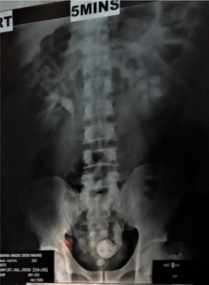
The intravenous urography showed a radiopaque stone in the bladder and a classical “cobra head” with multiple radiopaque stones in the dilated distal left ureter in keeping with multiple calculi in an ureterocele (Figure 1). Both renal units were functional but there was however left hydronephrosis and hydroureter. No stones were seen in the left kidney.
A diagnosis of left orthotopic ureterocele with multiple calculi and a bladder calculus was made.
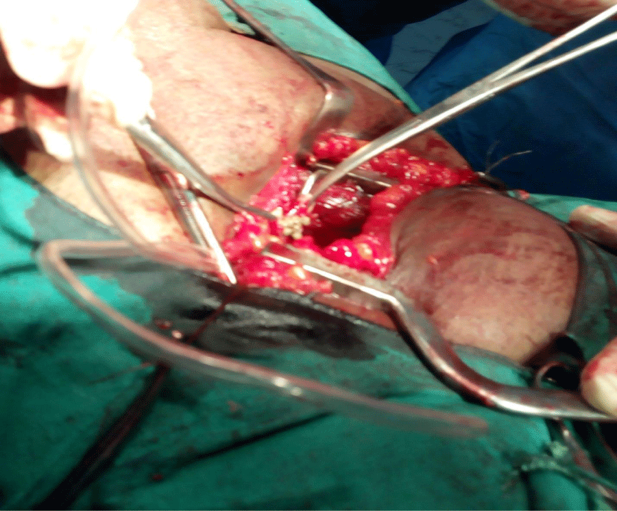
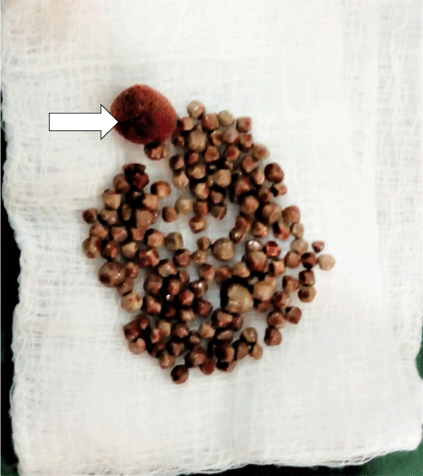
He was worked up for and had a cystolithotomy and open incision and marsupialization of the ureterocele with extraction of one hundred and thirty nine (139) stones from the ureterocele (Figures 2,3). A double J stenting of the left ureter was done.
The post operative recovery was uneventful and He was discharged on the eight post operative day. The double J stent was removed at cystoscopy six weeks post operatively. Follow up ultrasonography at three and six months showed resolved hydronephrosis and no residual stones were seen.
Discussion
An ureterocele is congenital cystic dilatation of the terminal ureter [1]. It is an uncommon anomaly [12]. The incidence is 1:4000 in Europe and North America with a female preponderance (4:1) and said to be an exclusive disease of caucasians [1,2], though a number of case reports have reported in Africans and Asians [3-11]. There is no established incidence in Africa. Eighty percent of ureteroceles arise from the upper poles of duplicated systems [13].
The most commonly accepted theory of its pathogenesis is incomplete dissolution of the schwalla’s membrane.
A simple (orthotopic) ureterocele is a very rare condition and is usually found in adults [12]. The index case is a 35yr old man with a left orthotopic ureterocele with multiple calculi and a bladder calculus. Calculi within ureteroceles are not uncommon [3-14]. Single unit ureteroceles are less likely to obstruct hence their late presentation leading to the term ‘’adult’’ ureterocele [2]. Muhammed A et al in Zaria, Nigeria established a median age of 31years in a report of ten consecutive patients correlating with the age of the index case.
Stasis in the distal dilated ureter leads to recurrent infection and predisposition to stone formation. The incidence of a single stone in single unit ureteroceles has been reported in adults to be 4- 39% [12]. Multiple stones have been reported in adults and even in children [5-13]. The highest number of stones ever extracted from a single ureterocele in world literature is two hundred and sixty five (265) by Kumar et al in a 34yr old Indian man [11]. Our index case with extraction of one hundred and thirty nine stones (139) is the highest number to be ever reported in sub Saharan Africa.
The diagnosis of ureterocele is commonly in prenatal or pediatric age, but occasionally can present in adult too [14]. Ureteroceles can be classified into either single system (having single kidney, pelvicalyceal system, and ureter) or duplex system ureterocele (in which completely duplicated ureteres are present): either orthotopic/intravesical, (when the whole ureterocele lies inside the bladder) or ectopic, (when ureterocele is fully or partially at or beyond bladder neck) [15,16].
An ureterocele may be asymptomatic or may present with symptoms of urinary tract infections, renal abnormalities and stone formation due to stasis of urine from obstruction [15]. Unilateral calculus tends to occurs in 4-39% of cases [10]. The index case presented with unilateral multiple caculi. The clinical presentation of ureteoceles varies widely depending on the age group with children having more acute and life threatening presentations than adults. These presentations have been attributed to the different types of ureteroceles with the ectopic variants and those arising from duplex systems presenting with more severe symptoms while the single unit orthotopic variants present in an unremarkable manner and may be detected incidentally at ultrasonography [17-20].
The myriad ways of presentation leads to many pitfalls in diagnosis of ureterocele. Ultrasonography and intravenous urography are diagnostic in 50% – 70% of cases [20]. Typical findings on ultrasonography is a cystic mass in the bladder neck that may extend to the posterior urethra while an intravenous urography and CT urography show the typical “cobra head” or “spring onion “sign is noted [21] (Figure 1). Cystoscopy is also diagnostic especially if diuresis is induced [20].
The goals of treatment of ureteroceles have been summarized by several authors to include preservation of renal function, elimination of obstruction, infection or reflux, and maintenance of urinary continence in women if the ectopic opening is beyond the external urinary sphincter [22].
Multiple treatment options abound for treating ureteroceles depending on the type, available technology and expertise and they range from minimal access procedures to open surgery [22].
Open incision and marsupialization as done for our patient was done for 30% of patients in a series of ten patients in Zaria, Nigeria [3]. Open surgeries are often reserved for complicated cases and often comes with longer hospital stay and an increased morbidity. Endoscopic treatment offers an advantage of early discharge from hospital; it is safe, minimally invasive, effective and standard approach where the facilities exist. Our patient could have had endoscopic incision and stone extraction of the stones as done by Kumar, et al. [11], but for the non availability of the facility in our resource poor centre.
Conclusion
Open surgery could be used safely with good outcome as an alternative to endoscopic approach in the treatment of ureterocele even in the presence of multiple stones.
- Stephens FD (1968) Aetiology of ureteroceles and effects of ureteroceles on the urethra. Br J Urol 40: 483-487. Link: https://bit.ly/2SSQwZI
- Aas TN (1960) Ureterocele. A clinical study of sixty-eight cases in fifty-two adults. Br J Urol 32: 133-144. Link: https://bit.ly/2SSZxSK
- Muhammed A, Hussaini MY, Ahmad B, Hyacinth MN, Garba KD (2012) Ureterocele in adults: Management of patients in Zaria, Nigeria. Arch int surg 2: 24-28. Link: https://bit.ly/3dx2qAo
- Stephens FD, Smith ED, Hutson JM (1996) Congenital anomalies of the kidney, urinary and genital tracts. Oxford: Isis Medical Media.
- Bhaskar V, Sinha RJ, Purkait B, Singh V (2017) Large bladder calculus masking a stone in single system ureterocele . BMJ Case Rep. Link: https://bit.ly/2xRrUsZ
- Prakash J, Goel A, Kumar M, Sankhwar S (2013) stone in Ureterocele peeping through ureteric orifice. BMJ Case Rep 2013. Link: https://bit.ly/3dBt0rU
- Sarsu SB, Koku N, Karakus SC (2015) Multiple stones in a single-system ureterocele in a child. APSP J Case Rep 6: 19. Link: https://bit.ly/2yMN3F7
- Yuksel MB (2015) Unilateral ureterocele presenting with multiple stones in an old lady: endoscopic incision and stone extraction. Med Sci and Discov 2: 231-232. Link: https://bit.ly/2WlsHfp
- Kaliskan S (2017) Cobra-head stone in single-system ureterocele Iran. J Med Sci 42: 221-222. Link: https://bit.ly/2zvJyTq
- Dominici A, Travaglini F, Maleci M, di Cello V, Rizzo M (2003) Giant stone in a complete duplex ureter with ureterocele. a case report. Urol Int 71: 336. Link: https://bit.ly/2YQJIPX
- Mishra KG, Garg A, Kumar S, Bharti PK, Goel K (2017) Right Sided Ureterocele Presented with Multiple Calculi: A Rare Case Report. J Nephrol Urol Res 5: 1-3. Link: https://bit.ly/3cmW8Di
- Sinha RK, Singh S, kumar P (2014) Prolapsed ureterocele, with calculus within causing urinary refentis in adult female. BMJ Case Rep 2014. Link: https://bit.ly/3bkrNEa
- Schlossel RN, Retick AB (2007) Ectopic ureter Ureterocele and other anomalies of the ureter. In wein AJ, ed Campbell –Walsh urology, 9th edn Philadelphia: Sauders Elsvier 3397-3398.
- Laird RM (2008) Prolapse of an ureterocele in an adult. Br J Urol 26: 72-74.
- Adesiyun OM, Oyenloye OI, Akande HJ. Atobatele MO, Adeniyi WA, et al. (2015) Bilataral giant orthotopic ureterocele appearing as kissing cobra in a Nigerian Child. West Afr J Radiol 22: 42-44. Link: https://bit.ly/3bl64Mf
- Shamsa A, Asadpour AA, Abolbashari M, Hariri MK (2010) Bilateral simple orthotopic ureteroceles with bilateral stones in an adult: A case report and review of literature. Urol J 7: 209-211. Link: https://bit.ly/2Lk8TTf
- Halachmi S, Pillar G (2008) Congenital urological anomalies diagnosed in adulthood. Management considerations. J Paediatr Urol 4: 2-7. Link: https://bit.ly/3coAePZ
- Sarsu SB, Koku N, Karakus SC (2015) Multiple stones in a single-system ureterocele in a child. APSP J Case Rep 6: 19. Link: https://bit.ly/3fFFiBt
- Atta ON<, Alhawari HH, Murshidi MM, Tarawneh E,m Murshidi MM(2018) adult ureterocele complicated by a large stone: A case report. Int J Surg Case Rep 44: 166-171. Link: https://bit.ly/2Luijf7
- Chowdhary SK, Kandpal DK, Sibal A, Srivastava RN (2014) Management of complicated ureteroceles: Different modalities of treatment and long-term outcome. J Indian Assoc Pediatr Surg 19: 156-161. Link: https://bit.ly/3bgDLyl
- Zagoria RJ (2004) Classic signs in uroradiology. Radiographics 24: S247-S280. Link: https://bit.ly/2YXr2hP
- Coplen D, Duckett JW (1995) The modern approach to ureteroceles. J Urol 153: 1-6-171. Link: https://bit.ly/2SVdOy3
Article Alerts
Subscribe to our articles alerts and stay tuned.
 This work is licensed under a Creative Commons Attribution 4.0 International License.


 Save to Mendeley
