Archives of Sports Medicine and Physiotherapy
Lower limb alignment in young female athletes is associated with knee joint moments during the drop vertical jump
Jacques Riad1*, Katarina Hjältman2 and Scott Coleman3
2Department of Orthotics and Prosthetics, Skaraborg Gait Analysis Laboratory, Skaraborg Hospital, Sweden
3Department of Orthopaedics, Baylor University Medical Center, Dallas, Texas, USA
Cite this as
Riad J, Hjältman K, Coleman S (2018) Lower limb alignment in young female athletes is associated with knee joint moments during the drop vertical jump. Arch Sports Med Physiother 3(1): 001-005. DOI: 10.17352/asmp.000008Background: Increased rotational forces and knee valgus forces puts strain on the anterior cruciate ligament, frequently ruptured in female athletes. Increased internal hip rotation and increased knee valgus alignment, possible risk factors for anterior cruciate ligament rupture, are more common in women than men.
Purpose: To study hip, knee and foot alignment related to forces in the knee joint during the drop vertical jump in young female athletes.
Methods: Seventy-one female athletes, mean age 17.1 years (range 14.3-25.7 years) were recruited from regional handball and soccer teams. Participants underwent a physical examination on passive range of motion of the hip, knee and ankle at the Gait Analysis Laboratory. They also performed three drop vertical jumps from 31 cm height onto force plates. A three-dimensional motion analysis system was used.
Results and main findings: Physical examination revealed more internal hip rotation, mean 51.0 degrees (SD 8.5) than external rotation, mean 42.2 (SD 6.8), (p=0.001) and mean 1.1 degree valgus (SD 1.9).
Maximal external knee rotation moments were higher, mean 0.18 Nm/kg, than maximal internal rotation moment, mean 0.13 Nm/kg (p=0.001). The difference between the maximal valgus and varus moments was significant (p=0.001) although equal in size (0.21 Nm/kg), respectively.
The degree of internal rotation of the hip on physical examination correlated positively with maximal external knee rotation moment, correlation coefficient=0.196 (p=0.019) in the drop vertical jump but correlated negatively with knee varus moment correlation coefficient = -0.175 (p=0.038). Thus, increased internal hip rotation is associated with higher external knee rotation moment and lower varus moment.
Conclusion and summary: On physical examination, increased internal hip rotation, often present in females, is associated with increased forces in the knee joint during the drop vertical jump. Increased internal rotation of the hip could be a risk factor for anterior cruciate ligament rupture in young female athletes.
Clinical Relevance: The physical examination of alignment is easy to perform and could help identify individuals at risk of anterior cruciate ligament rupture. These athletes could be informed and possibly undergo special muscle and motor control training.
Abbreviations
ACL: Anterior Cruciate Ligament; DVJ: Drop Vertical Jump
Background
During sport activities, increased knee motion and loading can lead to injury [1,2]. Anterior cruciate ligament (ACL) ruptures are frequently reported in female athletes participating in soccer, handball and basketball, and are mostly non-contact injuries [3]. Inadequate neuromuscular control, hormonal and anatomical factors have been suggested as possible risk factors for knee injuries in women [4].
Among the anatomical risk factors is malalignment of the lower extremity, where increased knee valgus is probably the finding most commonly associated with ACL rupture [5]. Increased knee valgus angle and valgus moment during landing in the drop vertical jump (DVJ) [6,7] and in landing in basketball players are associated with ACL injury [8]. In addition, increased internal rotation of the hip joint, with relative external rotation of the knee as a consequence, may form an injury mechanism, sometimes with combined knee valgus [9,10]. On the other hand, studies on cadavers have shown high stress on the ACL with internal rotation of the tibia relative to the femur [11], also in line with reported internal tibial rotation at the time of rupture [12]. More recently, increased internal tibial rotation forces were reported to place high strain on the ACL [13]. Ishida et al states the knee rotates externally during dynamic knee valgus, and the knee rotation is affected by toe direction [14]. They conclude that especially with the toe-out direction, the ACL may impinge on the femoral condyle in the case of dynamic valgus [14]. Furthermore, Beaulieu et al pointed out that limitation of the internal rotation of the hip joint causes increased strain on the ACL during landing [15].
Internal rotation of the hip joint is generally greater in women than in men, as is the valgus angle of the knee joint, both of which could contribute to the sex difference in ACL injuries [16-18].
Neither the exact mechanisms and forces responsible for ACL rupture during sport activities nor the risk factors in female athletes with a high frequency of injury are fully understood. Nevertheless, it is reasonable to presume that the anatomical alignment of the lower extremity (foot position and the range of motion in hip and knee joints) is associated with the motion and forces arising during dynamic physical activities, and could be important as a risk factor for ACL rupture. We found few reports on anatomical alignment of the lower extremity assessed by physical examination and the association with movements and forces in the knee joint in female athletes during jumping.
The aim was to assess hip, knee and foot alignment through direct physical examination and relate these to movements and forces in the knee joint during the drop vertical jump in young female athletes.
Methods
Participants
Seventy-one female athletes, mean age 17.1 years (range 14.3-25.7 years) participated. Mean weight was 63.6 kg (range 49.0–79.8 kg) and height 168.9 cm (156.0-181.0 cm). Participants were recruited from the main female handball club in the region, and from a female soccer club. The main sport activity was handball for 47 participants, and 15 played mainly soccer. Eight of the athletes participate in both handball and soccer, albeit not simultaneously since the two seasons do not overlap. Sixty-five trained at least 4 times per week and 6 had 3 training passes. Of the 71 participants, 66 were right foot dominant (i.e. preferred their right foot for kicking a ball), 3 were left foot dominant and 3 were unable to answer the question. Individuals with previous serious injury or surgery or any disease/condition influencing the hip and lower limb were excluded.
All participants were provided with oral and written information and gave their written consent to participate. In those younger than 18 years of age, caregivers signed the consent form.
Procedure
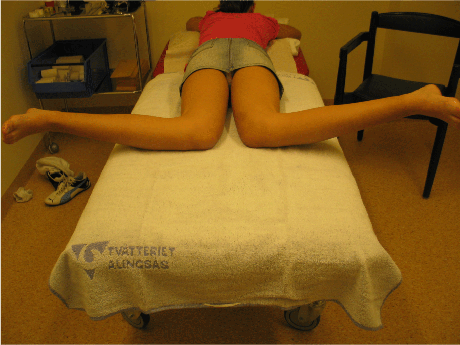
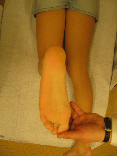
Participants made one visit to the Gait Analysis Laboratory, for a physical examination including passive range of motion of the hip, knee and ankle, using a goniometer at standardized positions [19]. The hip rotation was measured prone with extended hip joints (Figure 1). Bimalleolar angle and foot progression were also measured prone (Figure 2). Knee valgus/varus was measured supine. All goniometer measurements were performed by the same physiotherapist. A three-dimensional motion analysis system incorporating 62 retroflective markers and 12 digital cameras (Oqus 400 Qualisys medical AB, Gothenburg, Sweden) was used to obtain kinematic measurements. The markers were secured to specific anatomical locations on each subject in accordance with a modified Helen-Heyes Model [20,21]. Two force plates (Kistler, Winterthur Wulflingen, Switzerland) were used to obtain the kinetic measurements, the ground reaction force vectors.
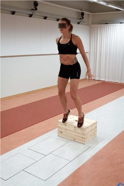
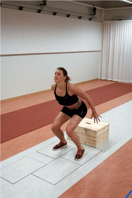
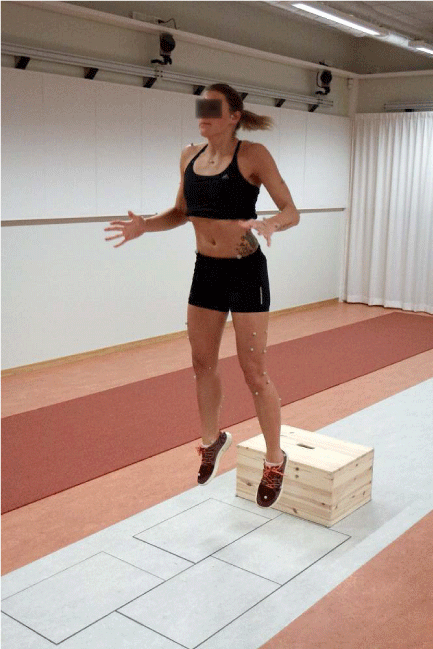
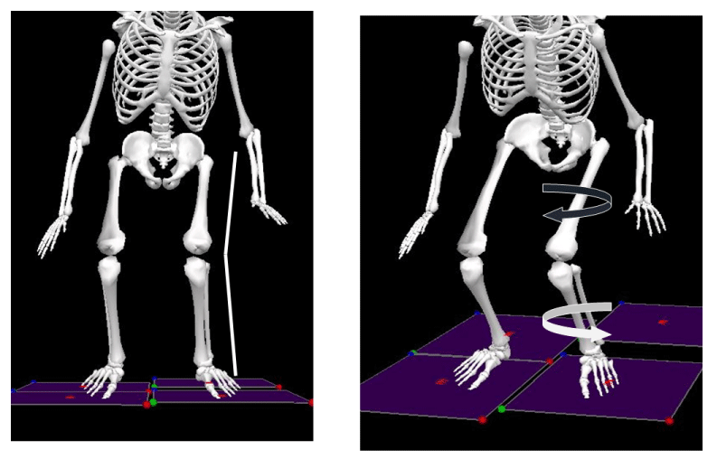
The participants performed 3 DVJs [22], with shoes after a standardized warm up of cycling 5 minutes and stretching. The participants received verbal instructions for the jump and tried 1 or 2 jumps to get familiar with the test. The drop jump starts with the participant standing on a 31-cm high box. She then drops to the floor and on landing immediately jumps as high as possible (Figure 3A, B and C). The right and left feet land on separate force plates positioned side by side. The mean value of the 3 jumps was used. Joint angles and force vectors were calculated using the Visual 3D software (C Motion Inc., USA). From the collected data, the hip and knee motion and moments as well as foot progression were calculated (Tables 1,2; Figure 4).
Statistical analyses
The data were assessed for normal distribution and were calculated accordingly with parametric analysis. The right and left side as well as the dominant and non-dominant sides were compared, and no difference was noted. Therefore, all 142 limbs from the 71 participants were used for further statistical analysis.
Results
Physical examination of the alignment of the hip revealed greater internal rotation, mean 51.0 degrees (SD 8.5), than external rotation, mean 42.2 (SD 6.8), p=0.001. It was noted that the younger the participants, the greater the internal hip rotation on the physical examination, Correlation Coefficient (CCO)=0.258 (p=0.002). The mean valgus/varus on physical examination was 1.1 degree valgus (SD 1.9) (Table 1).
The movements in the hip and knee joints during the drop vertical jump, as well as the foot progression varied considerably within the group.
The maximal valgus moment was 0.21 Nm/kg and the maximal varus moment was 0.21 Nm/kg, p=0.001. In the knee, maximal external rotation moments, were greater than internal rotation moments, mean 0.18 Nm/kg versus 0.13 Nm/kg, respectively, p=0.001 (Table 2).
Internal rotation of the hip on physical examination correlated with maximal external rotation moment in the knee, CCO=0.196 (p=0.019). Thus, greater internal rotation of the hip on physical examination was associated with increased external rotation moment in the knee during the DVJ.
Internal rotation in the hip had an inverse correlation with knee varus moment CCO= -0.175 (p=0.038). Thus, less internal rotation of the hip at physical examination was associated with greater varus moment in the DVJ. No correlation between internal rotation in the hip on physical examination and knee valgus moment was found.
Hip external rotation on physical examination did not correlate with either valgus- or rotational knee moments. Knee valgus/varus, bimalleolar angle and foot–thigh angle (tibial torsion) did not correlate with either valgus/varus- or rotational knee moments.
Discussion
The result from the physical examinations in this study revealed greater internal than external rotation of the hip in young female athletes. The degree of rotation correlated with knee moments measured in the Drop vertical jump.
The greater internal than external rotation in the hip joint, is in line with previous reports [16,18]. Skeletally mature women have greater internal rotation in the hip than skeletally mature men, even though the natural development in all children is that the femur rotation present at birth gradually decreases until skeletal maturity. Interestingly, even in this relatively small study group, the physical examinations revealed that with increasing age, internal hip rotation decreased and external hip rotation increased slightly. However, we found no age differences regarding the forces developed over the knee joint during jumping, probably owing to the small number of participants. We found scarcely any valgus or varus deformity on physical examination, and the bimalleolar angle and foot–thigh angle were within the normal range. In other words, we saw no signs of tibial torsional deformity.
When studying the forces (moments) over the knee joint during the DVJ we found high variability in both frontal plane moments (valgus/varus) and rotational plane moments (external and internal rotation moments). In addition, we noted that the external rotational moment over the knee joint was almost as high as the valgus and varus moments.
Our results showed that greater internal hip rotation on physical examination was associated with greater external rotation moment in the knee joint. This is in line with Amraee et al, who reported a higher risk of ACL rupture in male athletes with greater internal hip rotation of the hip [9]. McLean et al reported that increased valgus moment in female athletes performing sidestep cutting exercise was associated with internal rotation at the hip and knee valgus [10]. However, we did not find increased valgus moment associated with internal hip rotation in our jump study. On the contrary, internal rotation of the hip on the physical examination had a negative correlation with knee varus moment, meaning that lower internal rotation of the hip is associated with higher varus moment in the DVJ. Interestingly, Beaulieu et al reported that limitation of internal rotation of the hip increases the strain on the ACL in simulated pivot landing [15]. We only speculate, but this might be interpreted as indicating that there are different mechanisms to injury, depending on the task performed and the subsequent forces developed over the ACL.
Several previous assessments of strain on the ACL focus mainly on the frontal plane, when studying the risk of ACL rupture. The rotational plane is included in only 5% of publications on the mechanisms of ACL ruptures [22]. In a two dimensional study, Krosshaug et al found that female basketball players ran a higher risk of increased valgus moments in the knee joint than male players [8]. Meyer et al found that valgus angle of the knee is a risk factor for ACL rupture, but their study did not include rotational variables [5]. Hewett et al also reported no correlation with hip internal rotation or rotational moments in the knee but found that decreased knee flexion at landing influenced the risk of ACL rupture [7].
Knee valgus/varus were not associated with knee moments, although previous studies have revealed such an association [5,7,8]. Our negative finding could be explained by the fact that our participants essentially lacked valgus deformity of the knee joint.
In our study, bimalleolar angle and foot–thigh angle (tibial torsion) did not correlate with either valgus- or rotational knee moments. In an experimental study Ishida and co-workers reported association between knee rotations and foot progression [14]. However, since they did not include a physical examination, they had no information on the passive range of motion and the alignment of the knee and adjacent joints, which most likely influence the moments developed in the joint [11]. In addition, the influence of the hip on knee rotations was not addressed in their study.
Several factors are likely to play a role in ACL ruptures in female athletes, among them anatomical and alignment variables. One of the goals of this and previous studies has been to find a practical, easy-to-perform screening test that can be used by trainers and team leaders [5] to identify those with increased risk of ACL rupture. It is of importance not just to be able to identify those at risk of ACL injury, but also to develop strategies/training regimens to prevent injury or at least inform the individuals identified through such screening about the possible increased risk of injury associated with certain sport [22].
One limitation of this study was the small number of participants. Another is that hip rotation was assessed only with extended hips. It would have been useful to know the degree of possible rotation with flexed hips, since the hips are substantially flexed in landing.
Perhaps the most important limitation is the representativeness of the DVJ itself. This laboratory setting does not come close to replicating the forces over the knee joint in a game or training situation, where these injuries occur. Furthermore, there is obviously more movements of the soft tissue overlying the anatomical landmarks that the applied skin markers are representing. This means that the movement of the skin markers, in the high impact performance the DVJ is, makes it difficult to appreciate the exact accuracy of the measurements, even though we added more markers to the model as to increase the accuracy. This study is an attempt to better understand the anatomic risk factors and may hopefully in the future contribute towards studies that more realistically mirror the forces involved in competitive sport activities.
Conclusion
On physical examination, increased internal hip rotation, often present in females, is predictive of increased forces in the knee joint during the drop vertical jump. Although hip and tibial torsion are highly variable, increased internal rotation of the hip could be a risk factor for ACL rupture in young female athletes.
Our results indicate that hip joint motion is associated with knee moments and should be considered in the overall assessment in athletes. The physical examination is easily performed and could help identify individuals with increased risk of ACL rupture.
- Besier TF, Lloyd DG, Cochrane JL, Ackland TR (2001) External loading of the knee joint during running and cutting maneuvers. Med Sci Sports Exerc. 33:1168-1175. Link: https://goo.gl/H1i255
- Boden BP, Dean GS, Feagin JA, Garrett WE (2000) Mechanisms of anterior cruciate ligament injury. Orthopedics. 23:573-578. Link: https://goo.gl/6xuNiZ
- Agel J, Arendt EA, Bershadsky B (2005) Anterior cruciate ligament injury in national collegiate athletic association basketball and soccer: a 13-year review. Am J Sports Med. 33:524-530. Link: https://goo.gl/VQhYSn
- Hewett TE (2000) Neuromuscular and hormonal factors associated with knee injuries in female athletes. Strategies for intervention. Sports Med. 29:313-327. Link: https://goo.gl/Vrkt67
- Myer GD, Ford KR, Khoury J, Succop P, Hewett TE (2010) Development and validation of a clinic-based prediction tool to identify female athletes at high risk for anterior cruciate ligament injury. Am J Sports Med. 38:2025-2033. Link: https://goo.gl/RzcaVs
- Ford KR, Myer GD, Hewett TE (2003) Valgus knee motion during landing in high school female and male basketball players. Med Sci Sports Exerc. 35:1745-1750. Link: https://goo.gl/2uixFt
- Hewett TE, Myer GD, Ford KR (2005) Biomechanical measures of neuromuscular control and valgus loading of the knee predict anterior cruciate ligament injury risk in female athletes: a prospective study. Am J Sports Med. 33:492-501. Link: https://goo.gl/kp9Dyy
- Krosshaug T, Nakamae A, Boden BP (2007) Mechanisms of anterior cruciate ligament injury in basketball: video analysis of 39 cases. Am J Sports Med. 35:359-367. Link: https://goo.gl/2uCzkh
- Amraee D, Alizadeh MH, Minoonejhad H, Razi M, Amraee GH (2015) Predictor factors for lower extremity malalignment and non-contact anterior cruciate ligament injuries in male athletes. Knee Surg Sports Traumatol Arthrosc. 5:1625–1631 Link: https://goo.gl/76n6Md
- McLean SG, Huang X, van den Bogert AJ (2005) Association between lower extremity posture at contact and peak knee valgus moment during sidestepping: implications for ACL injury. Clin Biomech (Bristol, Avon). 20:863-870. Link: https://goo.gl/zc9PRQ
- Markolf KL, Burchfield DM, Shapiro MM, Shepard MF, Finerman GA, et al. (1995) Combined knee loading states that generate high anterior cruciate ligament forces. J Orthop Res. 13:930-935. Link: https://goo.gl/3dYkcd
- Arnold JA, Coker TP, Heaton LM, Park JP, Harris WD (1979) Natural history of anterior cruciate tears. Am J Sports Med. 7:305-313. Link: https://goo.gl/Je4oph
- Oh Y. K LDB, Ashton-Miller JA, Wojtys EM (2012) What Strains the Anterior Cruciate Ligament During a Pivot Landing? The American Journal of Sports Medicine. Link: https://goo.gl/NRP29D
- Ishida T, Yamanaka M, Takeda N, Aoki Y (2014) Knee rotation associated with dynamic knee valgus and toe direction. Knee. 21:563-566. Link: https://goo.gl/ihK53T
- Beaulieu M.L YKO, Bedi A, Ashton-Miller JA, Wojtys EM (2014) Does Limited Internal Femoral Rotation Increase Peak Anterior Cruciate Ligament Strain During a Simulated Pivot Landing? The American Journal of Sports Medicine. Link: https://goo.gl/V94hyD
- Shultz SJ, Nguyen AD, Schmitz RJ (2008) Differences in lower extremity anatomical and postural characteristics in males and females between maturation groups. J Orthop Sports Phys Ther. 38:137-149. Link: https://goo.gl/xVmr6R
- Simoneau GG, Hoenig KJ, Lepley JE, Papanek PE (1998) Influence of hip position and gender on active hip internal and external rotation. J Orthop Sports Phys Ther. 28:158-164. Link: https://goo.gl/ifTqzw
- Staheli LT, Corbett M, Wyss C, King H (1985) Lower-extremity rotational problems in children. Normal values to guide management. J Bone Joint Surg Am. 67:39-47. Link: https://goo.gl/WwYjVQ
- Norkin C WD (2003) Measurement of joint motion: a guide to goniometry. Philadelphia: FA Davis.
- Carson MC, Harrington ME, Thompson N, O'Connor JJ, Theologis TN (2001) Kinematic analysis of a multi-segment foot model for research and clinical applications: a repeatability analysis. J Biomech. 34:1299-1307. Link: https://goo.gl/QduquP
- Kadaba MP, Ramakrishnan HK, Wootten ME (1990) Measurement of lower extremity kinematics during level walking. J Orthop Res. 8:383-392. Link: https://goo.gl/qGMBnL
- Quatman CE, Quatman-Yates CC, Hewett TE (2010) A 'plane' explanation of anterior cruciate ligament injury mechanisms: a systematic review. Sports Med. 40:729-746. Link: https://goo.gl/KS6A34
Article Alerts
Subscribe to our articles alerts and stay tuned.
 This work is licensed under a Creative Commons Attribution 4.0 International License.

 Save to Mendeley
