Archives of Sports Medicine and Physiotherapy
Indications and Results of Ankle Arthroscopy in Vietnam
Dung Tran Trung1*, Manh Nguyen Huu2 and Thai Nguyen Hoang3
2Department of Orthopaedic Surgery, Saint Paul Hospital, Ha Noi, Vietnam
3Department of Orthopaedic Surgery, Hai Duong Hospital, Hai Duong, Vietnam
Cite this as
Dung TT, Huu MN, Hoang TN (2017) Indications and Results of Ankle Arthroscopy in Vietnam. Arch Sports Med Physiother 2(1): 018-020. DOI: 10.17352/asmp.000007Objectives: 1. Evaluate the result of ankle arthroscopy; 2. Evaluate the surgical indication and technique of ankle arthroscopy.
Patients and methods: retrospective research 40 patients underwent ankle arthroscopy in Saint Paul University Hospital and Hanoi Medical University Hospital.
Results: 70% male patients, average age is 41.9± 18.5. 50% impingement, 40% OCD lesion and 10% old ankle fracture. Evaluate the results with Olerud Molander with increasing score at least 10 points, all patients satisfied with the results.
Conclusion: Ankle arthroscopy give good result.
Objective
Ankle arthroscopy was first carried out by Burman in 1931 [1,2], however, because joint cavity is narrow and unimproved endoscopic equipment, Burman said “Ankle joint is not for endoscopic” [1,2]. In 1950s thanked to the development of endoscopic equipment ankle arthroscopy was cared again. However, ankle arthroscopy was carried out popularly in 1990s.
In Vietnam, knee arthroscopy and shoulder arthroscopy have developed popularly in many hospitals in every part. Nevertheless, ankle arthroscopy is used unpopularly, the main reason is that the indications and techniques of endoscopic ankle joints differ from the knee and shoulder joints. In addition, the main ankle arthroscopy difficulty is because of narrow ankle joint so that the development of ankle arthroscopy is slowly due to the need of for more specialized instruments. The report results of ankle endoscopic treatment of the authors domestic and abroad have shown the positiveness of the treatment result, which make the ankle endoscopic more developable.
The patients of ankle endoscopic were performed in Saint Paul University hospital in 2006 and then deployed at the Hospital of Hanoi Medical University. During the nearly 7 years (from 2006 to June 2013), we performed ankle endoscopy for 40 cases with different indications. This study aims at:
- Evaluation of results of surgery of ankle endoscopy
- Comment on indications and techniques of ankle endoscopy surgery.
Material and Methods
During the period from 2006 to 2013, at the Saint Paul University Hospital and Hanoi Medical University Hospital, we performed ankle endoscopy for 40 patients, in which the major lesions were Impingement ankle joint, OCD lesions (osteochondritis dissecan), deviated old ankle fractures. Beside the endoscopic support for the fracture of the ankle fracture, all remaining cases were evaluated for ankle function anterior and posterior surgery by Olerud Molander.
Surgery
Patient position: Patient lied on his back, spinal cord anesthesia, garo 1/3 under thigh, pressure 300mmHg.
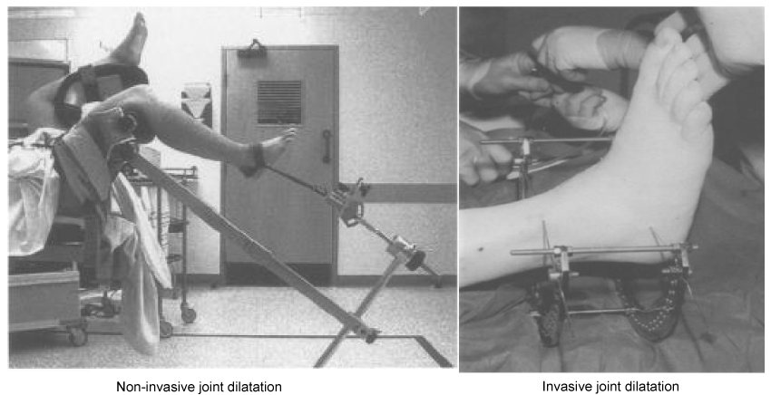
Joint restoration method: We used a non-invasive restoration procedure by using the surgeon’s body traction, which had the advantage of being able to enter the anterior cavity of the ankle joint very easily [1-6] (Figure 1).
Tools: We used a 4mm diameter camera and a 30° angle and a 4mm diameter plane, which is basically advantageous, but if we had had smaller tool, would have carried out more easily [1,2,4].
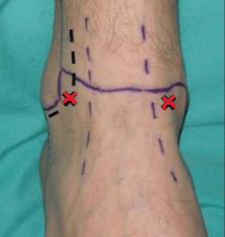
Incision: into the joints through the front internal and the front external anatomy (Figure 2). Assess joint damage, determine cause. For impingement lesions, proceed to cleanse the bone, remove the fibrous organ.
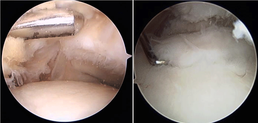
For OCD lesions, remove the damaged cartilage; drill the bone to stimulate blood vessel regeneration. A case of ankle fractures, traumatic retraction of the ankle, endoscopic supported to cleanse of the fibrous organ in the joint, and then a combination of bones through the external anatomy (Figure 3) (Table 1).
Discussion
Indications for ankle endoscopic
Today, ankle endoscopic is widely recommended, both in pathologies as well as trauma [1,2]. The major indications for ankle endoscopic are loose body, OCD, pathology Gout, rigid ankle joint surgery, endoscopic assist in combination of ankle bone and pylon…
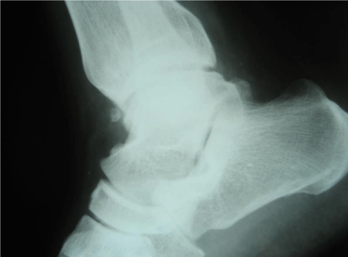
One reason for having a medical examination is usually painful persistent ankle joint that medical -surgical treatment and rehabilitation do not work. Beside the cases of physical injury diagnosed before surgery such as loose body, OCD, most cases are diagnosed as impingement of the ankle joint [7-10]. “Impingement syndrome of joints is a painful syndrome due to the friction between the components of the joint, possibly due to bones or soft tissue, which is the cause and also a consequence of the dynamic change of the joint [11]. This syndrome is common after injury or it can also be due to a condition such as osteoarthritis. The diagnosis of impingement of the ankle joint can be confirmed mainly by the front impingement syndrome based on the lesion on the X-ray [8,12]. Other cases are sometimes based on excluded diagnostic and confirmed by endoscopy [1]. All of our Impingement patients have a history of old, long-term injuries, with most of them type 2 and type 3 injuries, according to Scranton’s classification [9]. Treatment of ankle joint impingement syndrome is medical treatment and rehabilitation, surgical indication is placed when medical treatment and rehabilitation do not achieve the desired results [8]. Treatment of ankle joint impingement syndrome was published by many authors with positive results [2,4,7-9,12] (Figure 4).
OCD damage according to IRCS classification has four stages depending on the severity of the lesion, the purpose of the surgery is to remove the damaged cartilage and fill the gap in two ways: drill sponge bone in order to stimulate regeneration of blood vessels and transplant implantation, in Vietnam condition, we have only intervened to remove fragile cartilage and drill to stimulate regeneration of blood vessels. Clinically good results, but follow-up time has not been long enough to evaluate the outcome of regeneration of blood vessels at lesions, however, according to some authors, long-term evaluation has good results [10,12].
Joint arthroscopy combining with bone combination surgery has been applied to some joints, including ankle joints for lesions such as pylon fracture, double ankle fracture, the purpose of endoscopic is controlling joint when bones are combined [4,13]. Bone combination surgery for old fractures of the exterior ankle, sprain ankle, classically perform two incisions: internal and external incision, internal incision enters the joint to release the fibroid; external incision is to combine calf-bone. The use of an endoscope instead of an incision reduces the incision, minimizing interior joint intervention. The duration of surgery compared to conventional open surgery is slightly longer (90 minutes), showing more advantages than open surgery. Thus, the indication for endoscopic support in this case is absolutely right and the results of the surgery have proved it.
Technical
Method of the joint dilatation: Because of a small joint, narrow cavity, the ankle arthroscopy only really develops when different joint dilatation methods are found. There are two main types: non-invasive and invasion method [1,3,4,6] (Figure 5).
We use a non-invasive procedure, using the surgeon’s trunk, which has the advantage of being able to enter the anterior cavity of the ankle joint very easily.
There are two surgical postures associated with dilated joints, the first is the dilatation by folding knee posture with 90° of the surgeon’s stretching, and the second is the surgeon’s trunk with legs stretched straight posture. In general, both ways give good articulation results and convenient for conducting endoscopy.
Tools: We use tools for knee using to perform ankle arthroscopy, which are basically good, but with smaller tools it is easier to perform [1,2,4].
Incision: The ankle arthroscopy can be performed in many different ways, but the most common ones are front internal and the front external incision. We use these two incisions for all cases are convenient to joint intervention [1,2,4].
Conclusion
Ankle arthroscopy has been used more and more popularly in the world. Over 40 cases of surgery with positive results and can be indicated ankle arthroscopy joint for several other cases.
- Noyes ST (1998) Ankle arthroscopy, The Foot 8: 197-202.
- Niek van Dijk C, Dik Scholte (1997) Arthroscopy of the Ankle Joint, Arthroscopy: The Journal of Arthroscopic and Related Surgery 1997: 90-96. Link: https://goo.gl/FtX3D8
- Hedley D, Geary NPJ, Meda P (2001) Ankle arthroscopy: a new technique for non-invasive ankle distraction, Foot and Ankle Surgery 2001 7:137-139. Link: https://goo.gl/zTdZgn
- Kelly A, Winson I (1998) Recent advances in ankle arthroscopy, Foot and Ankle Surgery 4: 49-55. Link: https://goo.gl/EygzKk
- C N van Dijk, G Vuurberg, A Amendola, J W Lee (2016) Anterior ankle arthroscopy: state of the art, JISAKOS 1: 105-115. Link: https://goo.gl/mqJJpH
- Graham AJ, Hughes S, Cooke PH (2000) Ankle arthroscopy: the Half-Frame Fixator to use of an Ilizarov distract the ankle joint, Foot and Ankle Surgery 2000 6:55-58. Link: https://goo.gl/WhSfVe
- Thomas MD, Robert AA, Dean CT (1997) Arthroscopic Treatment of Soft-Tissue Impingement of the Ankle in Athletes, Arthroscopy: The Journal of Arthroscopic and Related Surgery 13: 492-498. Link: https://goo.gl/MqHJT1
- Kyle PL, Kevin JM, William HR, George T (2016) Ankle impingement ,Journal of Orthopaedic Surgery and Research. Link: https://goo.gl/QQHspH
- Richard DF, Van N, California, Pierce ES (1993) Current Concepts Review. Arthroscopy of the Ankle and Foot, The Journal of Bone and Joint Surgery 756: 199 – 208. Link: https://goo.gl/aFxxWW
- Siow HM, Darren Tay KJ, Mitra AK (2004) Arthroscopic treatment of osteochondritis dissecans of the talus, Foot and Ankle Surgery 10: 181-186. Link: https://goo.gl/cvHmRn
- Carlo M, Alessia C, Antonio B (1998) Ankle impingement syndromes, European Journal of Radiology 27: 70–73. Link: https://goo.gl/jwYcmT
- Ruben Z, Johannes IW, Christopher DM, Ethan JF, John GK, et al. (2015): Arthroscopic Treatment for Anterior Ankle Impingement: A Systematic Review of the Current Literature, Arthroscopy: The Journal of Arthroscopic and Related Surgery 31: 1585–1596. Link: https://goo.gl/A3o6zG
- Atsushi O, Shinji N, Akihiko N, Tomoyuki I, Motoshige S, et al. (2004) Arthroscopically Assisted Treatment of Ankle Fractures:Arthroscopic Findings and Surgical Outcomes, Arthroscopy: The Journal of Arthroscopic and Related Surgery 20: 627-631. . Link: https://goo.gl/1eLumF
Article Alerts
Subscribe to our articles alerts and stay tuned.
 This work is licensed under a Creative Commons Attribution 4.0 International License.


 Save to Mendeley
