Archives of Pulmonology and Respiratory Care
Spontaneous Haemomediastinum and Fatal Haemoperitoneum in woman with Vascular Ehlers-Danlos Syndrome
Isabel Cal1, Elena Fernández1, Jose V Méndez2 and Jose R Jarabo1*
2Department of Radiology, Hospital San Carlos, Profesor Martín Lagos s/n. 28040. Madrid, Spain
Cite this as
Cal I, Fernández E, Méndez JV, Jarabo JR (2017) Spontaneous Haemomediastinum and Fatal Haemoperitoneum in woman with Vascular Ehlers-Danlos Syndrome. Arch Pulmonol Respir Care 3(1): 048-049. DOI: 10.17352/aprc.000024Vascular Ehlers-Danlos Syndrome (EDS) presents with fragility of blood vessels, with high incidence of fatal hemorrhages in middle-age adults. We present a 38-year old female with vascular EDS presented to the emergency unit with spontaneous hemomediastinum and hemothorax. A selective arteriography showed a bronchial artery aneurysm that could be embolized. Coagulated hemothorax was cleared up using intrapleural instillation of urokinase. Acute massive hemoperitoneum happened a few hours later, due to spontaneous disruption of a splenic artery aneurysm. It could not be managed and led to the decease of the patient. Multifocal bleeding is a frequent threatening event in vascular EDS.
Introduction
Ehlers–Danlos syndrome (EDS) is an inherited rare group of disorders of collagen synthesis [1]. Type IV vascular EDS (EDS-IV), with mutated gene COL3A1, presents with fragility of medium-sized and large blood vessels, predisposing to life-threatening haemorrhages. Spontaneous hemomediastinum, with or without hemothorax, is an uncommon cause of thoracic pain. Dissection of a bronchial artery aneurysm (BAA) has been described. Relationship between these two infrequent entities has not been fully described.
Case
A 38-year-old female with known EDS-IV presented to the emergency room with dorsal chest pain, dyspnoea and low consciousness. Examination showed hypotension, tachycardia, tachypnea and right hypoventilation. Chest X-ray showed massive right pleural effusion.
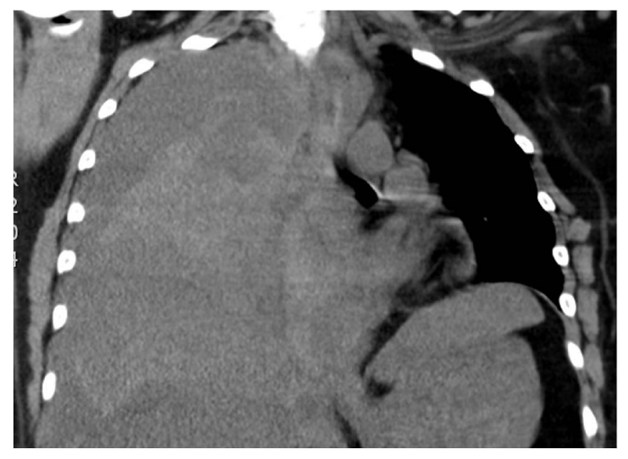
A thoracoabdominal computed tomography (CT) was performed, showing a massive right pleural effusion suggesting haemothorax. Posterior mediastinum was also occupied by blood (Figure 1), with a suggestive image of right BAA without evidence of bleeding.
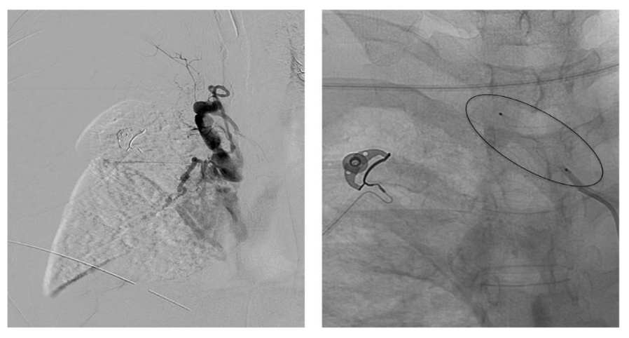
One litre of blood was drainaged trough a chest tube, achieving good lung reexpansion. One hour later she presented sudden hemodinamic instability and massive flow of blood through the chest tube, requiring orotracheal intubation. Chest X-ray showed right massive pleural effusion. An aortography showed extravasation of contrast into the chest cavity depending on a right bronchial artery with a 3 cm saccular aneurism. It was satisfactory embolized with an Amplatzer® vascular plug of 8mm in diameter (Figure 2).
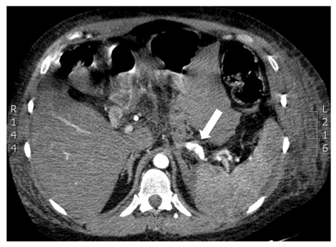
Intrapleural coagulated hemothorax was completely evacuated with pleural infusion of urokinase. Three hours later, again clinical data of acute bleeding were present, without an increasing on chest tube flow or intrathoracic collection. Thoraco-abdominal CT showed free abdominal fluid, along with an image of a disrupted splenic artery aneurysm (Figure 3). Due to the successful proceed in the thorax, another endovascular approach was indicated. However, within first steps of the procedure, the uncontrollable hemorrhagic shock led to electromechanical dissociation and patient’s death.
Discussion
EDS-IV accounts for 5% of EDS [1]. Spontaneous arterial rupture is the most common cause of sudden death, with the highest incidence in the third or fourth decade of life. Thoracic and abdominal midsized arteries are most commonly involved. EDS-IV first manifestations are neurovascular (10%) and rupture of hollow or solid organs (34%). Hemoptysis and recurrent spontaneous pneumothoraces, are the most frequent thoracic manifestations.
Spontaneous hemomediastinum is rare, and has been related with bleeding disorders, mediastinal organ hemorrhages and spontaneous idiopathic hemomediastinum. However it has not been fully described as a complication of EDS-IV. BAA as a source of massive mediastinal bleeding has been described within inflammatory disorders, infectious diseases or as a result of thoracic trauma. In our case its origin was the anomalous vascular histology associated to EDS-IV. Intrapulmonary BAA bleeding will present with hemoptysis. Mediastinal BAA hemorrhage will lead to mediastinal compression (chest pain, dyspnea, dysphagia, vena cava syndrome). Tearing of mediastinal pleura will move blood flood into the pleural cavity, as in our case. BAA is confirmed by CT with contrast. Profuse bleeding of pathologic bronchial arteries over 2mm usually requires an aggressive approach. Endovascular approach has become the first-line management strategy.
Embolization of aortic branch vessels and other medium sized-arteries has been successful in EDS patients presenting with mayor acute bleeding [2,3]. Current devices achieve complete obliteration of pathologic vessels with few complications. In case of failure, the use of endoprosthesis, fibrin glue or embolization with other substances (polyvinyl alcohol or N-butyl cyanoacrylate) has also been described. Complications include spinal chord infarction (1%), femoral rupture and pseudoaneurysm formation.
Embolization of aortic branch vessels and other medium sized-arteries has been successful in EDS patients presenting with mayor acute bleeding [2,3]. Current devices achieve complete obliteration of pathologic vessels with few complications. In case of failure, the use of endoprosthesis, fibrin glue or embolization with other substances (polyvinyl alcohol or N-butyl cyanoacrylate) has also been described. Complications include spinal chord infarction (1%), femoral rupture and pseudoaneurysm formation.
In our case, bleeding from an anomalous splenic artery occurred after the embolization of the BAA, resulting in massive intraperitoneal hemorrhage. Remote arterial injury during an angiography has been described, including intimal tear of the ascending aorta, spontaneous splenic arterial rupture or unknown intraperitoneal bleeding after the embolization of carotid cavernous fistula [4]. Remote injuries could be related to direct trauma to the arterial wall, either by the catheter itself or by the injection of intravenous contrast. Likewise, changes of blood pressure or flow on anomalous arteries could result in their rupture [5]. In conclusion, spontaneous hemomediastinum or hemothorax of an unknown origin in young or middle age people should make us think about non-diagnosed EDS-IV. Endovascular approach is the first therapeutic option. We must be aware of remote new acute severe hemorrhages needing new therapeutic maneuvers.
- Steinman B, Royce PM. Superti F, Ehlers DS, Royce P, et al. (2002) Connective tissue and its heritable disorders. 5th ed. New York, NY:Wiley-Liss,431-523.
- Oderich GS, Panneton JM, Bower TC, Lindor NM, Cherry KJ, et al. (2005) The spectrum, management and clinical outcome of Ehlers–Danlos-Syndrome type IV: a 30-year experience. J Vasc Surg; 42: 98–106. Link: https://goo.gl/ON7R73
- Brooke BS, Arnaoutakis G, McDonnell NB, Black 3rd JH (2010) Contemporary management of vascular complications associated with Ehlers–Danlos-Syndrome. J Vasc Surg; 51:131–138. Link: https://goo.gl/2kWS2V
- Horowitz MB, Purdy PD, Valentine RJ, Morrill K (2000) Remote vascular catastrophes after neurovascular interventional therapy for type 4 Ehlers–Danlos-Syndrome. Am J Neuroradiol 21: 974– 976. Link: https://goo.gl/KKIGlU
- Okada T, Frank M, Pellerin O, Primio MD, Angelopoulos G, Boughenou MF, et al (2014) Embolization of Life-Threatening Arterial Rupture in Patients with Vascular Ehlers–Danlos-Syndrome. Cardiovasc Intervent Radiol 37: 77–84. Link: https://goo.gl/yJaxhE
Article Alerts
Subscribe to our articles alerts and stay tuned.
 This work is licensed under a Creative Commons Attribution 4.0 International License.


 Save to Mendeley
