Archives of Pulmonology and Respiratory Care
What do we have to know about Cystic Lung Diseases?
Sánchez Díaz C and Díaz-Lobato S*
Cite this as
Sánchez Díaz C, Díaz-Lobato S (2017) What do we have to know about Cystic Lung Diseases? Arch Pulmonol Respir Care 3(1): 032-034. DOI: 10.17352/aprc.000021Introduction
Cystic lung diseases are a heterogeneous group of pathologies which differential diagnosis can be complicated [1]. We are going to comment some aspects that we should know about cystic lung diseases to facilitate a better understanding and clinical management of these entities.
It is necessary to know how to differentiate true cystic lung diseases from other pathologies
The improvement in computed tomography (CT) techniques, as the case of high resolution computed tomography, has allowed lung cysts and cysts-like lesions to be found more and more frequently. One important issue is how to differentiate true pulmonary cysts from others pathologies. We have to know that a cyst is a round area full of air, well delimited by normal lung, surrounded by a thin wall (<2 mm). Therefore, they are not cysts:
♦ Cysts that are not surrounded by wall. This is the case of patients with pulmonary emphysema (air space distal to the terminal bronchiole).
♦ Cysts that are not surrounded by healthy lung tissue. It is the case of the cavities: space full of gas inside a mass, a nodule or a consolidation.
♦ Cysts in which the wall is especially thin (<1 mm). This is the case of patients with bullas in the lung.
♦ Cysts that are not located within the pulmonary parenchyma as is the case of cysts in the visceral pleura or in the subpleural air (bleb) or the case of multiple subpleural cysts of 3-10 mm in diameter with well-defined walls (honeycomb).
Finally, in the true cysts an epithelial lining is observed in the staining of hematoxylin eosin. In pseudocysts it doesn´t exist.
Cystic lung diseases are a spectrum of diseases
Within the term of cystic lung diseases are included several diseases, mainly lymphangioleiomyomatosis (LAM), pulmonary Langerhans cell histiocytosis (PLCH), Birt-Hogg-Dube syndrome (BHD syndrome) and amyloidosis [2].
The localization of cysts in the HRCT (high resolution computerized tomography) can help guide the diagnosis
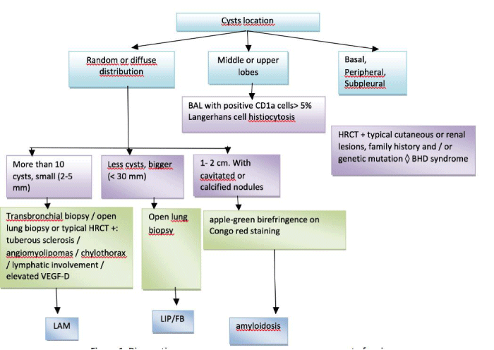
We can found three typical patterns attending to cysts localization (Table 1) [3]:
♦ Random or diffuse distribution: It is most likely to be a lymphangioleiomyomatosis, a lymphocytic interstitial pneumonia/follicular bronchiolitis or an amyloidosis. In order to differentiate these three pathologies in the usual clinical practice, we must pay attention to the size of the cysts and other characteristic features in the HRCT, the extrapulmonary manifestations of each single pathology and the abnormalities of the pulmonary function.
♦ Distribution in middle or upper lobes: If we found this typical distribution, it is most likely to be a pulmonary Langerhans cell histiocytosis. Other data supporting this diagnosis in the HRCT are the presence of nodules. These nodules often cavitates and converge as the disease progresses. Outbreaks of fibrosis can also be found, as well as sparing of the costophrenic angles. However pulmonary function may be normal, restrictive, or obstructive.
♦ Distribution in the basal, peripheral and subpleural lobes: In this case it is most likely to be a Brit-Hogg-Dube syndrome. The cysts are thin-walled, well defined and sometimes the cysts are blocked.
Pay attention: Cysts size may also help to guide the diagnosis
In the cases with diffuse cystic involvement, the smallest cysts are typically due to lymphangioleiomyomatosis (2-5 mm to 30 mm), followed by lymphoid interstitial pneumonia (<30 mm) and finally those of amyloidosis (1 to 2 cm). The size of cysts in X histiocytosis is highly variable (1-3 mm to 2 cm) and in Brit-Hogg-Dube syndrome they are usually 2 to 78 mm [4].
Can the morphology of cysts help?
The answer is yes. In pulmonary Langerhans cell histiocytosis cysts are bizarre, meanwhile in lymphangioleiomyomatosis and amyloidosis are round. In Birt-Hogg-Dube syndrome they are round or lentiform.
Can the patient´s symptoms be useful for diagnosis?
Although all the cystic lungs diseases share some data (cough, dyspnea of progressive effort, pneumothorax...), some symptoms and the characteristics of the systemic manifestations are typical of certain diseases, which can also guide the diagnosis.
In the case of lymphangioleiomyomatosis, the presence of hemoptysis and the evolution to respiratory failure is more frequent than in other cystic diseases. It is sometimes misdiagnosed as asthma or chronic bronchitis and it can produce chylothorax, and less frequently ascites, pleuropericardial effusion and chyloptisis. It is also possible to see angiomyolipomas, uterine and pancreatic involvement, and multisystemic hamartomas in tuberous sclerosis [5].
Is characteristic of pulmonary Langerhans cell histiocytosis that half of the patients can be asymptomatic. Other symptoms may be weight loss, skin rash and diabetes insipidus. Cystic bone lesions in the skull, long bones, ribs and pelvis, are very common, and are usually asymptomatic.
In other hand, Brit-Hogg-Dube syndrome usually presents cutaneous involvement (fibrofolliculomas, trichodisomas and acrochordons) and renal involvement (simple cysts, malignant tumors, which are usually bilateral and multifocal).
Finally, in the case of lymphoid interstitial pneumonia, subacute onset with cough, exertional dyspnea and systemic symptoms (arthralgias, fever and weight loss) it is typical. It is also possible to find anemia and hypergammaglobulinemia.
You have to note if the patient has recurrent pneumothorax
Pneumothorax is a common finding in patients suffering of cystic lung diseases. It is more frequent to see in Brit-Hogg-Dube syndrome (75%), followed by lymphangioleiomyomatosis (69%) and pulmonary Langerhans cell histiocytosis (25%).
When do we have to perform invasive tests to confirm the diagnosis?
In routine clinical practice, it is sometimes challenging to know when to perform invasive tests to confirm the diagnosis, as radiological findings are very common in asymptomatic patients.
In the lymphoid interstitial pneumonia, the chest X-ray and the HRCT are not diagnostic. In the bronchoalveolar lavage (BAL) lymphocytosis is evidenced, but it is not diagnostic either, so open biopsy is required to establish the diagnosis.
The diagnosis in lymphangioleiomyomatosis is established by transbronchial biopsy or open lung biopsy. In this case, the HMB-45 monoclonal antibody selectively stains the muscle proliferation of the lymphangioleiomyomatosis. Diagnosis it is also possible without perform a biopsy in the presence of a typical HRCT and any of the following findings: tuberous sclerosis, angiomyolipomas, chylothorax or elevated VEGF-D lymphatic involvement.
For the diagnosis of amyloidosis, it is necessary to demonstrate apple green birefringence and Congo red staining on lung biopsy.
Diagnosis of Brit-Hogg-Dube syndrome requires a typical HRCT, typical cutaneous or renal lesions, family history and / or genetic mutation (FLCN). In the pathological anatomy it is noted: cysts surrounded by normal lung tissue, without inflammatory cells.
In a patient with a typical HRTC, young and smoker, the presence of CD1a, S-100 cells> 5% in BAL, allow us to stablish the diagnostic of pulmonary Langerhans cell histiocytosis without biopsy.
Figure 1 shows an algorithm that indicates when to perform an invasive test and which test to perform, taking into account the radiological findings and the clinical suspicion.
How to treat cystic lung diseases
In the treatment of these pathologies, in addition to the specific treatment of each of them, complications must always be treated (pleurodesis in recurrent pneumothorax, specific treatment of pulmonary hypertension). Pulmonary transplantation is an option in advanced stages of the disease.
Ignorance of the pathogenesis of many of these diseases makes specific treatment difficult [6]. For example, in pulmonary Langerhans cell histiocytosis, since BRAF mutations have been described, vemurafenib treatment has been tested with promising results.
In the lymphangioleiomyomatosis, it is known that both sporadic and genetically modified mutations occur in tumor suppressor genes: TSC1 (encoding hamartin) and TSC2 (encoding tuberin), leading to abnormal activation of the signaling pathway of mammalian target of the rapamycin (mTOR), responsible for regulating cell growth, proliferation, migration and cell survival. That is why mTOR inhibitors (sirolimus, everolimus) are used in the treatment of this disease, demonstrating improving lung function and improving the quality of life, but further studies are needed to know the dose, duration and side effects of this treatment.
In the Brit-Hogg-Dube syndrome, numerous mutations have been described (in the FLCN gene) that also produce an alteration in TOR signaling, although it is not known whether the active or inactivated signaling, nor what is the role of mRT in the pathogenesis of the illness.
Summary
What do we not know about cystic lung disease?
In summary, we have to know how to differentiate true cystic lung diseases from other pathologies and the spectrum of diseases to be considered. It is important to know that the localization, size and cyst morphology as well as the clinic characteristics can help us to guide the diagnosis. We also have to know when we have to perform invasive tests to confirm the diagnosis and how to treat these diseases. Finally, it is essential what we do know about cystic lung diseases in order to a better management of the patients suffering these diseases.
- Park S, Lee JE (2017) Diagnosis and treatment of cystic lung disease. Korean J Intern Med 32: 229-238. Link: https://goo.gl/cmwlVg
- Gupta N, Vassallo R, Wikenheiser-Brokamp KA, McCormack FX (2015) diffuse cystic lung disease: part II. Am J Respir Crit Care Med 192: 17-29. Link: https://goo.gl/lT3e9o
- Seaman DM, Meyer CA, Gilman MD, McCormack FX (2011) Diffuse cystic lung disease at high-resolution CT. AJR Am J Roentgenol 196: 1305-1311. Link: https://goo.gl/OQlcrD
- Trotman-Dickenson B (2014) Cystic lung disease: achieving a radiologic diagnosis. Eur J Radiol 83: 39-46. Link: https://goo.gl/6xRmsc
- Harari S, Torre O, Cassandro R, Moss J (2015) The changing face of a rare disease: lymphangioleiomyomatosis. Eur Respir J 46: 1471-1485. Link: https://goo.gl/D60WFY
- Clarke BE (2013) Cystic lung disease. J Clin Pathol 66: 904- 908. Link: https://goo.gl/BExy6L
Article Alerts
Subscribe to our articles alerts and stay tuned.
 This work is licensed under a Creative Commons Attribution 4.0 International License.


 Save to Mendeley
