Archives of Otolaryngology and Rhinology
Clinical Tools to diagnose Eustachian tube Dysfunction in patients with a normal tympanic membrane exam and a type A tympanogram
Roya Azadarmaki*
Cite this as
Azadarmaki R (2019) Clinical Tools to diagnose Eustachian tube Dysfunction in patients with a normal tympanic membrane exam and a type A tympanogram. Arch Otolaryngol Rhinol 5(4): 099-101. DOI: 10.17352/2455-1759.000108Objective: Obstructive Eustachian tube dysfunction, a form of Eustachian tube dilatory dysfunction, can be a challenge to diagnose in individuals with classic Eustachian tube dysfunction symptoms but a normal tympanic membrane exam and tympanogram. The purpose of this article is to outline techniques that can be used by the otolaryngologist to evaluate patient’s under these circumstances.
Methods: Eustachian tube dysfunction was assessed combining findings from dynamic evaluation of the tympanic membrane with videoendoscopic evaluation of the Eustachian tube. The “Azadarmaki technique” of assessing the tympanic membrane for eustachian tube dysfunction involves evaluating the tympanic membrane live with a microscope or endoscope while the patient is performing the Valsalva maneuver followed by an immediate swallow.
Results: Patients with Eustachian tube dysfunction had lack of proper dynamic motion of the tympanic membrane noted using the Azadarmaki technique, combined with abnormal findings on videoendoscopy of the Eustachian tube based on criteria outlined in this article.
Conclusion: Poor tympanic membrane motion noted using the Azadarmaki technique combined with abnormal appearance and dynamic function of the cartilaginous Eustachian tube on videoendoscopy can be used to diagnose patient’s with obstructive Eustachian tube dysfunction in the setting of classic Eustachian tube dysfunction symptoms but a normal tympanic membrane exam and tympanogram. Establishing simple methods for the otolaryngologist to diagnosis obstructive Eustachian tube dysfunction is key in identifying patients who would benefit from balloon dilation of the Eustachian tube.
Introduction
Eustachian tube dynamic function serves as one of the 2 main mechanisms of establishing proper middle ear pressure. Mucociliary clearance of middle ear secretions and protection of the middle ear from nasopharyngeal hazards are 2 other purposes of this highly dynamic conduit between the nasopharynx and middle ear space [1].
In 2015, a clinical census statement defined 3 types of Eustachian tube dysfunction: Dilatory, Baro-challenge-induced, and Patulous [2]. The first type, Dilatory dysfunction, was further classified as functional, dynamic, and anatomic [2]. In the 2019 American Academy of Otolaryngology clinical consensus statement on balloon eustachian tuboplasty, the focus was on adults with obstructive eustachian tube dysfunction [3], which fits under the 2015 category of dilatory Eustachian tube dysfunction. This is reflective of the fact that the majority of adult patients referred to as having Eustachian tube dysfunction suffer from obstructive eustachian tube dysfunction.
Aural fullness, ear popping or crackling, ear discomfort or pain, pressure, clogged or ‘under water’ sensation, ringing, autophony, and muffled hearing, are all symptoms a patient with obstructive Eustachian tube dilatory dysfunction may present with. “Silent Eustachian tube dysfunction” can be seen in patients who do not have any of the above symptoms, however they have tympanic membrane retraction, secondary cholesteatoma formation, or tympanic membrane atelectasis with and without ossicular chain erosion.
In the patient with symptoms of Eustachian tube dilatory dysfunction, the diagnosis can be made using tympanometry, otoscopy (tympanic membrane retraction, middle ear effusion, cholesteatoma formation, …), pneumatic otoscopy, audiometry, tunning fork exam, and videoendoscopy of the Eustachian tube.
Ruling out superior canal dehiscence, temporomandibular joint disorder, cochlear hydrops, hearing loss manifesting as aural fullness, and otologic migraines, are key in avoiding a misdiagnosis of obstructive Eustachian tube dysfunction.
The purpose of this article is to guide the reader on techniques used to diagnose obstructive Eustachian tube dysfunction in a patient with classic symptoms but a normal tympanic membrane exam, normal conductive hearing, and a type A tympanogram. In the author’s experience, many patients that fit into this category have had a prolonged journey of doctors’ visits with associated frustration without a diagnosis and with persistent symptoms.
Combining a proper history with the two techniques outlined below, can identify the patient with obstructive Eustachian tube dysfunction that does not meet classic diagnostic criteria but may benefit from Eustachian tube balloon dilation. Including information from the validated Eustachian Tube Dysfunction Questionnaire–7 (ETDQ-7) can help define the severity of Dilatory Eustachian tube dysfunction symptoms.
Microscopic evaluation of the tympanic membrane and assessment for tympanic membrane motion while the patient is actively performing the Valsalva maneuver followed by an immediate swallow is a highly useful measure that can be used to assess for obstructive Eustachian tube dysfunction in a patient with an otherwise normal exam. Using this method, the patient is asked to pinch their nose with their mouth closed, perform the Valsalva maneuver gently, and immediately swallow. If the patient is confused about how to perform the Valsalva maneuver, they are asked to mimic the maneuver of popping their ears while they are on a plane with their nose pinched and lips sealed. This technique needs to be performed without significant head motion so the physician can properly focus on the tympanic membrane under the microscope. An endoscope can also be used for direct visualization of the tympanic membrane using this technique. The gentle Valsalva maneuver has to be followed by an immediate swallowing maneuver while the physician has a live view of the tympanic membrane.
The second useful technique to establish obstructive eustachian tube dysfunction is by performing a video-endoscopy of the eustachian tube. Proper numbing of the nasal cavity is beneficial. Ideally a rigid 30 or 45 degree scope should be used to assess the eustachian tube function, as the Eustachian tube is positioned laterally in the nasopharynx, however this can be challenging to perform due to poor patient tolerance. A flexible scope is an alternative method of assessing the Eustachian tube. Once along the floor of the posterior nasal cavity, adjacent to the most posterior aspect of the inferior turbinate, eustachian tube function can be evaluated. The factors below should be assessed and documented (Table 1).
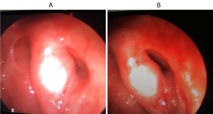
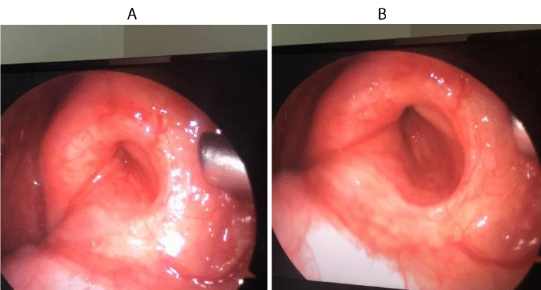
An example of a normal videoendoscopic evaluation of the Eustachian is given to provide the reader with an idea of how a comprehensive videoendoscopic evaluation of the Eustachian tube can be documented. An example of a normal Eustachian tube videoendoscopic evaluation should be documented as follows: There is no significant Eustachian tube peritubal or intraluminal edema, the lumen of the cartilaginous Eustachian tube can be nicely visualized with proper dilatory function of the valve, there are no abnormal secretions within the Eustachian tube lumen or adjacent to the Eustachian tube opening, there is no mass effect on the posterior cushion of the Eustachian tube from nasopharyngeal disease, and the posterior tail of the inferior turbinate does not extend beyond the level of the choana.
With the “Azadarmaki Technique” of visualizing the tympanic membrane with the microscope or endoscope while performing the Valsalva maneuver followed by an immediate swallow without significant head motion, combined with an abnormal videoendoscopic evaluation of the Eustachian tube with one or more abnormal criteria, a patient can be diagnosed with obstructive Eustachian tube dysfunction in the presence of classic symptoms despite a normal tympanic membrane exam and type A tympanogram.
There is ample room for establishing solid measures to define Eustachian tube dysfunction. Properly diagnosing Eustachian tube dysfunction in a patient with the right symptoms but normal tympanic membrane exam and tympanometry is very important in identifying the proper candidate for balloon dilation of the Eustachian tube.
Future measures should include comparing the diagnostic criteria established in this article with patient reports on the validated Eustachian Tube Dysfunction Questionnaire–7 (ETDQ-7) (Figures 1-3) (Videos 1,2).
Video 1 (click and view): A videoendoscopy of a right Eustachian tube is demonstrated by the author.
- Bluestone CD (2005) In Eustachian Tube: Structure, Function, Role in Otitis Media, New York. 1-9. Link: http://bit.ly/2NhJcoh
- Schilder AG, Bhutta MF, Butler CC (2015) Eustachian tube dysfunction: consensus statement on definition, types, clinical presentation and diagnosis. Clin Otolaryngol 40: 407-411. Link: http://bit.ly/2WEEG6h
- Tucci DL, McCoul ED, Rosenfeld R (2019) Clinical Consensus Statement: Balloon Dilation of the Eustachian Tube. Otolaryngol Head Neck Surg 161: 6-17. Link: http://bit.ly/34s7PUL
Article Alerts
Subscribe to our articles alerts and stay tuned.
 This work is licensed under a Creative Commons Attribution 4.0 International License.


 Save to Mendeley
