Archives of Otolaryngology and Rhinology
Cystic cervical lymph nodes metastasis revealing a papillary carcinoma of the thyroid gland: A case report
Diouf MS*, Thiam A, Maïga S, Deguenonvo REA, Diop A, Barry MW and Diouf R
Cite this as
Diouf MS, Thiam A, Maïga S, Deguenonvo REA, Diop A, et al.(2019) Cystic cervical lymph nodes metastasis revealing a papillary carcinoma of the thyroid gland: A case report. Arch Otolaryngol Rhinol 5(2): 040-042. DOI: 10.17352/2455-1759.000094Cystic cervical lymph nodes are a rare anatomo-clinical form. They are a typical presentation of metastasis of cancers of the waldeyer ring in particular those of the palatine tonsil. Nevertheless, they may reveal a papillary carcinoma of the thyroid even though the thyroid gland is clinically normal. In this case the combination of ultrasound and fine needle aspiration cytology (FNAC) with intranodal thyroglobulin dosage allows to differentiate lymphadenopathies of thyroid origin from other causes. We report the case of a 40-year-old patient received in December 2015 in our hospital for cystic cervical lymph nodes without clinical thyroid mass. FNAC was for a metastasis of an adenocarcinoma.
Pan endoscopy was normal. A lymph node biopsy was performed and the histology returned in favor of metastasis of papillary adenocarcinoma of the thyroid gland. The patient had undergone a cervicotomy with total thyroidectomy, left modified radical neck dissection and right functional lymphadenectomy. The histology of the surgical specimen confirmed the diagnosis of papillary carcinoma of the thyroid gland with cystic lymph node metastasis.
This clinical case highlights the diagnostic approach of cystic cervical lymph node metastasis.
Introduction
Cystic cervical lymph nodes are a rare mode of detection for papillary carcinoma of the thyroid gland, and account for about 40% of their lymph-nodes metastasis [1]. The cystic character is a radiological criterion of malignancy that can also be confused with a benign cervical cystic mass [2]. In the absence of obvious thyroid mass, cystic cervical lymph nodes raises the problem of their etiological diagnosis, which is based on a rigorous clinical and paraclinical investigation. Their early detection allows more effective surgery and radiotherapy.
Case Report
We report the case of a 40-year-old patient with no particular pathological history. He was received in December 2015 in our department for a bilateral lateral cervical mass gradually increasing in volume, without signs of compression or thyroid dysfunction. The whole evolving since approximately 3 months with a stability of the general state.
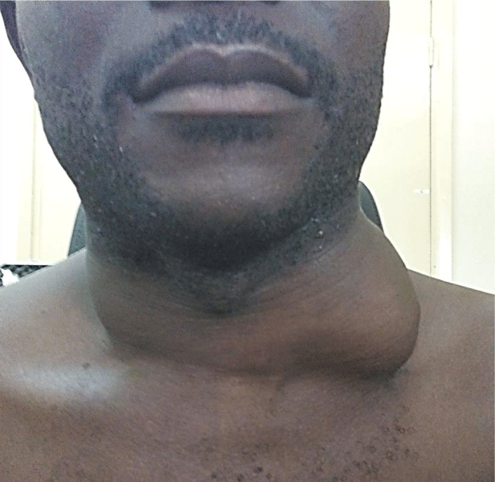
The physical examination revealed a bilateral lateral cervical mass, looking like a ganglion of approximately 10 cm of large axis on the left and 4cm of large axis on the right (Figure 1). The mass was firm, painless and mobile in relation to the shallow and deep plans. There was no palpable thyroid mass. Nasofibroscopy did not show pharyngolaryngeal lesion and the chest X-ray was normal.
Cervical CT scan showed two large cystic cervical lymph nodes, the left located being larger than the right one and the thyroid gland had multiple hypodense nodules (Figure 2).
FNAC was in favour of metastasis of adenocarcinoma.
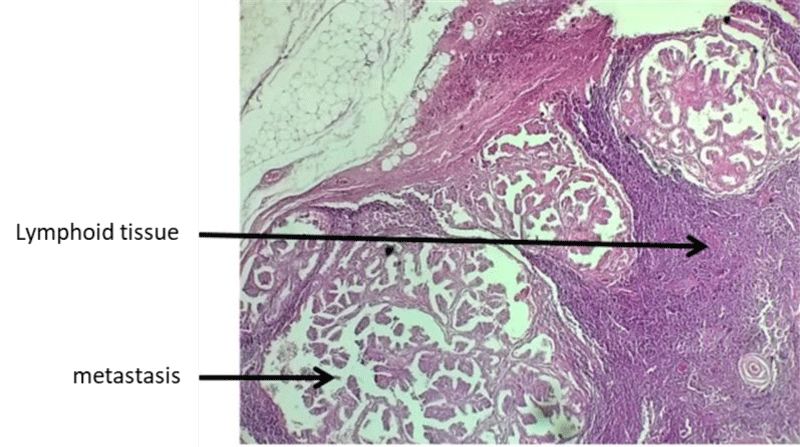
Panendoscopy was without particularity. A Lymph node biopsy was performed and the histology returned in favor of metastasis of papillary adenocarcinoma of thyroid origin (Figure 3a). The patient had undergone a cervicotomy with total thyroidectomy, left modified radical neck dissection and right functional lymphadenectomy.
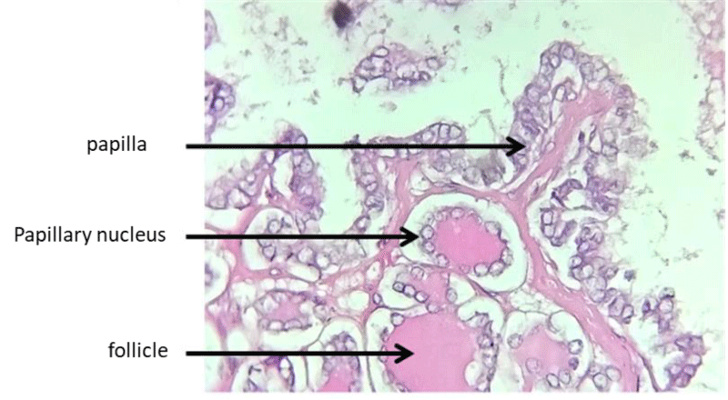
The histology of the operative specimen confirmed the diagnosis of papillary carcinoma of the thyroid (Figure 3b) with cystic lymph nodes metastasis.
The postoperative follow-up was simple, an hormone replacement therapy based on L-thyroxine was instituted at a frenetic dose for tumor growth (150 μg / day).
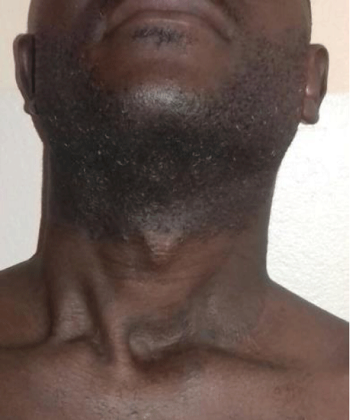
Adjuvant treatment with iodine-131 was performed. With a follow-up of 36 months, there was no recurrence or metastasis (Figure 4).
Discussion
Cystic cervical lymph nodes are a rare anatomo-clinical form. According to Regauer, they are characterized by the presence of transformed (malignant) keratinocytes with intrinsic properties of cystisation [2]. This would result from subcortical liquefaction of the keratin they include or sudden obstruction of the lymphatic flow that passes through them [3,4]. This lymphatic flow fills a potential space with tumor cells at its periphery.
Cervical lymph node formation with cystic internal reshaping may be in order of decreasing frequency: metastatic of squamous cell carcinoma of the Waldeyer's ring, particularly the palatine tonsil, metastatic of a papillary carcinoma of the thyroid gland, secondary to lymph node involvement in tuberculosis or lymphoma [5]. Cystic cervical lymph node is a typical revelation of squamous cell carcinoma of the Waldeyer's ring: palatine tonsils, tongue base, nasopharynx [3]. In these cases, the primary tumor is solid whereas lymph node metastasis has one or more cystic areas [3]. The incidence of metastatic cystic lymph nodes of squamous cell carcinoma of the Waldeyer's ring is estimated to be between 33% and 50% [4].
There is a close relationship between cystic ganglion metastasis, human papilloma virus (HPV) and squamous cell carcinoma of the tongue base or palatine tonsil. Indeed, Yasui et al demonstrated that HPV-positive oropharyngeal (palatine tonsil or the base of tongue) squamous cell carcinoma and cancer of unknown primary site tend to form cystic node metastasis; as compared with necrotic and/or solid node metastasis they also found that cystic node metastasis was more likely to be HPV-positive 33 versus 75% [6].
Lymph node metastasis indicative of papillary carcinoma of the thyroid are rare, estimated between 10% and 21% [1]. Cystic lymphadenopathies account for about 40% of all these metastasis [7]. As our case illustrates, the greater aggressiveness of papillary carcinoma of the thyroid in men would explain the predominance of cystic ganglion metastasis in the male population.
According to Zhang, an age of less than 40 years, male sex, multifocality and bilaterality of papillary carcinoma of the thyroid are risk factors for cystic ganglion metastasis [8].
Thus, the diagnosis of metastasis of papillary carcinoma should always be mentioned in any patient with cystic cervical lymphadenopathy even if the thyroid is of normal size [1]. The amygdaloid cyst represents their main differential diagnosis [9]. The proportion of initially presumed cystic metastatic lymph node of branchial origin varies between 11 and 21% [4]. The following echographic features, namely the existence of internal septa, internal nodules and a thick outer wall, characterize cystic ganglion metastasis and are rarely found in branchial cysts [7]. However, in younger patients their appearance may be purely cystic [10].
The combination of radiology and FNAC was the key to the diagnosis, of which the confirmation remains histological.
FNAC is an essential examination for determining the cytological nature of a cervical mass, particularly its metastatic origin [9]. It remains difficult in cystic lymph nodes due to hypocellularity and cellular changes caused by inflammation [4]. In these cases, the determination of intra-ganglionic thyroglobulin dosage may help to distinguish thyroid lymph nodes from other masses [5,9]. Cervical ultrasound is the imaging method of choice for the detection and characterization of metastatic cervical lymph nodes. It makes it possible to select the ganglion to be punctured, to guide the FNAC and to look for signs of malignancy at the thyroid level by the TI-RADS score, but also at the ganglion level by the existence of microcalcifications or cystisation. About 40% of ganglionic metastasis of papillary thyroid carcinoma tend to have complete cystic degeneration [7]. Cervical CT scan is involved in ganglionic staging.
Pan-endoscopy with ipsilateral tonsillectomy to lymphadenopathy, plus or minus lymph node biopsy, should be systematically performed to eliminate palatal tonsil cancer, which is the most common cause of cystic ganglion metastasis [11]. Some advocate in addition a systematic biopsy of the tongue base. In our case, the tonsillectomy was not performed because of the results very suggestive of the FNAC and the CT scan. The exploratory cervicotomy by the extemporaneous examination confirms the diagnosis, thus making it possible to carry out a suitable treatment. In the case of cystic lymph nodes revealing papillary carcinoma of the thyroid, the best treatment consists of a total thyroidectomy with ipsilateral and / or contralateral cervical lymph node dissection [1, 10].
Post-surgical radioiodine therapy, which completes the treatment, improves prognosis and facilitates monitoring. Whole body scintigraphy allows early detection of metastasis [1,5].
The prognosis remains good despite the fact that lymph nodes metastasis are a predictor of recurrence [1].
Conclusion
Cystic cervical lymph node is a typical presentation of metastasis of palatine tonsil cancers. Nevertheless, papillary carcinoma of the thyroid should always be mentioned even if the thyroid gland is normal. FNAC with determination of intra-ganglionic thyroglobulin dosage confirms the thyroid origin. When non-contributory, panendoscopy with ipsilateral tonsillectomy to lymph node, biopsy of the tongue base and lymph node should be systematically performed.
- Norman OM, Pradeep JC, Aisha AH (2009) Papillary carcinoma of the thyroid presenting primarily as cervical lymphadenopathy an approach to management. SQU Med J 9: 328-332. Link: https://tinyurl.com/yxefhlqk
- Regauer S, Mannweiler S, Anderhuber W (1999) Cystic lymph node metastasis of squamous cell carcinoma of waldeyer’s ring origin. Br J Cancer 79: 1437-1442. Link: https://tinyurl.com/y3myre95
- Sepideh M (2012) Mechanisms of cyst formation in metastatic lymph nodes of head and neck squamous cell carcinoma. Diagn Pathol 7: 6. Link: https://tinyurl.com/yyyx3z3n
- Goldenberg D, Begum S, Westra WH (2008) Cystic lymph node metastasis in patients with head and neck cancer: an hpv-associated phenomenon. Head Neck 898-903. Link: https://tinyurl.com/y2l3zmmp
- Marcy PY (2009) Imagerie thyroïdienne du diagnostic au traitement. Sauramps Médical 259. Link: https://tinyurl.com/y3wpv9jq
- Yasui T, Morii E, Yamamoto Y, Yoshii T, Takenaka Y, et al. (2014) Human Papillomavirus and Cystic Node Metastasis in Oropharyngeal Cancer and Cancer of Unknown Primary Origin. PLoS ONE 9: e95364. Link: https://tinyurl.com/y6kmc3g2
- Mišeikytė-Kaubrienė E, Trakymas M, Ulys A (2008) Cystic lymph node metastasis in papillary thyroid carcinoma. Medicina (Kaunas) 44: 455-459. Link: https://tinyurl.com/y4qcv74t
- Zhang L, Wei WJ, Ji QH (2012) Risk factors for neck nodal metastasis in papillary thyroid microcarcinoma: a study of 1066 patients. J Clin Endocrinol Metab 97: 1250-1257. Link: https://tinyurl.com/yygrt4oc
- Athanasios DA, Stergios AP, Makras P (2012) Papillary thyroid microcarcinoma presenting as lymph node metastasis A diagnostic challenge: case report and systematic review of literature. Hormones (Athens) 11: 419-427. Link: https://tinyurl.com/y28rrcgl
- Wunderbaldinger P, Harisinghani MG, Hahn PF (2002) Cystic Lymph Node Metastases in Papillary Thyroid Carcinoma. American Journal of Roentgenology 178: 693–697. Link: https://tinyurl.com/yxe3rylf
- Granström G, Edström S (1989) The relationship between cervical cysts and tonsillar carcinoma in adults. J Oral Maxillofac Surg 47:16-20. Link: https://tinyurl.com/yyo25lry
Article Alerts
Subscribe to our articles alerts and stay tuned.
 This work is licensed under a Creative Commons Attribution 4.0 International License.


 Save to Mendeley
