Archives of Otolaryngology and Rhinology
Clinical prognostic index for tympanoplasty (PRIT) in Pediatric patients
Sevilla DY1*, Rivas RR2, Mendoza SM3, Hernandez AM4, Boronat EN4, Aguirre MH4 and Mendoza SA5
2Neonatologist, Center of Health Research and Documentation, IMSS, México
3Medical Intern, Universidad Anáhuac Norte, Huixquilucan, México
4Pediatric Otorhinolaryngology Department, High Specialty Medical Unit, Pediatric Hospital, National Medical Center Siglo XXI, IMSS, City of Mexico
5Biomedical Engineering Internship, Universidad Anáhuac Norte, Huixquilucan, México
Cite this as
Sevilla DY, Rivas RR, Mendoza SM, Hernandez AM, Boronat EN, et al. (2019) Clinical prognostic index for tympanoplasty (PRIT) in Pediatric patients. Arch Otolaryngol Rhinol 5(1): 008-013 DOI: 10.17352/2455-1759.000088Objetive: Pediatric myringoplasty surgical failure reported is generally attributed to different factors. The purpose of this study is to develop a clinical index based on some of these factors, which will allow surgical prognosis to be predicted.
Methods: This was a cohort study of 148 patients who underwent myringoplasty and received a 6-month follow-up during the period from January 2005 to March 2107 Variables with risk for failure (RR 95%) were introduced into a logistic regression, with those with significance being selected. The following were included for the index: otorrhea, contralateral ear status and marginal perforation (clinical significance), which were assigned values of 6, 3, and 1, respectively, with adjustments being made for age.
The PRIT > 30 or < 30 cutoff point was obtained by means of ROC.
Results: A success rate of 59.6% was reported. Otorrhea, with an OR of 11.859 (95% CI: 2.441-57.626) and an assigned value of 6, contralateral ear abnormal status, with an OR of 2.484 (1.181-5.223) and an assigned value of 3, and marginal perforation, with an OR of 2.717 (.857-8.611) and an assigned value of 1, were included in the PRIT index. Adjustment was made for age, and the cutoff point was established at > 30 (p < 0.001).
Conclusions: Construction of the index was achieved. One shortfall of the study is that only one third of patients were < 7.5 years of age, which might represent a bias. All results correspond to a 6 months follow-up period. Validation will be carried out in a subsequent study.
Introduction
Factors commonly associated with otologic surgery outcome in the pediatric patient are directly related to cranial dimensions of the child and adequate function of the middle ear [1,2], which directly depends on the eustachian tube (ET) and is even further compromised if the patient has any craniofacial malformation such as cleft lip and palate (CLP) [3], or syndromic disorders such as trisomy 21 [4], just to mention some examples [5].
Assessment of the eustachian tube in a perforated ear can be difficult [6] and we can therefore resort to the contralateral ear function to assess both tubes function and their relationship with the nasopharynx [7,8].
Myringoplasty success in children ranges from 35% to 92% [9] and these results are generally attributed to different patient selection criteria [10] and/or surgical success definition by the author [11]. Some define myringoplasty success solely as integration of the tympanic membrane graft [12]. A more complete definition of success is: 1) Tympanic membrane or graft without evidence of perforation at the last clinic visit. 2) Hearing improvement of at least 20 dB or no auditory threshold decrease, and 3) aerated middle ear space, expressed by a tympanic membrane in anatomic position without atelectasis, retraction or lateralization [13,14]. For the purposes of this study, the latter will be the definition of success.
In 2012, Boronat et al. reported a retrospective cohort of 44 patients with 53.6% of tympanoplasty success, and associated seven logistic regression-obtained variables that intervene in surgical outcome; [15] they proposed these variables as a prognostic model for surgical outcome, but the results were not entirely conclusive owing to the sample size. Dornhoffer [16], analyzed a retrospective cohort of 1000 patients, out of which 129 were pediatric patients, and found discouraging differences between myringoplasty success and patient age at the moment of surgery [17]. Manning found that ET adequate function was a predictive factor for good surgical outcome [18]. Other factors such as perforation type and size, presence of otorrhea, the surgeon, the surgical technique, etc. have been studied as risk factors for surgical outcomes, but the reports are heterogeneous.
There is no clinical preoperative assessment method that allows for tympanoplasty outcome to be predicted in pediatric patients. We believe that having a tool such as the proposed index, the Prognostic Index for Pediatric Tympanoplasty (PRIT), will be useful for the otorhinolaryngologist, since it is a practical instrument.
Material and Methods
This was an ambispective cohort study carried out at the otorhinolaryngology department of a pediatric tertiary care hospital from January 2005 to May 2017.
Sample size
The calculation is based on events per variable for multivariate analyses, with at least 10 patients per variable in the logistic regression.
Criteria for sample selection
All patients with tympanic membrane perforation for any cause who underwent type I tympanoplasty or myringoplasty were included.
Inclusion criteria
Patients aged 5-16 years, with or without craniofacial alterations, with or without otorrhea, with or without contralateral ear altered status, with or without adenoidectomy, with lateral or medial technique, preoperatively assessed with tonal audiometry or brainstem auditory evoked potentials with latency-intensity curve, with hypoacusis of up to 40 dB were included. Participants also had to have 6-month postoperative audiometric evaluations available, in addition to complete medical records.
Statistical analysis
One hundred and sixty-one patients were included. Descriptive statistics were used for baseline dichotomous variables (gender, time of evolution, perforation size, perforation type, surgeon, cause of perforation, previous adenoidectomy, history of craniofacial malformation and status of contralateral ear), with values expressed as simple frequencies and percentages, and for quantitative variables (age in years), medians and IQR were reported owing to their free distribution.
Bivariate analysis
The gender, perforation site, contralateral ear status, cause of perforation, craniofacial malformation, otorrhea, mucosal status, etc. variables were analyzed using the 2 test or Fisher’s exact test contrasted with anatomo-functional success. In the cases where the variable was quantitative or ordinal (age, degree of perforation, pre- and postsurgical auditory threshold), receiver operating characteristics (ROC) curves were constructed to look for cutoff points and make them dichotomous. In all cases, the clinical relevance measure was weighted using the odds-ratio (OR) with the corresponding 95% confidence interval (CI).
Multivariate analysis
Logistic regression was used, with anatomo-functional success as the dependent variable. In this model, those variables that achieved statistical significance in the bivariate analysis were included and variables that in clinical practice constituted a risk factor, such as age and perforation type or size, were added.
Prognostic index model
Possible risk factors were weighed, and those with the highest statistical relevance were assigned a value. Adjustment was made for age, since craniofacial development is regarded by many authors as an important factor for prognosis [19] and a ROC curve was constructed with the purpose to find an optimal cutoff point for the PRIT scale.
The PRIT significance was analyzed using the [2], test, contrasting the resulting cutoff point vs. failure. In all cases, a p-value < 0.01 was considered to be significant. The analyses were carried out using the SPSS 21.0 software.
According to the 2008 Declaration of Helsinki and its subsequent amendments for biomedical research studies involving human subjects [2021], as well as to local regulations on health research [22], this work is classified as minimal risk research.
Results
General characteristics of the population
The cohort included 161 patients of 5 to 16 years and 11 months of age, with tympanic membrane perforation for any cause and superficial hypoacusis, who were programmed for myringoplasty or type I tympanoplasty between January 2005 and March 2017. Thirteen patients were excluded due to some extension of the surgical technique.
To construct the PRIT index, 148 patients were analyzed, out of which 2 (1.42%) were lost to followup at six months. Of the remaining 146 patients, 65.9 (45.20%) were females, the most common cause of perforation was otitis media (75.34%) , 47.8% had inferior localization, in 49.3%, the perforation size was 25-50 %, and most perforations (89%) were of the central type. No craniofacial malformations were observed in 91.1% of the population and the remaining 8.9% had CLP sequels; adenoidectomy prior to tympanoplasty had only been performed in 50 patients (34.2%).
Fourteen patients with otorrhea were intervened, which accounted for 9.8%, and approximately half of intervened patients (75, 52.36%) had some type of contralateral ear abnormality, generally tympanic membrane perforation or serous otitis media (Table 1).
Anatomo-functional success
Anatomo-functional success was obtained in 59.6% of cases. For the bivariate analysis of all qualitative variables [2], was calculated, and surgical outcome was observed to be directly affected by otorrhea (p < 0.0001), as well as by contralateral ear abnormal status (p < .024), with no other variable being significant for surgical outcome. For quantitative variables (mean auditory threshold and age), ROC curves were constructed in order to establish cutoff points. Once the cutoff point was available, quantitative variables were analyzed similarly to the other variables, and were not significant when contrasted with the surgical outcome; however, the age variable remained for the rest of the analysis owing to its biological importance (Table 2).
Factors related to failure
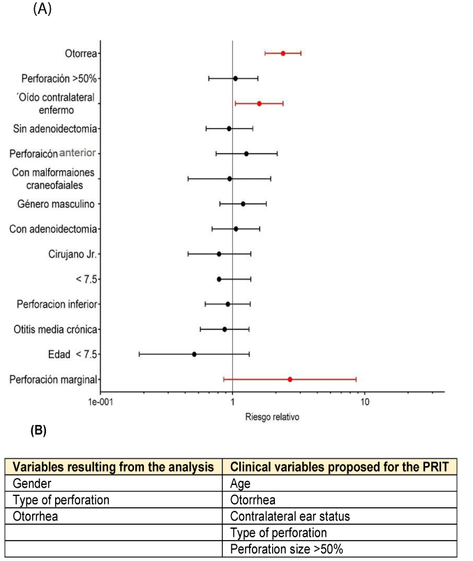
For the multivariate analysis, the selected variables were those that have been commonly reported by other authors as probable factors for failure, including the work that was previously carried out in our department by Boronat et al. in 2012. All variables were analyzed, contrasting them with anatomo-functional success (success of all 3 variables), with the OR being calculated with the corre sponding 95% CI (Figure 1).
According to data shown in table 2, failure occurs 2.4 fold more frequently in patients with otorrhea than in those without it (95% CI: 1.76.3.29); patients whose contralateral ear has inflammatory pathology, such as chronic su ppurative otitis media or serous otitis media have 1.59fold more failure than patients with a healthy contralateral ear (95% CI: 1.05-2.41) (Figure 1).
Marginal perforation shows that patients with this type of perforation have 1.27 fold higher risk for experiencing failure than those with central perforation (95% CI: 0.752.18) ; in spite of the CI crossing the value of 1 , this variable was taken as the third one because, from the clinical point of view, it is regarded as a dangerous perforation.
Integration of variables to the index model
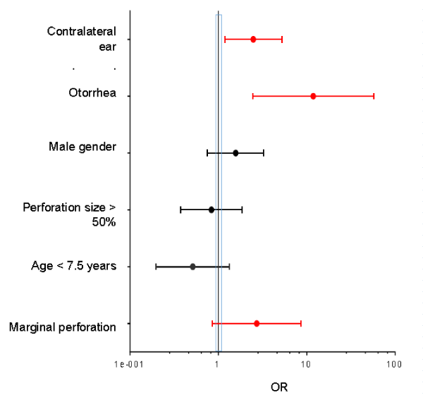
All variables introduced to the regression satisfy the minimum sample size (n > 10), and are therefore considered to have statistical power. According to table 3 results, gender, with an OR of 1.570 (95% CI: 0.754 -3.266), perforation size > 50%, with an OR of 0.834 (95% CI: 0.373-1.861) and the age < 7.5 years variable, with an OR of 0.512 (95% CI: 0.196-1.337), are no risk factors for surgery failure (Figure 2). Although dichotomous age has been shown not to be a risk factor, the PRIT index model was adjusted for the age in years.
Elaboration of the PRIT index
With logistic regression-associated values and clinical variables, a PRIT model is proposed. This model weighs potentially predictive variables according to the ORs significance and, in this case, otorrhea was assigned a value of 6 (OR: 11.59; 95% CI: 2.441-57.626), 3 points were assigned to the contralateral ear abnormal status variable (OR: 2.484; 95% CI: 1.181-5.223) and 1 point to marginal perforation ( OR: 2.717; 95% CI: .857-8.611) (Table 3), with a possible score of 0 to 10 points resulting when added up, according to the assessed clinical characteristics. For the creation of the index, this value was adjusted for the age in years, that is, total score was multiplied by the patient’s age in years (Table 4).
The PRIT score was calculated for each patient and subsequently, by means of a ROC curve, 30 points was established as the cutoff point, with a sensitivity of 80% and specificity of 80%.
Clinical use of the PRIT index is hypothetically exemplified above; on both examples, a 5-year-old patient is assessed.
Risk associated to the PRIT value
Age-adjusted clinical significance of the proposed PRIT (< 30 and > 30 points) was analyzed against the failure outcome variable using Pearson’s [2], test with a pvalue < 0.001.
The values expressed in table 5 clearly show that patients who obtained a PRIT score > 30 points have a 2.3-fold higher risk for experiencing failure than those with < 30 points.
Discussion
There are numerous articles [23] that try to reach a consensus on the risk factors tympanoplasty failure might be attributed to in the pediatric population [24]. Surgical success obtained in the present analysis is 59.6%, which is similar to that reported by Boronat et al. in 2012, but with regard to risk factors that produce failure there is considerable controversy or lack of homogeneous evidence.
One of the most controversial factors is patient age. The cutoff point in our study was 7.5 years, similar to that reported by Splete [25], where the age to consider surgery is suggested to be above 7 years. By itself, age had no statistical significance, as opposed to findings in a meta-analysis reported in 2015 [26], but when interacting with other variables such as otorrhea, contralateral ear status and type of perforation or marginal perforation [27] (the PRIT model), age was able to significantly predict failure (p < 0.001).
Otorrhea has no statistical significance in multiple reports [28], such as the one by Onal in 2005; however, in our study, otorrhea was significant since the beginning of the analysis (p < 0.001), and at risk estimation, it was shown to be an important factor for failure (RR: 2.41; 95% CI: 1.76 – 3.29) [29]. Otorrhea was mentioned as a factor that can directly affect surgical outcome by Lin et al. in 2008, but many other studies report having practiced this type of surgeries in patients with otorrhea with good results being obtained. In the multivariate analysis [30], otorrhea was clearly the most relevant variable in the outcome of failure with an OR of 11.859 (95% CI: 2.441-57.626), followed by the contralateral ear abnormal status variable (OR: 2.484; 95% CI: 1.181-5.223).
Craniofacial postnatal development apparently should be taken into account when programming any ear surgery since, at younger ages, the ET function is not fully adequate and, in addition, at pediatric age, its anatomical situation seems to put the patient in disadvantage prior to 7.5 years of age, which is consistent with Boston pediatric hospital experience, where only the age younger than 8 years was reported to be significant for failure [31]. In contrast, other authors claim that age has nothing to do with tympanoplasty outcomes in children [32,33]. On the other hand, Hassman concluded that contralateral ear abnormal status must be taken into account, since it is an indicator of nasopharyngeal pathology that might affect ET correct functioning [34].
The type of perforation is also important. In some textbooks of the specialty, marginal perforations [35] have been referred to as dangerous or insecure perforations, since the remnant eardrum in the tympanic perforation acts as a barrier that prevents squamous epithelium of the duct from migrating to the middle ear [35]. In the logistic regression carried out in this study, the marginal perforation type was found to be the third most relevant variable, in spite of the CI crossing the null hypothesis value of 1, with an OR of 2.717 (95% CI: .857-8.611). This type of perforations have also been associated with failure by Schraff and by Mendes R [35].
Conclusions
Tympanoplasty success was obtained in 59.6% of pediatric patients. The construction of the PRIT model for clinical use in tympanoplasty planning for pediatric patients was achieved (p < 0.001). Tympanoplasty failed in 40.4% of patients, and the variables that influenced on the outcome were otorrhea and contralateral ear status. All results correspond to a 6 months follow-up period.
Perspective
Validation of this index is necessary making sure there is a balance, especially in the sample of groups by age, in order to assess if age really represents a risk for surgical outcomes.
- Habesoglu T, Habesoglu M (2011) Effect of Type I Tympanoplasty on the Quality of Life of Children. Ann Otol, RhinolLaryngol 5: 114-119.
- Tos M (2000) Reasons for Reperforation after Tympanoplasty in Children. Acta Otilaryngol 120: 143-146. Link: https://goo.gl/1X6V8V
- Gardner E, Dornhoffer JL (2002) Tympanoplasty results in patients with cleft palate: An ageand procedure-matched comparison of preliminary results with patients without cleft palate. Otolaryngol Head Neck Surg 126: 518-523. Link: https://goo.gl/mUe6Tu
- Bluestone CD, Eustachian Tube Function and Dysfuction (2003) In: Rosenfel. EvidenceBased Otitis Media. 2° Edición.Canada: BC Decker 163-175.
- Magerian CA (2000) Pediatric Tympanoplasty and the role of preoperative Eustachian tube evaluation. Arch Otolaryngol Head Neck Surg 126: 1039-1041. Link: https://goo.gl/TszgtW
- Carlson LH, Carlson RD (2003) Diagnosis. In: Rosenfel. Evidende-Based Otitis Media. 2da edición: BC Decker 140-142.
- Collins WO, Telischi FF, Balkany TJ (2003) PedyatricTympanoplasty: effect of contralateral ear status on outcomes. Arch Otolaryngol Head Neck Surg 129: 646-651. Link: https://goo.gl/8YVZb1
- Hassman-Poznaska (2012) The status of the contralateral ear in children with acquired cholesteatoma; Acta Otolaryngol 132: 404-408. Link: https://goo.gl/R7vdM5
- Ribeiro JC, Rui C, Natercia S, Jose R, Antonio P (2011) Tympanoplasty in children: A review of 91 cases. Auris Nasus Larynx 38: 21-25. Link: https://goo.gl/ZPPJU7
- Bajaj Y, Bais A (1998) Tympanoplasty in children - A prospective study. J of Laryngol Otol 28: 121-132.
- Sarkar S, Roaychoudhury A (2009) Tympanoplasty in children. Eur Arch of Otorhinolaryngol 266: 627-633. Link: https://goo.gl/gu22zj
- Eredità D, Lens MB (2009) Anterior tab flap versus Estándar Underlay Myringoplasty in children. Otology 30: 777-781. Link: https://goo.gl/xHmX4p
- Kumar S, Acharya A, Hadjihannas E (2010) Pediatric Myringoplasty:Definicion de Succes and factors afecting Outcome. Otol Neurotol 31: 1417-1420.
- Takahashi TE, MishiroY,Katsura H (2014) Longitudinal follow-up after pediatric myringoplasty:long-term outcome is defined at 12 months. Otol Neurotol 35: 126-128. Link: https://goo.gl/Wczd5H
- Boronat-Echeverría N (2012) Prognostic factors of successful tympanoplasty in pediatric patients: a cohort study. BMC Pediatrics 12: 67. Link: https://goo.gl/QRKEuc
- Dornhoffer J (2003) CartilageTympanoplasty: Indications,Techniques and outcomes in a 1000patient series. Laryngoscope 113: 1844-1856. Link: https://goo.gl/kv8TSP
- Umapathy N, Dekker PJ (2003) Myringoplasty Is it Worth Performing in Children?. Arch OtolaryngolHead Neck Surg 129: 1053-1055. Link: https://goo.gl/UBqWgp
- Manning SC, Cantekin EI, Kenna MA, Bluestone CD (1987) Prognostic Value Of Eustachian Tube Function In Pediatric Tympanoplasty. Laryngoscope 97: 1012-1016. Link: https://goo.gl/iK7VYL
- Westerberg J, Harder H, Magnuson B (2010) Ten-year myringoplasty series: Does the cause of perforation affect the success rate? Laryngoscope 58: 712-718.
- Helsinki Declaration 2008. Link: https://goo.gl/9XC63y
- Rosato J (2000) The ethics of clinical trials: A child's view; J Law, Med Ethics 28: 228-234.
- Health law in research 2007.
- Emir H, Ceylan K, Kizilkaya, Zeynep (2007) Success is a matter of experience: type 1 tympanoplasty: Influencing factors on type 1 tympanoplasty. Eur Arch Otolaryngol 264: 645-656.
- Novas (2010) Failure of Tympanoplasty in Children: Predictive FactorsOtolaryngol - Head Neck Surg 143: 114-121.
- Splete H (2003) Tympanoplasty works as well in children as adults: age 7 and older fare best. Pediatric News 37: 117-123.
- Hardman J, Muzaffar J (2015) Tympanoplasty for Chronic Tympanic Membrane Perforation in Children: Systematic Review and Meta-analysis. OtolNeurotol 36: 796-804. Link: https://goo.gl/3r3wFR
- Westerberg J, Harder H Magnuson B (2011) Ten-year myringoplasty series: Does the cause of perforation affect the success rate?.JLaryngol Otol 125: 126-132. Link: https://goo.gl/h6d3ss
- Onal K, Uguz MZ, Kazikdas KC (2005) A multivariate analysis of otological, surgical and patient related factors in determining success in myringoplasty. Clin Otolaryngol 30: 115-120. Link: https://goo.gl/8iPF16
- Lin AC, Messner AH (2008) Pediatric tympanoplasty: Factors affecting success. Otolaryngol Head Neck Surg 16: 64-68. Link: https://goo.gl/7MSTJ6
- Onal K, Uguz MZ (2005) A multivariate analysis of otological, surgical and patient related factors in determining success in myringoplasty.ClinOtolaryngol 30: 115-120. Link: https://goo.gl/c2TWg6
- Koch WM, Friedman EM (1990) Tympanoplasty in children. The Boston Children's Hospital experience; Arch Otolaryngol Head Neck Surg 116: 34-40. Link: https://goo.gl/dpFPhz
- Tos M, Lau T (1989) Stability of tympanoplasty in children. OtolaryngolClin of N Am 22: 228-236.
- Attallah MS (1994) Tympanoplasty in children--our experience in Riyadh, Saudi Arabia Otolaryngol 48: 441-444. Link: https://goo.gl/nDLPpa
- Hassman-Pozna SKA, Bieta El, Kurzyna, Agnieszka, Trzpis, et al. (2012) The status of the contralateral ear in children with acquired cholesteatoma. Acta Otolaryngol 132: 404-408. Link: https://goo.gl/rRKaa9
- Schraff, Scott (2005) “Window shade” or tympanoplasty for anterior marginal perforations. Laryngoscope 115: 1655-1659. Link: https://goo.gl/DvUJFz
Article Alerts
Subscribe to our articles alerts and stay tuned.
 This work is licensed under a Creative Commons Attribution 4.0 International License.


 Save to Mendeley
