Archives of Otolaryngology and Rhinology
A surgical challange for primary hyperparathyroidism: Intravagal parathyroid adenoma
Adem Binnetoglu*, Adem Binnetoglu, Yavuz Gundogdu, Tekin Baglam and Murat Sari
Cite this as
Binnetoglu A, Binnetoglu A, Gundogdu Y, Baglam T, Sari M (2018) A surgical challange for primary hyperparathyroidism: Intravagal parathyroid adenoma. Arch Otolaryngol Rhinol 4(3): 057-060. DOI: 10.17352/2455-1759.000078A missed parathroid adenomas are the most common cause of surgical failure in persistent primary hyperparathyroidic patients. Abnormalities in the normal migration of the parathyroid glands during embryological development of the head and neck may result in considerable variability in the location of parathyroid tissue. Imaging studies were crucial in localizing the neoplasms in these patients. It is important to develop a strategy to systematically locate these glands either by preoperative investigations or surgical exploration. We describe a patient with persistent primary hyperparathyroidism who underwent three unsuccessful surgical procedures due to an intravagal parathyroid adenoma.
Introduction
The incidence of primary hyperparathyroidism (PHPT) is 4–30 per 100,000 population [1,2]. A solitary parathyroid adenoma(PTA) is present in 75–80% of cases of PHPT, hyperplasia involving multiple parathyroid glands is found in 15–20% of cases, and parathyroid carcinoma is present in <1% of patients. The incidence of PHPT peaks in the fifth–sixth decade, and a female-to-male ratio of 3:2 has been reported [2].
Clinical manifestations of hyperparathyroidism are dependent upon the severity of hypercalcemia. Most common presentation of PHPT is asymptomatic hypercalcemia. However, complications of prolonged and severe hypercalcemia are nephrolithiasis, reduced bone mineraldensity, bone pain, constipation, and severe fatigue [3,4].
A total of 90–95% patients with PHPT are cured after initial surgery [5]. For the remaining 5–10% of patients, persistent hyperparathyroidism may be the result of unrecognized multiglandular disease or the inability to identify an ectopic parathyroid adenoma (EPTA). Ectopic parathyroid glands may be located anywhere along the path of their embryologic descent [6,7].
“True” intravagal PTAs are extremely rare. Herein, we describe an intravagal PTA found at a fourth explorative surgical procedure for an EPTA.
Case
A 57-year-old female presented with asymptomatic hypercalcemia. Investigations revealed primary hyperparathyroidism, with increased serum-corrected calcium of 10.7 (normal range, 8.8–10.6) mg/dL and parathyroid hormone (PTH) of 1303 (normal range, 12–88) pg/mL. Our patient had undergone three unsuccessful surgical procedures for hyperparathyroidism.
During the first procedure, a biopsy specimen was obtained from the right superior parathyroid gland. Histology of the specimen showed normal parathyroid tissue. After the first procedure, the results of technetium-99m sestamibi scintigraphy suggested a right inferior PTA. At the second procedure, right-neck exploration was negative for an entopic and ectopic PTA. A biopsy specimen were obtained from a mass (2 cm × 1cm) inferoposterior to the left thyroid gland. A right thyroid lobectomy was done. Histology of the specimen showed normal thyroid tissue and a lymph node. The third procedure was done because a left superior PTA was suspected. A biopsy specimen was obtained from the left superior parathyroid gland. Histology of this specimen showed mild and diffuse hyperplasia in the left upper parathyroid gland.
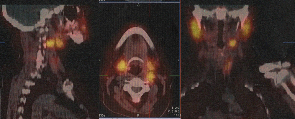
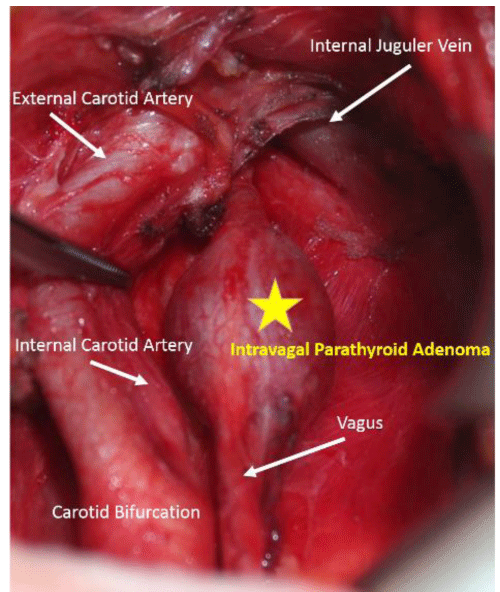
Before the fourth procedure, magnetic resonance imaging(MRI) of the neck and mediastinum revealed a mass (2 cm × 1 cm) on the left side of the neck at level 2. Computed tomography (CT) confirmed the neck mass to be a lymph node. Scintigraphy showed a suspicious accumulation at left level 2, posterior to submandibular gland (Figure 1). Ultrasonography of the neck was unremarkable. On the basis of these findings, a fourth surgical procedure was carried out: the left thyroid lobe was mobilized and explored, but entopic parathyroid glands were not discovered. The left paratracheal–paraesophageal groove and upper mediastinum were cleared. The left recurrent laryngeal nerve was identified and preserved. The carotid sheath was explored and a fusiform swelling (2 cm × 1cm) was identified in the vagus nerve at the level of the carotid bifurcation. A longitudinal incision was made in the perineurium. A soft, tan-colored nodule was identified and enucleated from the vagus nerve (Figures 2-4). This enucleation did not give rise to intraoperative or postoperative complications such as bradycardia, hoarseness, or dysphagia. Pathology confirmed a PTA in the specimen.
On postoperative day-1, levels of serum-corrected calcium and PTH had dropped to 8.5 mg/dL and 7 pg/mL, respectively. Her voice was normal, she had no difficulty in swallowing, could tolerate a normal diet, and was discharged home on postoperative day-3. The patient is asymptomatic and eucalcemic.
Discussion
During embryogenesis, the parathyroid glands migrate from the third and fourth pharyngeal pouches to their respective entopic inferior and superior locations on the dorsal surface of the thyroid gland [6]. Normal parathyroid tissue have been identified at any point from the parapharyngeal skull base to the middle mediastinum [6]. The inferior parathyroid gland has a long embryologic migration tract. Therefore, it is more likely to descend to an ectopic locations. Up to 16% of PTAs are found in ectopic locations, which include the thymus gland(24–38%), retro-esophageal space(22–31%), thyroid gland(7–18%), mediastinum(6–20%), carotid sheath(3–9%), undescended glands(2–7%) [5]. PTAs rarely seen in the lateral neck, adjacent to the hyoid bone, submandibular gland or in the paranasopharyngeal space, pyriform fossa or intravagal [6,8-10].
Only 13 cases of supernumerary intravagal parathyroid glands have been reported in the English literature. However, those case reports may not reflect the true incidence because intravagal parathyroid tissue has been documented in autopsy reports with a prevalence of ≤6%. Part of the vagus nerve is derived from the fourth pharyngeal arch, which is bordered on either side by the third and fourth pharyngeal pouches. This close proximity may explain the presence of intravagal parathyroid tissue [8,11]. In a postmortem study, Lack et al. carried out serial sections of both vagus nerves from the up perneck of 32 individuals aged ≤1 year. Four (6%) of the 64 nerves contained solitary microscopic collections of parathyroid chief cells, which were confirmed by their positive immunoreactivity for chromogranin and PTH [8].
In all cases except one, patients had previously undergone 1–4 neck explorations involving thymectomy and total thyroidectomy, as well as bilateral carotid-sheath explorations. Multiple explorations are associated with an increased risk of complications, including damage to the recurrent laryngeal nevre [12], bleeding [6], and a prolonged surgical procedure [13]. An increased morbidity and decreased success rate, primarily due to fibrous tissue in the previous operative side and distortion of normal tissue planes that results in difficulty in identification of parathyroid tissue and other cervical structures (most notably the recurrent laryngeal nerve), has been noted [14].
Imaging studies before exploration are crucial. Radionuclide imaging allows extensive examination of a large area, thereby affording less difficult localization of the lesion. For efficient investigation of ectopic lesions, radionuclide imaging should be undertaken first. If accumulation of radioactive tracer is found in the neck, it should be confirmed by ultrasonography [15]. Often, ectopic glands are not identified by preoperative ultrasound due to their unexpected locations, and are commonly mistaken for salivary glands with increased uptake on scintigraphy [16]. If the site of accumulation is far from the neck, CT and/or MRI should be used to confirm the lesion. Single-photon emission CT offers low-dose radiation but lends itself to low-resolution images that do not necessarily give the anatomic detail provided by high-resolution CT. Repeat imaging can facilitate successful surgery, but some patients require invasive preoperative localization studies, including angiography and venous sampling [17].
Reported cases suggest that these intravagal neoplasms are usually situated at the level of the carotid bifurcation, anterior to or within the carotid sheath, just as in our patient. Excision of an intraneural PTA without transection of at least some of the nerve fibers can be extremely difficult. Pawlik et al. suggested that, with careful dissection, nerve fibers adherent to the capsule of the adenoma can be freed, whereas those coursing through the lesion are sacrificed eventually [18].
Intraoperatively, a systematic diagnosis can minimize the prevalence with which ectopic lesions are missed during clinical care and maximize their accurate localization. First, the surface of the thyroid gland (including the subcapsular plane) should be inspected for an EPTA. The surface of the tubercle of Zuckerkandl, posterior surface of the upper pole and vessels, the para-esophageal space as far inferioras the posterior mediastinum, and the posterior surface of the inferior pole vessels must be cleared. The recurrent laryngeal nerve must be identified and protected when the tracheo-esophageal groove and paraesophageal space are explored. Then, a cervical thymectomy should be carried outfor a possible PTA in the thymic horn. Next, inspection of the posterior surface of the thyroid gland, as well as access to the cricothyroid and parapharyngeal/retropharyngeal spaces, should be carried out. Then, a mediastinal or maldescended PTA should be considered. First, the parapharyngeal and retropharyngeal spaces should be explored superiorly up to the level of the pyriform fossa. The carotid sheath should be explored from the carotid bifurcation to the level of the inferior thyroid pole. The vagus nerve should be examined for a “bulge” because the EPTA may be contained within its fibers. Finally, a thyroid lobectomy can be carried outto inspect for intrathyroid PTA if the latter remains elusive after the explorations described above. Intraoperative measurements of PTH levels can confirm PTA removal, but do not aid EPTA localization. However, adequate reduction in intraoperative PTH levels may give the surgeon confidence to stop further exploration after removal of one adenoma [19].
Conclusions
Ectopic PTAs can be located anywhere along the path of their embryologic descent. Hence, development of a strategy to locate these glands in a systematic manner by preoperative investigations or surgical exploration is important. Though extremely rare, an intravagal PTA is another type of ectopy that surgeons must keep in mind to increase the chance of success at the initial surgical procedure.
This work presented as electronic poster presentation at the 38th Politzer society meeting, 26-30 November 2016, Antalya, Turkey (EP-012).
Financial Disclosure
This research did not receive any specific grant from funding agencies in the public, commercial, or not-for-profit sectors this material has never been published and is not currently under evaluation by any other peer-reviewed publication
- Mundy GR, Cove DH, Fisken R (1980) Primary hyperparathyroidism: changes in the pattern of clinical presentation. Lancet 1:1317–20. Link: https://goo.gl/Ab1ja4
- Wermers RA, Khosla S, Atkinson EJ (1997) The rise and fall of primary hyperparathyroidism: a populationbased study in Rochester, Minnesota, 1965–1992. Ann Intern Med 126:433–40. Link: https://goo.gl/hJnohr
- Khan A, Bilezikian J (2000) Primary hyperparathyroidism: pathophysiology and impact on bone. Can Med Assoc J 163:184-7. Link: https://goo.gl/bSXvxa
- Bilezikian JP, Potts Jr JT (2002) Asymptomatic primary hyperparathyroidism: new issues and new questions–bridging the past with the future. J BoneMiner Res 17 Suppl 2;N57–67. Link: https://goo.gl/6NNcEB
- Delbridge L, Younes N, Guinea A (1998) Surgery for primary hyperparathyroidism 1962–1996: indications and outcomes. Med J Aust 168:153-6. Link: https://goo.gl/P4brDc
- Wang C (1976) The anatomic basis of parathyroid surgery. Ann Surg 183;271–75. Link: https://goo.gl/iKm6uY
- Brennan MF, Norton JA (1985) Reoperation for persistent and recurrent hyperparathyroidism. Ann Surg 201:40–4. Link: https://goo.gl/tFd6xM
- Lack EE, Delay S, Linnoila RI (1988) Ectopic parathyroid tissue within the vagus nerve. Incidence and possible clinical significance. Arch Pathol Lab Med 112:304–6. Link: https://goo.gl/zo2Bim
- Simeone DM, Sandelin K, Thompson NW (1995) Undescended superior parathyroid gland: a potential cause of failed cervical exploration for hyperparathyroidism. Surgery 118:949–56. Link: https://goo.gl/V2VLuq
- Chan TJ, Libutti SK, McCart JA (2003) Persistent primary hyperparathyroidism caused by adenomas identified in pharyngeal or adjacent structures. World J Surg 27:675–9. Link: https://goo.gl/vEr9cf
- Benson MT, Dalen K, Mancuso AA (1992) Congenital anomalies of the branchial apparatus: embryology and pathologic anatomy. Radiographics 12:943–60. Link: https://goo.gl/CTZRHE
- Reiling RB, Cady B, Clerkin EP (1972) Aberrant parathyroid adenoma within the vagus nerve. Lahey Clin Bull 21:158-62.
- Chan TJ, Libutti SK, McCart JA (2003) Persistent primary hyperparathyroidism caused by adenomas identified in pharyngeal or adjacent structures. World J Surg 27:675–9. Link: https://goo.gl/8mmjbu
- Jaskowiak N, Norton JA, Alexander HR (1996) A prospective trial evaluating a standard approach to reoperation for missed parathyroid adenoma. Annals of surgery 224:308-21. Link: https://goo.gl/83EABC
- Feingold DL, Alexander HR, Chen CC (2000) Ultrasound and sestamibi scan as the only preoperative imaging tests in reoperation for parathyroid adenomas. Surgery 128:1103-10. Link: https://goo.gl/48H33y
- Fraker DL, Doppman JL, Shawker TH (1990) Undescended parathyroid adenoma: an important etiology for failed operations for primary hyperparathyroidism. World J Surg 14:342–8. Link: https://goo.gl/Mgz4Cq
- Okuda I, Nakajima Y, Miura D (2010) Diagnostic localization of ectopic parathyroid lesions: developmental consideration. Jpn J Radiol 28:707–13. Link: https://goo.gl/EEbiQM
- Pawlik TM, Richards M, Giordano TJ (2001) Identification and management of intravagal parathyroid adenoma. World J Surg 25:419–23. Link: https://goo.gl/JJY4DU
- Lee JC, Mazeh H, Serpell J (2015) Adenomas of cervical maldescended parathyroid glands: pearls and pitfalls. ANZ journal of surgery 85:957-61. Link: https://goo.gl/bDpABw
Article Alerts
Subscribe to our articles alerts and stay tuned.
 This work is licensed under a Creative Commons Attribution 4.0 International License.


 Save to Mendeley
