Annals of Musculoskeletal Medicine
Surgical Cure of Foot Macrosyndactyly: A Case Report
Leopoldo Maizo1*, Rafael Arcia2 and Julio Trillo1
2Specialist in Lower Reconstructive Surgery, Hospital Ortopédico Infantil de Caracas, Venezuela
Cite this as
Maizo L, Arcia R, Trillo J (2017) Surgical Cure of Foot Macrosyndactyly: A Case Report. Ann Musculoskelet Med 1(1): 019-021. DOI: 10.17352/amm.000004Macrodactyly is a congenital disease characterized by an increase in the volume of one or more fingers disproportionately relative to normal fingers. It is a rare congenital, non-hereditary disease that can occur in the hands or feet. It is characterized by an increase in all mesenchymal elements particularly fibro-adipose tissue. The vast majority of patients with macrodactyly require surgical treatment. Numerous techniques have been described each with different peculiarities and methods, and with very variable results what makes a pathology of great complexity when deciding its treatment.
Clinical Case: A 3-month-old male patient, his mother referred to the onset of current illness from his birth, when we noticed increased volume and progressive deformity on the left foot, we observed clinical and radiographic signs compatible with second and third finger macrosyndactyly So it is decided surgical treatment for macrosyndactyly cure with placement of surgical wires. We performed the removal of the wires to the month with satisfactory evolution and definitive fisiodesis.
Introduction
Macrodactyly is a congenital disease characterized by an increase in the volume of one or more fingers disproportionately relative to normal fingers. It was first described by Feriz in 1925 as partial gigantism of the lower limb [1-3]. Other terms used to name this condition are: local gigantism, lipomatous macrodistrophy, localized hypertrophy, finger fibrolipomatosis and digital gigantism [4].
It is a rare congenital, non-hereditary disease that can occur in the hands or feet [5-7]. It is characterized by an increase in all mesenchymal elements particularly fibro-adipose tissue [1,8,9].
According to Barsky, there are two forms of clinical manifestation: the static form in which the finger is larger from birth, but the growth from here is proportional to the rest of the fingers, and the progressive form, in which the finger is normal at birth, but begins to grow faster than the rest of the fingers and can cause its angular deviation, describes this form as the most frequent [10]. Unlike De Laurenzi who described the less frequent progressive form for cases documented in the hand [11].
In 95% of the patients the disease presents unilaterally. It is slightly more frequent in males and appears in decreasing direction from the index to the fifth finger. In the progressive form the growth of the finger ceases when the epiphyses are closed, usually the sensitivity is normal, the mobility decreases with time and the ulcers in the bud are frequent.
The etiology of macrodactyly is still unknown [1,2,4,8], as already mentioned has not been proven to be hereditary, but when it is associated with other syndromes it is inherited in an autosomal dominant way. Some authors claim that it is a frustrating form of Von Reclinghausen syndrome or neurofibromatosis.
The causes can be: abnormal innervation, abnormal humoral mechanism and abnormal irrigation of the finger (the last two causes are not well demonstrated, the first one because the nerves exert great control over the growth).
Macrodactyly is associated with syndactyly in 10% of patients [2] and polydactyly and cryptorchidism in a lower percentage. It may be associated with Klipper Syndrome Trenaunag Weber (hemangiomatosis, varicose veins and limb hypertrophy), Maffuci Syndrome (multiple hemangiomatosis), Proteus Syndrome (hamartomatous dysplasia, pigmented nevi and subcutaneous tumors), hemangiomas, arteriovenous malformations, congenital lymphedema, lipomas, osteoid osteoma and melorrestosis, which constitute “non-true” forms of macrodactyly [5]. Macrodactyl may be associated with hernia.
The vast majority of patients with macrodactyly require surgical treatment. Numerous techniques have been described each with different particularities and methods, and with very variable results what makes a pathology of great complexity when deciding its treatment; Reason that has led to the elaboration of this work which pretends to describe a different technique in these patients.
Clinical Case
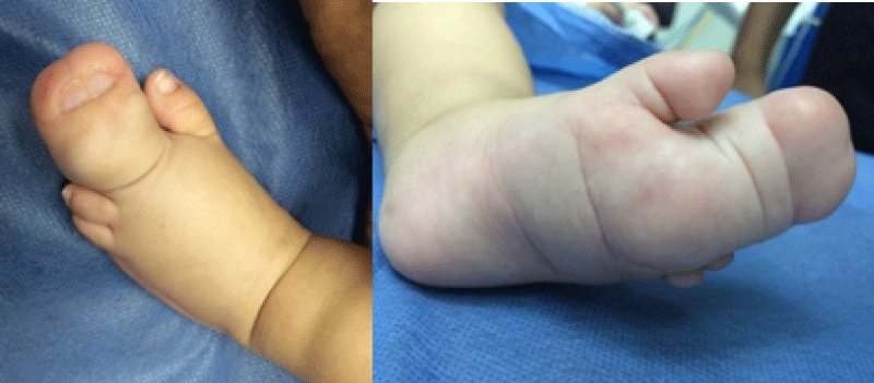
A 3-month-old male patient, whose mother refers to the onset of the current illness from birth, when she appreciates an increase in volume and progressive deformity in the left foot of her child, which is why she visits a specialized lower limb service where we value. Previous History: His mother is 18 years of age, first pregnancy, pregnancy of 6 controls without complications. He was born by cesarean section with 38 weeks of gestation, with no peri and post-natal complications (Figure 1).
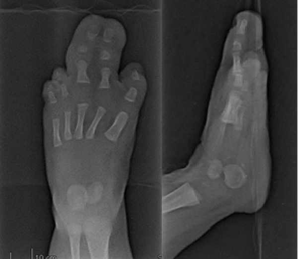
On physical examination, we observed an increase in volume and non-painful deformity with fusion of the second and third toes. The rest of the physical examination showed no pathological alterations. This is the reason why we indicate radiographs in the frontal and sagittal plane where we appreciate bone overgrowth of second and third finger phalanges (Figure 2).
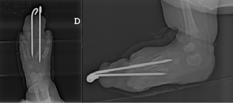
In April 2016, we performed surgery where by percutaneous technique we performed curettage on growth nuclei of all phalanges of the second and third toe of the left foot, with subsequent stabilization with Kirchner wires 2.0 mm after direct visualization with fluoroscope (Figure 3).
Subsequently, we performed removal of wires from Kirchner after one month of placement, we observed consolidation bone magma, with closure of the growth nuclei of phalanges of second and third rays of left foot, retaining their orientation and alignment.
He returned to the control clinic in June 2016 where we observed radiologically the longitudinal growth of the phalanges with persistence and increased thickness at the expense of soft tissue, so we decided to re-control in 3 months for definitive surgical behaviour.
Discussion
As pathologies for surgical treatment, with the tendency to deformity and progressive evolution, there are numerous techniques described for its handling and resolution. In children, soft tissue reductions, epiphysiodesis, epiphysectomies, osteotomies and other techniques are recommended [4,12-17]. In adults, shortening techniques and arthrodesis are more commonly used [4,10]. The technique applied in our case was epiphysectomy, not usually described in the literature.
In 1996, Kostakoglu published a series of 8 patients with macrodactyly who underwent proximal phalangeal excision and soft tissue mass reduction. Functional and aesthetic results were poor in most cases with limited range of motion and frequent joint contractures [14]. Ishida operated on 23 patients diagnosed with macrodactyly and 65% of them needed to perform more than two operations. The average thickness of the finger operated at the level of the proximal interphalangeal joint was 121% and in the distal interphalangeal 124% with respect to the contralateral healthy finger. The average range of motion at the level of the three finger joints was approximately 50% of normal. The techniques used were epiphysiodesis and epiphysectomy that had the same effect on the longitudinal growth arrest, but not the thickness, which was not effectively reduced by the resection of the digital peripheral nerves [18]. Kotwal operated on 21 cases and after 9 years of follow-up, the aesthetic aspect of the finger was good only in 57% of patients. The techniques used were two-stage soft tissue reduction and phalangectomy [19].
Complications are very common after reconstruction techniques in macrodactyly. Cutaneous necrosis is the most frequent and can sometimes be severe and compromise the viability of the finger [4]. Kostakoglu reported cutaneous necrosis in 25% of patients after soft tissue reduction [14]. Dell also found this complication frequently after reconstruction techniques [20].
Despite the experience accumulated over the years in the treatment of this complex congenital anomaly, most patients require multiple surgical interventions during infancy and in a high percentage of them, the result is an unsightly and dysfunctional finger [21,22].
Conclusion
We can conclude that macrodactyly is a complex condition of varied treatment and frequent complications. We achieved during the first treatment the satisfactory reduction of the longitudinal growth of the phalanges, although with persistence of the transversal growth of the same ones, reason why the reintervention of the patient to correct future complications is considered essential to avoid the resections and dysfunctional results and unsightly.
- Feriz H. Macrodystrophia lipomatosa Progressive. Virchows Arch Pathol Anat Physiol Klin Med. 1925; 260: 308-68.
- Ozturk A, Baktiroglu L, Ozturk E, Yazgan P (2004) Macrodystrophia lipomatosa: A case report. Acta Orthop Traumatol Turc 38: 220-223. Link: https://goo.gl/PBMNv2
- Blacksin M, Barnes FJ, Lyons MM (1992) MR diagnosis of Macrodystrophia lipomatosa. AJR 158: 1295-1297. Link: https://goo.gl/MyWbpH
- Wood VE. Macrodactyly. En: Green DP. Operative hand surgery. Vol 1. New York: Churchill-Livingstone; 1993.p. 497-509.
- Krengal S, Morales AF, Carrcasco P, Vazquez M, Mickinster CD, et al. (2000) Macrodactyly: Report of eight cases and review of the literature. Pediatric dermatology 17: 270-276. Link: https://goo.gl/LCGNtT
- Sharma S, Vyas S, Sood RG, Auupam, Kaushik NK (2006) Macrodactyly: Report of report of 3 cases. Ind J Radiol Imag 16: 583-584. Link: https://goo.gl/nYeqHT
- Pearu J, Bloch CE, Nelson MM (1986) Macrodactyly simplex congenita: A case series and considerations of diff erential diagnosis and etiology. S Afr Med J 70: 755-758. Link: https://goo.gl/I0Hcvo
- Yuksel A, Yagmur H, Kural BS (2009) Prenatal diagnosis of isolated macrodactyly. Ultrasound Obstet Gynecol 33: 360-362. Link: https://goo.gl/BhSDl1
- Wahab S, Khan RA, Ahmed I (2008) Congenital localized limb hypertrophy: Macrodystrophia lipomatosa. JBR-BTR 91: 209-210. Link: https://goo.gl/nKH989
- Barsky AJ (1967) Macrodactyly. J Bone Joint Surg 49:1255-1266. Link: https://goo.gl/Vt3GKa
- The hand, part II. In: May JW, Hillter JW, editors (1990) McCarthy plastic surgery. Philadelphia: WB Saunders 8: 5362-5365.
- Tsuge K (1967) Treatment of macrodactyly. Plast Reconstr Surg 39: 590-599. Link: https://goo.gl/HPtS5N
- Kelikian H (1974) Congenital deformitie of the hand and forearm. Philadelphia: WB Saunders. Link: https://goo.gl/Y5RTdk
- Kostakoglu N, Kayikcioglu A, Safak T, Ozcan G, Kecik A, et al. (1996) Macrodactyly: report of eight cases of a rare anomaly. Turk J Pediatr 38: 73-79. Link: https://goo.gl/oCC8JQ
- Bertilli JA, Pigozzi L, Pereima M (2001) Hemidigital resection with collateral ligament transplantation in the treatment of macrodactyly: a case report. J Hand Surg 26: 623-627. Link: https://goo.gl/mPPjuC
- Tsuge K (1985) Treatment of macrodactyly. J Hand Surg 10: 968-969. Link: https://goo.gl/nJumGs
- Chen SH, Huang SC, Wang JH, Wu CT (1997) Macrodactyly of the feet and hands. J Formos Med Assoc 96: 901-907. Link: https://goo.gl/u3OcVM
- Ishida O, Ikuta Y (1988) Long-term results of surgical treatment for macrodactyly of the hand. Plast Reconstr Surg 102: 1586-1590. Link: https://goo.gl/7PYGNG
- Kotwal PP, Farooque M (1988) Macrodactyly. J Bone Joint Surg 80: 651-653.
- Dell PC (1985) Macrodactyly. Hand Clin 1: 511-524. Link: https://goo.gl/nF6U1d
- Álvarez R, Peña L, López H, Hernández R, Hernández O, et al. (2002) Transposición digital en el tratamiento de la macrodactilia. Rev Cubana Ortop Traumatol 16: 65-69. Link: https://goo.gl/2MLbhH
- Isik A, Gursul C, Peker K, Aydın M, Fırat D, et al. (2017) Metalloproteinases and Their Inhibitors in Patients with Inguinal Hernia. World J Surg 41: 1259-1266. Link: https://goo.gl/SX4BCt
Article Alerts
Subscribe to our articles alerts and stay tuned.
 This work is licensed under a Creative Commons Attribution 4.0 International License.


 Save to Mendeley
