Archives of Hematology Case Reports and Reviews
Chronic myelomonocytic leukemia: A rare hematologic malignancy that needs due consideration
Temesgen Assefa1* and Yosef Alebel2
2Internal Medicine Resident, College of Medicine & Health Sciences, Bahirdar University, Ethiopia
Cite this as
Assefa T, Alebel Y (2020) Chronic myelomonocytic leukemia: A rare hematologic malignancy that needs due consideration. Arch Hematol Case Rep Rev 5(1): 025-027. DOI: 10.17352/ahcrr.000026Introduction
Chronic Myelomonocytic Leukemia (CMML) is a malignant hematopoietic stem cell disorder with clinical and pathological features of both a myeloproliferative neoplasm and myelodysplastic syndrome [1]. According to the 2016 WHO classification of hematologic malignancies it is categorized as MyeloDysplastic /Myeloproliferative Neoplasm [2].
CMML is a rare aggressive Chronic Leukemia with a tendency to progress to Acute Leukemia.It occurs most commonly in older adults, with a median age at diagnosis is ~ 65–75 years. There is a moderate male predominance in its incidence [3].
The 2016 World Health Organization (WHO) criteria for the diagnosis of CMML include:
- Persistent (≥3 months) peripheral blood monocytosis; absolute monocyte count >1000/microL and greater than 10 percent of the entire white blood cell differential
- Not meeting WHO criteria for BCR-ABL1 positive chronic myeloid leukemia, primary myelofibrosis, polycythemia vera, or essential thrombocytosis
- <20 percent myeloblasts + monoblasts + promonocytes in peripheral blood and bone marrow
- Dysplastic changes in one or more myeloid lineages. If myelodysplastic changes are absent or minimal, the diagnosis of CMML can be made if the above three criteria are met and:
- An acquired clonal cytogenetic (or mutational abnormality) is present in bone marrow cells or
- Persistent monocytosis for ≥3 months and all other causes of monocytosis have been excluded. If myelodysplastic changes are absent or minimal, the diagnosis of CMML can be made if the above three criteria are met and an acquired clonal cytogenetic (or mutational abnormality) is present in bone marrow cells or Persistent monocytosis for ≥3 months and all other causes of monocytosis have been excluded [4].
It needs a high index of suspicion to diagnose CMML in patients presenting with a complaint of clinical symptoms of Anemia, abdominal swelling & early satiety because of its similarity with other Myeloproliferative & Myelodysplastic syndromes in clinical presentation especially in resource limited setups where utilization of cytogenetic analysis is limited.
Case report
History
This is a 45 years old female patient from Hamusit, a rural area around 580 KM North west of Adiss Ababa, a capital of Ethiopia Presented with a complaint of worsening of left sided abdominal swelling of 04 months with associated dragging sensation & early satiety for the last 4 months which was there for the past 10 months, She also has symptom complex of Anemia but no fever or bleeding tendency.
She also has Anorexia & significant amount of weight loss during the period of illness.
The patient visited different health institutions at different times since the onset of the illness but no definitive diagnosis has been made.
She didn’t report previous malaria or kalazar treatment history, Has No family history of similar illness.
She is living in rural area & has no history of Exposure to therapeutic radiations or Industrial dusts.
Physical Examination/at presentation/
General appearance
- Chronically sick looking
Vital sign
Bp – 100/70mmHg PR – 80 beatsper minute RR -20bpm Temperature - 37.30 C
Head,Ear,Eye,Nose & Neck – Pink Conjunctiva & Non Icteric sclera
No other abnormality
Lymphoglandular system – No palpable Lymphnodes
Chest - clear & resonant
Cardiovascular system – S1 & S2 are well heared
Abdomen
- Liver – 6cm below right costal margin
- Spleen – 20 cm below left costal margin
Genitourinary Examination
- No abnormality
Musculoskeletal & Integumentary
- No abnormality
Central Nervous system
- No abnormality
Work up of the patient /at presentation / - 01/07/2020
Complete Blood count
- Total WBC count – 143.4×10³ Cells /µl
- Neutrophil count -125.1×10³/ µl
- Lymphocyte count - 14.9×10³/ µl
- Monocyte count – 7.8 ×10³/ µl
- Eosinophil -1.3×10 3/ µl
- Basophil count – 0.04×10³/ µl
- RBC Count –3×10³/ µl
- Hemoglobin -7.9 g/dl
- Platelet count -109×10 3/ µl
Liver Enzymes & Renal function tests - July 01 2020
- Creatinine -1mg/dl
- Alaninine amino transferase – 28 U/L
- Aspartate amino transferase – 25 U/L
- Creatinine – 1 mg/dl
- Negative for RVI ,Hepatitis B & C screening
Uric acid
- 2.62mg/dl
- Bone Marrow Evaluation – Report by pathologist
- The smear shows sheets of myeloid series cells with maturation with a conclusion of CML
- Cytogenetics test from whole blood
-BCR –ABL T (99,22) by FISH - NEGATIVE for t (9,22)
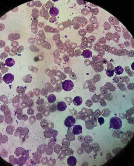
After the Negative BCR – ABL test the Peripheral Morphology of the patient was reevaluated again considering the possibility of alternative diagnosis, it is found as shown below:
Peripheral Morphology done on 12/8/2020 & evaluated with oil immersion 100X magnification microscopy shows:
Monocytes with with well-defined nuclear lobes & Increased Nucleus to cytoplasm ratio with clinical course after diagnosis.
The patient was started on Hydroxyurea 500mg Po Bid and supportive care with Hematinics, on subsequent follow up the patient improved symptomatically & her subsequent CBC after is as follows:
- On 16/9/2020
Total WBC count – 48.68×10³ Cells/µl
Neutrophil count – 28.6×10³/µl
Monocyte count – 9.05×10³/µl
Hemoglobin – 10.2 g/dl
Platelet count – 372×103/µl
- On 8/10/2020
WBC – 35.8×10³ Cells/µl
Neutrophil – 27.1×10³/µl
Monocytes – 2.01×10³/µl
Hemoglobin – 9.6 g/dl
Platelet – 73×103
Discussion
A presumptive diagnosis of CML is usually done with a clinical presentation of patients that is similar to our patient until molecular tests are done to confirm or exclude the diagnosis of CML this is because CML is the commonest Leukemia in Ethiopian patients accounting for 57.8 % of Leukemia admitted in hospital [5]. With the availability of BCR-ABL genetic studies it is possible to easily made the diagnosis of CML. patients with a Negative BCR-ABL translocation pose a diagnostic challenge ,the Physician need to think of the possible alternative differential diagnosis & evaluate the patient carefully with all the available laboratory tests as the setup allows, & CMML is one of these differential diagnosis that need due attention while evaluating patients with similar presentation.
CMML is a rare Chronic Myeloid Neoplasm/Myelodysplastic syndrome which has a diagnostic challenge specially in conditions where patients are strongly suspected to have Chronic Myeloid Leukemia but the BCR-ABL genetic analysis is negative. The diagnosis of CMML should be considered when persistent, unexplained peripheral monocytosis is present in an older adult Presenting with Anemia & hepatosplenomegally. Evaluation of the peripheral blood smear and a bone marrow biopsy and aspirate to document the dysplastic cytologic features identifiable in any or all of the hematopoietic lineages are key components to the diagnosis of CMML ,If resources allow the bone marrow evaluation should include karyotype analysis by conventional cytogenetics or FISH [6].
In patients with similar presentation like this we need to consider all the possible differential diagnosis based on the clinical presentation & CBC profile , evaluate the Bone Marrow Aspirate & peripheral morphology thoroughly with experienced Hematopathologist if possible with cytogenetic analysis.
In this Particular patient Initially the diagnosis of Chronic Myelogenous Leukemia was considered based on the Bone Marrow aspirate report by the pathologist & the patient was ordered to have BCR-ABL analysis which was negative then the Clinical presentation was discussed with experienced Hematologist & the Peripheral morphology was also revised as shown above then the diagnosis of CMML was considered & the patient is being managed accordingly.
Patient perspective
- Verbal Informed consent was obtained from the patient
- The patient is fully volunteer for the reporting of the case & to continue the care.
Conclusion
In patients with a clinical presentation & hematologic profile similar to our patient consideration of all the possible differential diagnosis & a thorough evaluation of the peripheral morphology & Bone marrow aspiration by an experienced Hematopathologist is very crucial for diagnosis of rare malignancies like CMML ,It will also avoid unnecessary cost to the patient & will avoid delay in the treatment of the patient.
- Itzykson R,Solary E (2013) An evolutionary Perspective on chronic Myelomonocytic leukemia. Leukemia 27: 1441-1450. Link: https://bit.ly/38itJ2a
- Arber DA, Orazi A, Hasserjian R, Thiele J, Borowitz MJ, et al, (2016) The 2016 revision to the World Health Organization classification of myeloid neoplasms and acute leukemia. Blood 127: 2391-2405. Link: https://bit.ly/3l4CkZB
- Rollison DE, Howlader N,Smith MT,et al. (2008) Epidemiology of Myelodysplastic syndromes & myeloproliferative disorders in UnitedStates: 2001 -2004, Blood.
- Arber DA, Orazi A, Hasserjian R, Thiele J, Borowitz MJ, et al, (2016) The 2016 revision to the World Health Organization classification of myeloid neoplasms and acute leukemia. Blood 127: 2391-2405. Link: https://bit.ly/3l4CkZB
- Shamebo M (1990) Leukemia in Adult Ethiopians. Ethiop Med J 28: 31-37. Link: https://bit.ly/36dlX6K
- Swerdlow SH, Campo E, Harris NL, Jaffe ES, Pileri SA, et al. (2008) WHO classification of Tumors of Haematopoietic and Lymphoid Tissues. IARC Press, Lyon. Link: https://bit.ly/3l3aFZh

Article Alerts
Subscribe to our articles alerts and stay tuned.
 This work is licensed under a Creative Commons Attribution 4.0 International License.

 Save to Mendeley
