Archives of Hematology Case Reports and Reviews
Secondary Breast Lymphoma: A Case Report
Jana Taron1*, Sabrina Fleischer1, Sonja Bahrs1, Heike Preibsch1, Valerie Hattermann1, Markus Hahn2 and Benjamin Wiesinger1
2Department for Obstetrics and Gynecology, University Hospital of Tuebingen, Tuebingen, Germany
Cite this as
Taron J, Fleischer S, Bahrs S, Preibsch H, Hattermann V, et al. (2017) Secondary Breast Lymphoma: A Case Report. Arch Hematol Case Rep Rev 2(1): 019-021. DOI: 10.17352/ahcrr.0000107914 Nevertheless, multimodal imaging and subsequent histopathology are crucial for diagnosis and treatment. To draw attention to this very rare entity, we present a case of a 74 year old female patient with a tumorous mass in the left maxillary sinus. The subsequent whole-body staging CT revealed an additional multicentric lesion of the right breast suggesting breast cancer. Histopathology of both lesions resulted in the diagnosis of a diffuse large B cell lymphoma (DLBCL).
Introduction
Breast lymphomas are a rare sighting accounting for about 0.5% of all breast malignancies and 1% of non-Hodgkin lymphoma (NHL) [1,2]. They can be categorized as primary (PBL) or secondary breast lymphoma (SBL). In case of PBL, the breast is the primary/only site of manifestation, whereas in SBL further manifestations of lymphoma are present [3], which has been reported as more common [2]. In general, breast lymphomas can be differentiated into B-cell (with 94% the most common type) and T-cell types (6%) [4], with the diffuse large B-cell lymphoma (DLBCL) representing the predominant entity with 40 - 70% of all cases [5,6].
Clinical findings in breast lymphoma are non-specific making it difficult to differentiate from benign or malignant breast lesions. The most common clinical finding in B-cell lymphoma of the breast is a painless palpable mass, less frequently associated with retraction of the nipple [2,7,8]. Skin changes or edema, on the other hand, have been reported to occur more often in T-cell breast lymphoma [4,9]. Similarly, image findings have been described as non-specific with a broad variety of features leaving an uncertainty to distinguish between breast carcinoma or hematological malignancy [2,4,7,8,10-12].
Due to its rare occurrence and its non-specific findings, these ambiguities can lead to misinterpretations of clinical and radiological features which may result in a delayed or inaccurate diagnosis. Yet, the differentiation between a malignant breast disease (i.e. invasive cancer or metastases) and a lymphoma affecting the breast is essential for treatment [4]. In B-cell lymphoma the therapy of choice consists of rituximab combined with an anthracycline-based CHOP (cyclophosohamide, doxorubicin, vincristine, prednisone) chemotherapy (and frequently additional radiation therapy) [1,13], moreover, excision - as opposed to breast cancer - is not recommended [4,5,14].
To avoid delayed or unnecessary treatment, radiologists and clinicians need to be aware of this rare entity and especially its ambiguous findings. The initial diagnostic concept to evaluate breast masses includes a radiological work-up consisting of ultrasound (US), mammography (MG) and magnetic resonance imaging (MRI) to determine characteristics of a lesion and - if needed - identify a potential location for biopsy, which is the gold standard for the final diagnosis [14]. Computed Tomography (CT) is the standard diagnostic tool to detect additional lesions for disease staging and hybrid imaging (Positron-Emission-Tomography/Computed Tomography, PET/CT) is able to provide additional information on chemosensitivity and metabolic activity of the lesion under study [15,16].
The purpose of this report is to display the imaging features in a case of secondary breast lymphoma in multimodal imaging and review the key imaging findings in published literature to raise awareness for this rare but, nevertheless, important-to-diagnose entity.
Clinical Report
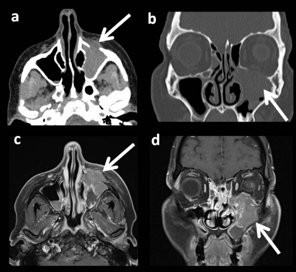
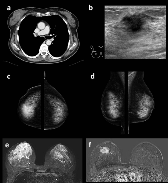
A 74 year old patient with left-sided facial drooping and numbness as well as disturbed vision was referred for a cranial computed tomography (cCT) scan with a tentative diagnosis of stroke. In the native cCT images a stroke was excluded, though, revealing a soft tissue mass in the left maxillary sinus with destruction of the left orbital floor and infiltration of the inferior muscle and inferior oblique muscle (Figure 1) suggestive of a carcinoma of the nasal cavity. On these grounds a biopsy of the soft tissue tumor was gained endoscopically. In a second step cranial magnetic resonance imaging and whole body CT were performed for staging. In addition to the known tumor in the nasal cavity, the CT scan presented contrast-enhancing masses in all four quadrants of the right breast suspect of multicentric breast cancer. Based on these findings the patient was scheduled for ultrasound-guided breast biopsy as well as mammography and breast MRI in our breast cancer center. Mammography revealed an asymmetrical increase in opacity in the right breast with suspicious polymorphous calcifications in the upper outer and inner quadrant as well as in the lower inner quadrant, categorized as BI-RADS 5® lesion, highly suspicious of breast cancer. The following breast MRI showed corresponding multicentric contrast-enhancing masses in the right breast with wash-out curve in the dynamic images. (Multimodal imaging of the breast (Figure 2). The patient had no known risk-factors, no known co-morbidities and a negative family history regarding breast cancer. The final histopathological result of the soft tissue tumor presented a diffuse large B cell lymphoma (DLBCL) with CD45-, CD20-, MUM1-, Bcl2- and Bcl6-expression. Histopathology of the breast revealed DLBCL with CD20 expression confirming the diagnosis of secondary breast lymphoma. Positron-Emission-Tomography (PET) -CT showed no further lesions and no affection of the bones (Figure 3).
Discussion
In our case, the patient presented with a secondary breast lymphoma. Due to its rare occurrence, only few studies on radiological findings have been published, unanimously reporting radiologic features as non-pathognomonic [2,8,12,10]. Nevertheless, radiological work-up is usually the foundation on the way to diagnosis using mammography, ultrasound and MRI of the breast [5].
Mammographic findings have been described as various ranging from diffuse attenuation of the breast parenchyma to solitary masses without calcifications [4,11]. Literature reports the absence of calcifications, spiculations and architectural distortions, which are most common among breast carcinomas, in breast lymphoma [16,17]. Interestingly, in our case the patient presented with a diffuse increase in opacity in all four quadrants associated with suspicious polymorphous calcifications initially mimicking ductal carcinoma in-situ (DCIS). This finding can be interpreted in two ways: The microcalcifications may have developed based on a similar mechanism as in DCIS (i.e. calcification of endoluminal debris due to tumor proliferation and/or secretion) [18]. Alternatively, the microcalcifications could be an incidental finding due to underlying benign changes (i.e. adenosis) and not associated to the hematologic malignancy.
Ultrasound likewise has been reported to deliver unspecific results with hyperechoic or hypoechoic masses and well-defined to spiculated margins [4,11,19]. Corresponding to these experiences, US findings in our patient presented three hypoechoic masses with ill-defined margins, one of which was biopsied.
On MRI lesions are most commonly hypointense on T1 weighted- images, intermediate grade hyperintense on T2 weighted-images with strong and early contrast enhancement [2,4,11] and wash-out or plateau phase in the dynamics [4,8]. These findings were also present in our case in which we found multicentric contrast-enhancing masses in the right breast with wash-out type curves in the dynamic series.
PET/CT plays an important role in lymphoma staging. As a hybrid imaging technique, it is able to combine standard information assessed in a CT-scan with information on metabolic activity of the present lesion. In staging of lymphoma, PET/CT has been proven superior in identifying nodal/extra-nodal involvement and treatment response as compared to CT alone. Especially in DLBCL, it has been described as favorable in the planning of radiation therapy [15,16].
Conclusion
In summary, radiologic features of breast lymphomas are non-pathognomonic and may mimic different forms of invasive cancer. Yet, radiologists and clinicians need to be aware of this rare entity to avoid misinterpretation. Multimodal imaging may - at the most - deliver a tendency towards the diagnosis of breast lymphoma, but certainly pave the way for further diagnostics as biopsy and histopathological evaluation remain the gold-standard for diagnosis.
- Sun Y, Joks M, Xu L-M, Chen X-L, Qian D, et al. (2016) Diffuse large B-cell lymphoma of the breast: prognostic factors and treatment outcomes. OncoTargets and therapy 9: 2069-2080. Link: https://goo.gl/3rqikX
- Surov A, Holzhausen HJ, Wienke A, Schmidt J, Thomssen C, et al. (2012) Primary and secondary breast lymphoma: prevalence, clinical signs and radiological features. Br J Radiol 85: e195-e205. Link: https://goo.gl/rUi8tD
- Aviv A, Tadmor T, Polliack A (2013) Primary diffuse large B-cell lymphoma of the breast: looking at pathogenesis, clinical issues and therapeutic options. Ann Oncol 24: 2236-2244. Link: https://goo.gl/b6Hjb1
- Shim E, Song SE, Seo BK, Kim Y-S, Son GS (2013) Lymphoma Affecting the Breast: A Pictorial Review of Multimodal Imaging Findings. J Breast Cancer 16: 254-265. Link: https://goo.gl/d5M8AJ
- Jabbour G, El-Mabrok G, Al-Thani H, El-Menyar A, Hijji IA, et al. (2016) Primary Breast Lymphoma in a Woman: A Case Report and Review of the Literature. Am J Case Rep 17: 97-103. Link: https://goo.gl/NikPiV
- Hugh JC, Jackson FI, Hanson J, Poppema S (1990) Primary breast lymphoma: An immunohistologic study of 20 new cases. Cancer 66: 2602-2611. Link: https://goo.gl/9nCnUF
- Arizono S, Isoda H, Maetani YS, Hirokawa Y, Shimada K, et al. (2008) High-spatial-resolution three-dimensional MR cholangiography using a high-sampling-efficiency technique (SPACE) at 3T: Comparison with the conventional constant flip angle sequence in healthy volunteers. J Magn Reson Imaging 28: 685-690. Link: https://goo.gl/Ymqmof
- Yang WT, Lane DL, Le-Petross HT, Abruzzo LV, Macapinlac HA (2007) Breast Lymphoma: Imaging Findings of 32 Tumors in 27 Patients. Radiology 245: 692-702. Link: https://goo.gl/ZqJPJK
- Gualco G, Chioato L, Harrington WJ, Weiss LM, Bacchi CE (2009) Primary and secondary T-cell lymphomas of the breast: clinico-pathologic features of 11 cases. Appl Immunohistochem Mol Morphol 17: 301-306. Link: https://goo.gl/czqm8S
- Liberman L, Giess CS, Dershaw DD, Louie DC, Deutch BM (1994) Non-Hodgkin lymphoma of the breast: imaging characteristics and correlation with histopathologic findings. Radiology 192:157-160. Link: https://goo.gl/vk9Eit
- Demirkazık FB (2002) MR imaging features of breast lymphoma. Eur J Radiol 42 : 62-64. Link: https://goo.gl/52nCHk
- Sabate JM, Gomez A, Torrubia S, Camins A, Roson N, et al. (2002) Lymphoma of the breast: clinical and radiologic features with pathologic correlation in 28 patients. Breast J 8: 294-304 Link: https://goo.gl/peqcg3
- Mato AR, Feldman T, Goy A (2012) Proteasome Inhibition and Combination Therapy for Non-Hodgkin's Lymphoma: From Bench to Bedside. Oncologist 17: 694-707. Link: https://goo.gl/WDvw3M
- Zhang B-N, Cao X-C, Chen J-Y, Chen J, Fu L, et al. (2012) Guidelines on the diagnosis and treatment of breast cancer (2011 edition). Gland Surg 1: 39-61 Link: https://goo.gl/QkRksA
- Gallamini A, Borra A (2014) Role of PET in lymphoma. Current treatment options in oncology 15: 248-261. Link: https://goo.gl/D1FF85
- Irshad A, Ackerman SJ, Pope TL, Moses CK, Rumboldt T, et al. (2008) Rare Breast Lesions: Correlation of Imaging and Histologic Features with WHO Classification. RadioGraphics 28:1399-1414. Link: https://goo.gl/81nwpq
- Lyou CY, Yang SK, Choe DH, Lee BH, Kim KH (2007) Mammographic and sonographic findings of primary breast lymphoma. Clin Imaging 31 : 234-238. Link: https://goo.gl/vN3T5r
- Henrot P, Leroux A, Barlier C, Genin P (2014) Breast microcalcifications: the lesions in anatomical pathology. Diagnostic and interventional imaging 95: 141-152. Link: https://goo.gl/mxiSXt
- Nicholson BT, Bhatti RM (2016) Glassman L Extranodal Lymphoma of the Breast. Radiologic Clinics 54: 711-726. Link: https://goo.gl/hYcEYa
Article Alerts
Subscribe to our articles alerts and stay tuned.
 This work is licensed under a Creative Commons Attribution 4.0 International License.


 Save to Mendeley
