Archive of Gerontology and Geriatrics Research
A rare cause of acute abdomen: Isolated cecum necrosis; A case report
Feridun Suat Gokce1 and Aylin Hande Gokce2*
2Depatment of General surgery, Istanbul Atlas Univercity Faculty of medicine, Istanbul, Turkey
Cite this as
Gokce FS, Gokce AH (2020) A rare cause of acute abdomen: Isolated cecum necrosis; A case report. Arch Gerontol Geriatr Res 5(1): 010-011. DOI: 10.17352/aggr.000017Isolated cecum necrosis is a very rare condition. The most common symptom is pain in the right lower quadrant. We are presenting a patient who visited the emergency clinic with right lower quadrant pain and subsequently was diagnosed with cecum necrosis during the operation and underwent a right hemicolectomy
Background: Isolated cecum necrosis is a very rare condition [1]. While not precise in etiology, it is mostly detected in patients with chronic renal failure [2]. In the literature, it has been reported that cecum necrosis, which is rarely detected, was also detected in patients with chronic heart diseases, cardiopulmonary bypass and aortitis apart from dialysis patients [3].
The most common symptom is pain in the right lower quadrant. It can be confused with acute appendicitis and cecum tumor during diagnosis [4]. Therefore, we should consider cecum necrosis in differential diagnosis, especially with elderly patients, where we plan operations due to acute abdomen [5].
Case presentation
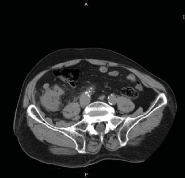
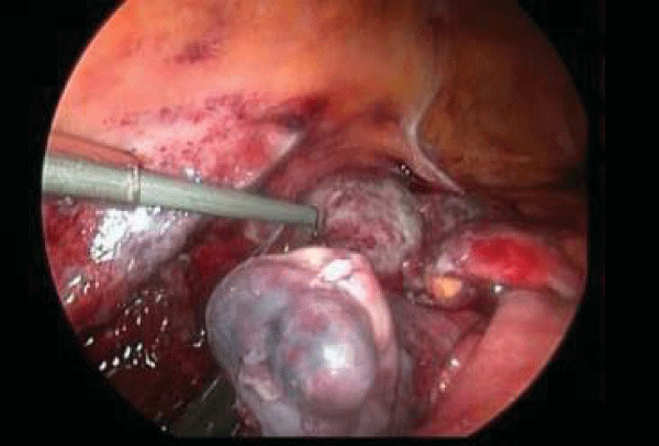
An 87-year-old male patient visited the outpatient clinic for abdominal pain that had been ongoing for 2 days. The patient with a history of trauma 2 days ago had no systemic disease other than hypertension. After physical examination, rebound was found in the right + left lower quadrant of the abdomen. Laboratory values were normal except for the value of C reactive protein of 7 mg / L. In all abdominal ultrasonography it was thought that there could be a perforated appendicitis, although in the entire abdominal tomography it was thought that fl uid and perforated appendicitis could be a possibility (Figure 1). The case was operated with the diagnosis of acute abdomen. Laparoscopically necrosis was detected in the cecum. The cecum and other bowel loops were highly adherent to the surrounding tissues (Figure 2). It was decided to switch to open operation because exploration could not be done properly due to adhesion. The cecum and appendix were necrosis. Right hemicolectomy was performed. Histopathology revealed necrosis in the entire cecum and appendix. No tumor or other pathology were detected. The patient was discharged on the 6th postoperative day without any complications.
Conclusion
The diagnosis of isolated cecum necrosis is diffi cult. In differential diagnosis, acute abdominal causes such as appendicitis, cecum tumor, and diverticulum perforation should be considered [6]. In our case, it was thought that there was a hypoxic nutritional disorder that developed in the colon wall due to trauma. In conclusion, patients with acute abdominal symptoms, anamnesis should be taken well, clinical fi ndings should be evaluated correctly and rare pathologies such as cecum necrosis should be considered. With early diagnosis and early surgery, we wanted to emphasize that isolated cecum necrosis can be healed in most cases.
- Beck DE, de Aguilar-Nascimento JE (2011) Surgical management and outcomein acute ischemic colitis. Ochsner J 11: 282-285. Link: https://bit.ly/2zCrrMb
- Cakar E, Ersoz F, Bag M, Bayrak S, Colak S et al. (2014) Isolated cecal necrosis: our surgical experience and a rewiew of the literatüre. Ulus Cerrahi Derg 30: 214-218. Link: https://bit.ly/2M7Vhed
- Dirican A, Unal B, Bassulu N, Tatlı F, Aydin C, et al. (2009) Isolated cecalnecrosis mimicking acute appendicitis: a case series. J Med Case Rep 3:7443-7447. Link: https://bit.ly/2Aho11c
- Atay A, Doğ ru O, Arslan K, Erenoğ lu B, Kö kç am S, et al. (2012) Partial necrosis of caecum mimicking acute appendicitis Genel Tıp Derg 22: 28-30
- Kılınc G,Balcı B, Tuncer K, Ogucu H, Emiroglu M, et al. (2019) Isolated cecalnecrosis in a patient with chronic renal failure. Tepecik Eğit ve Araşt HastDergi 29: 95-98. Link: https://bit.ly/2ZMdANY
- Perko Z, Bilan K, Vilovic K, Druzijanić N, Kraljević D, et al. (2006) Partial cecalnecrosis tre ated by laparosopic partial cecal resection. Coll Antropol 30: 937–939. Link: https://bit.ly/2ZIt85m
Article Alerts
Subscribe to our articles alerts and stay tuned.
 This work is licensed under a Creative Commons Attribution 4.0 International License.


 Save to Mendeley
