Archives of Clinical Nephrology
Pyonephrosis Presenting as Lumbar Abscess-An Uncommon Clinicoradiological Entity
Rajul Rastogi1*, Neha, Vijai Pratap1 and Gulzari Lal Meena2
2S.N Medical College & Hospital, Jodhpur, Rajasthan, India
Cite this as
Rastogi R, Neha, Pratap V, Meena GL (2020) Pyonephrosis Presenting as Lumbar Abscess-An Uncommon Clinicoradiological Entity. Arch Clin Nephrol 6(1): 017-019. DOI: 10.17352/acn.000041Pyonephrosis secondary to an obstructing calculus in renal pelvis, pelviureteric junction or ureter usually presents clinically with fever, loin pain and other signs of urinary tract infection. Rarely, severe thinning of renal parenchyma in pyonephrosis may allow direct forniceal rupture into the retroperitoneum producing retroperitoneal abscess that might be the only presenting complaint without any complaints related to renal system. Prompt and aggressive management including optimal imaging and correct diagnosis is needed in such cases to prevent further complication. Hence, this article focuses on the rare spontaneous rupture of calculus pyonephrosis in to ipsilateral psoas & quadratus lumborum muscle to clinically present as a lumbar pain & abscess, presentation not described before in medical literature.
Introduction
Complete obstruction of renal collecting system causing moderate to severe hydronephrosis can uncommonly undergo spontaneous decompression following a caliceal rupture [1-2]. This spontaneous rupture of collecting system is usually secondary to an obstructing calculus but less commonly may be due to posterior urethral valves, benign prostatic hyperplasia, pelvic tumors and rarely abdominal aortic aneurysm & retroperitoneal fibrosis [3-4]. Occasionally, the urine in collecting system gets infected forming pus (pyonephrosis) with parenchymal destruction rarely rupturing in to retroperitoneum in to perinephric space, psoas muscle and quadratus lumborum muscle [5,6]. In this article, we present a rare case of spontaneous rupture of pyonephrotic kidney secondary to pelviureteric junction calculus presenting as a lumbar abscess.
Case report
A 27-year old female presenting to outpatient department complained of fever with chills and pain in left lumbar region. Patient was visiting local doctors in rural area for fever for few weeks prior to present visit and wad receiving antibiotics without any laboratory or radiological investigations. Clinical examination revealed a soft & tender bulge in left lumbar region with signs of inflammation. Laboratory test revealed raised total leucocyte count (16000 cells/microliter of blood) with predominant neutrophilia (>90%). Blood urea nitrogen and serum creatinine values were within normal limits being 18mg/dL and 1.1mg/dL respectively. Urine microscopy revealed 10-11 pus cells/high-power field. Chest radiograph in posteroanterior view did not reveal any significant abnormality.
Ultrasonography of abdomen revealed grossly-enlarged, hydronephrotic left kidney measuring up to 170*98 mm [Superoinferior*Anteroposterior] with severe thinning of renal parenchyma, low-level internal echoes in collecting system and a large, calculus measuring up to 23-24 mm in maximum dimension located at the pelviureteric junction. In addition, a septate collection measuring up to 110*110*70 mm (corresponding approximately up to 400-450 ml in volume) was noted in the ipsilateral psoas and quadratus lumborum muscles; latter corresponding to the clinically palpable lumbar swelling. The above-described collection was extending in to the pelvis up to the left iliacus muscle causing its thickening.
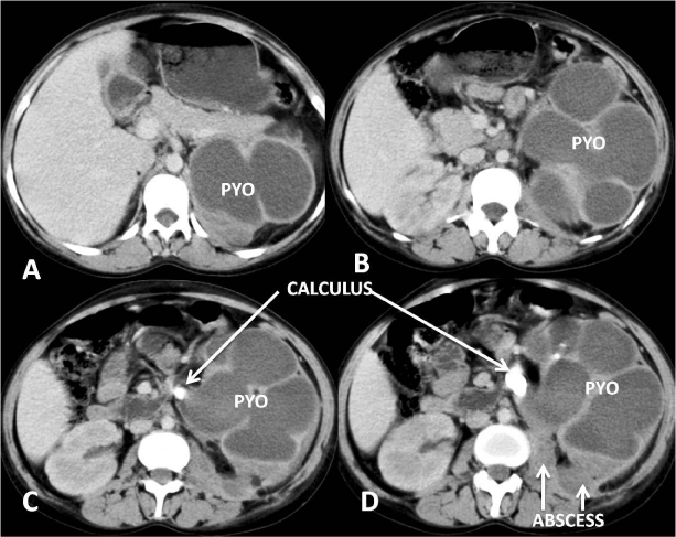
A contrast-enhanced, computed tomography (CECT) of abdomen revealed a large, calculus of 26*11*9 mm in maximum dimensions with 900-1000 HU attenuation value causing a gross hydronephrosis in left kidney which reveal very thin but enhancing, rim-parenchyma and high-attenuating fluid (25-30 HU) along with few subcentimeter secondary caliceal calculi in anterior midpolar calices without aeroceles (Figure 1). In addition, there was a fairly-defined, thick-walled, septate collection without obvious internal calcification/air in left psoas & quadratus lumborum muscles extending up to left iliacus muscle measuring 500-550 ml in volume (Figure 2). The above-described myofascial collection was apparently communicating with posterior inferior-polar calices of left kidney associated with soft-tissue stranding in the visualised pararenal / retroperitoneal fat. No obvious abnormality was noted in the bony spine. No evidence of any free fluid was noted in the peritoneal cavity.
Based on the radiological findings, enlarged left kidney with Grade-III hydronephrosis causing by PUJ calculus showing secondary caliceal calculi, signs of pyonephrosis and left iliopsoas & quadratus lumborum collection secondary to spontaneous caliceal rupture was made. Fine-needle aspiration was performed from both the collecting system of left kidney and quadratus lumborum muscle not only to confirm the presence of pus & detect etiological agent but also to establish the cause and effect relationship. Culture of pus revealed Escherichia coli from both sites.
The patient was treated nephrostomy with pyelolithotomy, placement of ureteral stent and drainage of myofascial collection. Significant clinicoradiological improvement in form of near-complete resolution of retroperitoneal & pararenal pus without any other complication was noted on one-month follow-up of patient. Due to financial constraints, CECT abdomen with urography phase was postponed to a later date.
Discussion
Retroperitoneal abscess especially psoas abscess secondary to caliceal rupture in calculus-induced pyonephrosis is a very rare phenomenon [5]. The psoas abscess thus formed may be detected more sensitively with computed tomography rather than with ultrasonography [7]. Rarely, it may further extend in to the muscles of the posterior abdominal wall especially quadratus lumborum when it may present as a lumbar abscess as in our index case. Management is usually with broad-spectrum antibiotics in addition to image-guided percutaneous or surgical drainage of pus collection by nephrostomy & ureteral stent [5]. Surgical removal of affected kidney is the treatment of choice with non-functioning, affected and normal-functioning contralateral kidney [8-9].
Thorough search in to the existing English medical literature revealed less than five reported cases of spontaneous rupture of pyonephrosis secondary to urolithiasis with psoas abscess formation [5]. Posterior abdominal wall abscess in lumbar region was an additional finding in our case not reported in literature. However, one case showing lumbar panniculitis with subcutaneous abscess secondary to pyonephrosis has been reported [10]. Rarely, peritoneal rupture of pyonephrosis causing peritonitis & splenic abscess has been also described in the literature [11-12].
Conclusion
Spontaneous retroperitoneal rupture in pyonephrosis forming a psoas & quadratus lumborum abscess is a rare occurrence. Furthermore rarer is the presentation of pyonephrosis as a lumbar abscess. Contrast-Enhanced Computed Tomography (CECT) abdomen is superior to ultrasonography abdomen in demonstrating the retroperitoneal rupture of pyonephrosis and its extent. Hence, pre-procedural CECT abdomen should form part of usual protocol for evaluation of all lumbar abscesses not only to rule out this rare entity by detecting renal cause of lumbar abscess but also to minimize morbidity by early diagnosis and management.
- Koktener A, Unal D, Dilmen G, Koc A (2007) Spontaneous rupture of the renal pelvis caused by calculus: A case report. J Emerg Med 33: 127-129. Link: https://bit.ly/2SF0X39
- Patel N, Thanvi R, Jain A, Sathvara P (2015) Devastating complication due to rupture of obstructive perinephric urinoma with secondary pyonephrosis necessitating nephrectomy of nonfunctional kidney in a child. Archives of Medicine & Health Sciences 3: 314-316. Link: https://bit.ly/2Yyzt2X
- Singh I, Joshi M, Mehrotra G (2009) Spontaneous renal forniceal rupture due to advanced cervical carcinoma with obstructive uropathy. Arch Gynecol Obstet 279: 915-918. Link: https://bit.ly/3d8GL11
- Lien WC, Chen WJ, Wang HP, Liu KL, Hsu CC (2006) Spontaneous urinary extravasation: An overlooked cause of acute abdomen in ED. Am J Emerg Med 24: 347-349. Link: https://bit.ly/3aUAZPk
- Patil V, Ichalakaranji R, Patil SB, Pattar R, Ashrith IM (2014) Spontaneous rupture of a calculus pyonephrotic kidney into the retroperitoneal cavity presenting as psoas abscess-A Case Report. International Journal of Biomedical and Advance Research 5: 262-263.
- Erol A, Coban S, Tekin A (2014) A giant case of pyonephrosis resulting from nephrolithiasis. Case Rep Urol 2014: 161640. Link: https://bit.ly/35jGs0Z
- Nabil M, Riyad YM, Salem MA, Nur A (2003) Pyogenic Psoas Abscess: discussion of epidemiology, etiology, bacteriology, diagnosis, treatment and prognosis. Kuwait Medical Journal 35: 44-47.
- Eroğlu M, Kandıralı E (2007) Akut Pyelonefrit ve pyonefroz. Turkiye Klinikleri Journal of Surgical Medical Sciences 3: 24-28. Link: https://bit.ly/2YkKZyE
- Rabii R, Joual A, Rais H, Fekak H, Moufid K, et al. (2000) Pyonephrosis: diagnosis and treatment: a review of 14 cases. Annales d'Urologie 34: 161-164. Link: https://bit.ly/3bUJoDw
- Pauwels C, Bulai-Livideanu C, Chiavassa H, Lamant L, et al. (2009) Lumbar panniculitis with subcutaneous abscess revealing pyonephrosis. Ann Dermatol Venerol 136: 727-729. Link: https://bit.ly/2VTjYka
- Hendaoui MS, Abed A, M’Saad W, Chelli H, Hendaoui L (1996) A rare complication of renal lithiasis: peritonitis and splenic abscess caused by rupture of pyonephrosis]. J Urol (Paris) 102: 130-133. Link: https://bit.ly/2VWlulP
- Jalbani IK, Khurrum M, Aziz W (2019) Spontaneous rupture of pyonephrosis leading to pyoperitoneum. Urol Case Rep 31; 26:100928. Link: https://bit.ly/2xnFQL4
Article Alerts
Subscribe to our articles alerts and stay tuned.
 This work is licensed under a Creative Commons Attribution 4.0 International License.


 Save to Mendeley
