Archives of Clinical Gastroenterology
Anal adenosquamous carcinoma on perineal fistula: A case report
Hajri Amal, Lamnaouar Abderrahmane, Falousse Kabira*, Erreguibi Driss, Boufettal Rachid, Saad Rifki El Jay and Farid Chehab
Cite this as
Amal H, Abderrahmane L, Kabira F, Driss E, Rachid B, et al. (2023) Anal adenosquamous carcinoma on perineal fistula: A case report. Arch Clin Gastroenterol 9(1): 004-007. DOI: 10.17352/2455-2283.000116Copyright License
© 2023 Amal H, et al. This is an open-access article distributed under the terms of the Creative Commons Attribution License, which permits unrestricted use, distribution, and r eproduction in any medium, provided the original author and source are credited.Introduction: Adenosquamous Carcinoma (ASC) of the anus presenting both glandular and squamous histopathologic features is a rare colorectal neoplasm.
Case presentation: A 63-year-old man presented with a one-year history of intermittent rectal pain and bleeding on a perineal fistula. Pathological analysis of the surgical specimen revealed adenosquamous carcinoma of the anus, The patient received neoadjuvant treatment with chemotherapy and radiotherapy over a period of five weeks. then he underwent abdominoperineal resection and a permanent colostomy. Histology revealed invasive adenosquamous carcinoma on the resection specimen and the patient was started on adjuvant therapy with FOLFIRINOX.
Discussion: Primary ASC of the colon and rectum are extremely rare clinical entities with poor prognosis. Most of the data come from individual case reports and a few small series, making it a challenge to understand the histogenesis of the disease. And the treatment modalities are not yet well codified, which has an impact on overall survival.
Conclusion: Early diagnosis and radical surgery with other available therapeutic modalities can improve clinical outcomes.
Introduction
Adenosquamous carcinoma is a tumor characterized by the presence of both squamous cell carcinoma and adenocarcinoma.
The first reported case of ASC was documented in 1907 by Herxheimer [1]. The data concerning adenosquamous carcinoma of the anus is scarce due to its extremely low incidence. In a single United States population-based study published, from 244,410 colorectal and anal cancers, only six cases of primary anal adenosquamous carcinomas were registered (incidence of 0.002%). with the most common location being the right and transverse colon [2]. We report a 63-year-old patient with anal adenosquamous carcinoma of the anus.
The work has been reported in line with the SCARE criteria [3].
Case report
A 63-year-old man, chronic smoker and alcoholic, with a one-year history of rectal pain and intermittent bleeding. His past medical history revealed cholecystectomy for biliary lithiasis, without a remarkable family history. Clinical examination found a posterior renittent perianal swelling with intermittent discharge onto the perineum, including blood and pus, with no palpable mass in the rectal examination.
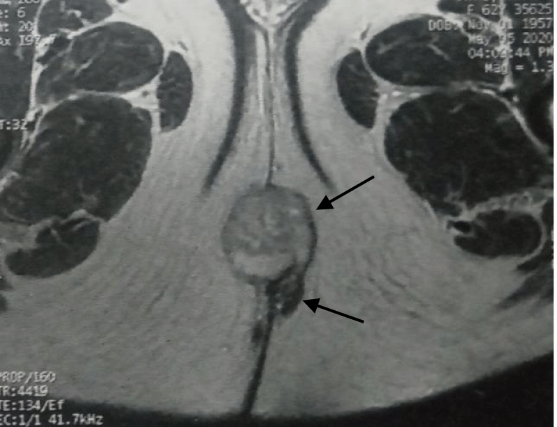
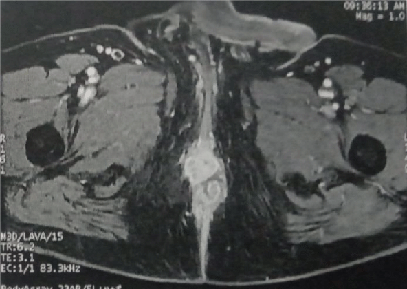
Pelvic MRI showed a simple fistula of the left posterior quadrant Inter-sphincteric, whose secondary orifice is located at the level of the intergluteal fold at 5 o’clock and the primary orifice at 2 o’clock. It shows the contrast of its margins. This fistula communicates with a small collection, enhanced in the periphery after injection, measuring 15 x 10 mm. with a heterogeneously enhanced right inguinal lymph node measuring 27 mm (Figure 1).
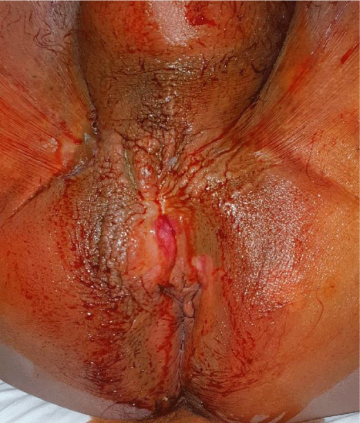
The patient had benefited from a fistulotomy with flattening of the collection and biopsy. With in the pathological examination : Tubular adenocarcinomatous tumoral process was revealed (Figure 2).
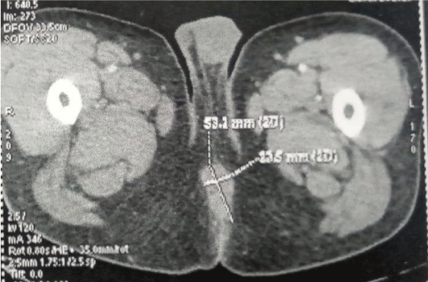
The CT scan showed a normal-sized liver with regular contours, with a small hypodense nodule in segment VI, measuring about 8 mm. The sections through the pelvis show the existence of an infiltrating process involving the distal part of the anal canal with infiltration and left lateral extension. This infiltration is extended over approximately 5 x 2.5 cm in the axial plane with Integrity of the mesorectum. The external sphincters appear to be respected as well as the levator ani and the recti fascia with the right inguinal lymph node measuring 28 mm (Figure 3).
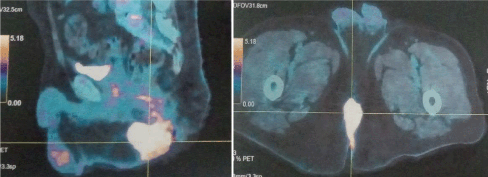
A PET scan performed 2 weeks after the fistulotomy showed a hypermetabolic tumor process of the anal canal extended over 55 mm in the axial section. Two right inguinal hypermetabolic adenopathies, the most voluminous one measuring 24 mm of the minor axis. With hypermetabolic left inguinal adenopathy. And hypodense nodule of the segment VI of the liver, non-hypermetabolic measuring 7 mm (Figure 4).
Subsequently, the patient was treated with capecitabine and external beam radiation therapy (50 Gy) over a five-week time course.
Control pelvic MRI shows a tumoral thickening of the anal canal measuring 37 x 32 mm. With the stable aspect of the known sub-levator Inter-sphincteric fistula, non-active. And stable appearance of the right inguinal adenopathy measuring 13 mm, with multiple bilateral inguinal adenopathies, the most voluminous on the left, measuring 10 mm (Figure 5).
Colonoscopy does not reveal any abnormality and the chest CT shows a lack of detectable secondary thoracic lesions.
The tumor markers were normal except for the CEA which was elevated to 27.53 ng/ml.
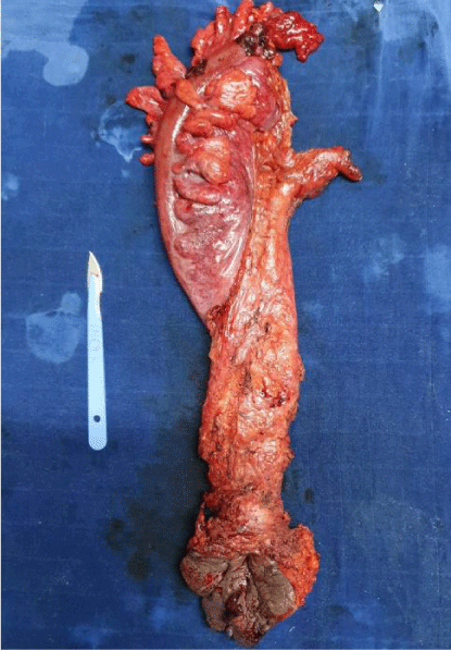
Then, 4 months after fistulotomy the patient benefited from an abdominoperineal resection and definitive colostomy, whose intraoperative findings reveal infiltration of the soft tissues of the anterior perineum, which was biopsied (Figure 6).
Histology found on the resection specimen an invasive adenosquamous carcinoma measuring 4.6 cm invading the perirectal adipose tissue. The colonic border was clean and the anal border was less than 1 mm. Also, the infiltration of the soft tissues of the anterior perineum biopsy was tumoral and the p16 status was negative. Then, the patient began adjuvant FOLFIRINOX.
He developed oedema and ulceration of the scrotum and soft tissue of the perineum 4 months later. The patient underwent a soft tissue biopsy which demonstrated the presence of a previously documented adenosquamous carcinoma. It was a tumour residual, the case was rediscussed in the multidisciplinary consultation meeting, they indicated adjuvant chemotherapy with reassessment, which is still followed in oncology.
Discussion
Adenosquamous carcinoma is a malignant tumor that has both glandular and squamous histological components. Both components are malignant and have the ability to metastasize. And metastases can be glandular, squamous, or both.
There are various theories about the histogenesis of adenosquamous carcinoma in the rectum. Three hypotheses have been proposed:
- The existence of embryonic rests of ectodermal cells
- The squamous metaplasia of the intestinal mucosa
- The presence of pluripotent stem cells of endodermal origin with multidirectional differentiation [4].
ASC is an aggressive histologic subtype of rectum cancer. A 1999 study by Cagir et al.was one of the first to report the demographic and clinicopathological features of 145 cases of colorectal and anal ASC, and a 2020 paper by Nasseri, et al. . analyses data of the National Cancer Database (NCDB) registry from 2004 to 2016 comparing 332 ASC patients 496,950 AC patients. All results confirm that ASC is comparable to AC in sex distribution, age and race; but unlike adenocarcinoma, adenosquamous carcinoma most often presents at an advanced stage, as poorly differentiated lesions and its overall survival is worse [5].
The clinical presentation of rectal or anal adenosquamous carcinoma is identical to that of other histological types in these regions [6].
In our case, the ASC was revealed by a perineal fistula whose initial biopsy revealed an adenocarcinoma, which was treated according to the known guidelines. But after the radical treatment, the histological diagnosis showed the presence of the squamous component.
Because adenosquamous carcinoma of the anus is an extremely rare cancer, the management approach is also difficult [7]. The National Comprehensive Cancer Network guidelines for anal carcinomas are followed with surgical resection being the mainstay of treatment in non-metastatic disease. The operation performed depends on the location of the lesion and follows the same decision-making process as that for adenocarcinoma.
Adjuvant therapy has been used in the treatment of adenosquamous carcinomas; however, its benefit is unknown [2,4-8].
Our case delineates difficulties encountered in the treatment of this locoregionally advanced cancer, the progression of disease on treatment, and suspected poor prognosis.
Conclusion
Primary ASC of the colon and rectum are extremely rare clinical entities with poor prognoses. Most of the data come from individual case reports and a few small series, making it a challenge to understand the histogenesis of the disease. And the treatment modalities are not yet well codified, which has an impact on overall survival.
Written informed consent was obtained from the patient for the publication of this case report and accompanying images. A copy of the written consent is available for review by the Editor-in-Chief of this journal on request.
- Masoomi H, Ziogas A, Lin BS, Barleben A, Mills S, Stamos MJ, Zell JA. Population-based evaluation of adenosquamous carcinoma of the colon and rectum. Dis Colon Rectum. 2012 May;55(5):509-14. doi: 10.1097/DCR.0b013e3182420953. PMID: 22513428; PMCID: PMC3330249.
- Cagir B, Nagy MW, Topham A, Rakinic J, Fry RD. Adenosquamous carcinoma of the colon, rectum, and anus: epidemiology, distribution, and survival characteristics. Dis Colon Rectum. 1999 Feb;42(2):258-63. doi: 10.1007/BF02237138. PMID: 10211505.
- Agha RA, Franchi T, Sohrabi C, Mathew G, Kerwan A; SCARE Group. The SCARE 2020 Guideline: Updating Consensus Surgical CAse REport (SCARE) Guidelines. Int J Surg. 2020 Dec;84:226-230. doi: 10.1016/j.ijsu.2020.10.034. Epub 2020 Nov 9. PMID: 33181358.
- Frizelle FA, Hobday KS, Batts KP, Nelson H. Adenosquamous and squamous carcinoma of the colon and upper rectum: a clinical and histopathologic study. Dis Colon Rectum. 2001 Mar;44(3):341-6. doi: 10.1007/BF02234730. PMID: 11289278.
- Nasseri Y, Cox B, Shen W, Zhu R, Stettler I, Cohen J, Artinyan A, Gangi A. Adenosquamous carcinoma: An aggressive histologic sub-type of colon cancer with poor prognosis. Am J Surg. 2021 Mar;221(3):649-653. doi: 10.1016/j.amjsurg.2020.07.038. Epub 2020 Aug 22. PMID: 32862977.
- Petrelli NJ, Valle AA, Weber TK, Rodriguez-Bigas M. Adenosquamous carcinoma of the colon and rectum. Dis Colon Rectum. 1996 Nov;39(11):1265-8. doi: 10.1007/BF02055120. PMID: 8918436.
- Johnson M, Casillas M Jr, Orucevic A. Adenosquamous Carcinoma of the Anus: An Uncommon Malignancy Presenting a Treatment Challenge. Am Surg. 2018 Jul 1;84(7):e248-e250. PMID: 30454337.
- Lan YT, Huang KH, Liu CA, Tai LC, Chen MH, Chao Y, Li AF, Chiou SH, Shyr YM, Wu CW, Fang WL. A Nation-Wide Cancer Registry-Based Study of Adenosquamous Carcinoma in Taiwan. PLoS One. 2015 Oct 7;10(10):e0139748. doi: 10.1371/journal.pone.0139748. PMID: 26445240; PMCID: PMC4596803.
Article Alerts
Subscribe to our articles alerts and stay tuned.
 This work is licensed under a Creative Commons Attribution 4.0 International License.


 Save to Mendeley
