Archives of Clinical Gastroenterology
Cecal Volvulus: Etiology uncommon of intestinal occlusion acute
A Elbakouri, A Elwassi, Y Eddaoudi*, M Bouali, K El Hattabi, FZ Bensardi and A Fadil
Cite this as
Elbakouri A, Elwassi A, Eddaoudi Y, Bouali M, Hattabi K El, et al. (2022) Cecal Volvulus: Etiology uncommon of intestinal occlusion acute. Arch Clin Gastroenterol 8(2): 025-028. DOI: 10.17352/2455-2283.000108Copyright License
© 2022 Elbakouri A, et al. This is an open-access article distributed under the terms of the Creative Commons Attribution License, which permits unrestricted use, distribution, and r eproduction in any medium, provided the original author and source are credited.Caecum volvulus is, in frequency, the second part of the colon concerned by volvulus after the sigmoid and before the transverse colon. This pathology occurs in the cecum with abnormal mobility The mechanism of volvulus can be summarized in 2 either by torsion or tilting. The clinical picture is that of an acute intestinal occlusion by strangulation. The abdomen without preparation (ASP) and the abdominal CT are the radiological examinations of the first choice for the diagnosis. It is a surgical emergency, the conduct of which is to make a resection of the cecum and the terminal ileum. We report the case of a cecal volvulus admitted to the emergency room with an acute intestinal obstruction, the diagnosis was confirmed by an abdominopelvic CT scan and the treatment consisted of an ileocecal resection with immediate restoration of continuity, the postoperative follow-up was simple.
Introduction
The first description of cecal volvulus was made by Rokitansky in 1837 [1].
The volvulus of the cecum is an axial torsion of the cecum, involving the terminal ileum due to abnormal fixation of the cecum [2].
It is responsible for 25-40% of all colonic volvuli in adults. It represents the second most frequently affected part of the colon by volvulus after the sigmoid [3]. The clinical signs are of a similar intestinal obstruction and they are not specific to cecal volvulus, and it is difficult to differentiate cecal volvulus from other forms of intestinal obstruction [4]. The diagnosis may be delayed
Acute caecal volvulus may progress to caecal gangrene with necrosis and then to perforation leading to acute peritonitis. Abdominal radiography and abdominal CT are the essential radiological procedures for the diagnosis of the volvulus of the cecum [5]. The only effective treatment for cecal volvulus is surgical resection [6].
Patient and observation
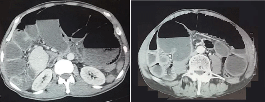
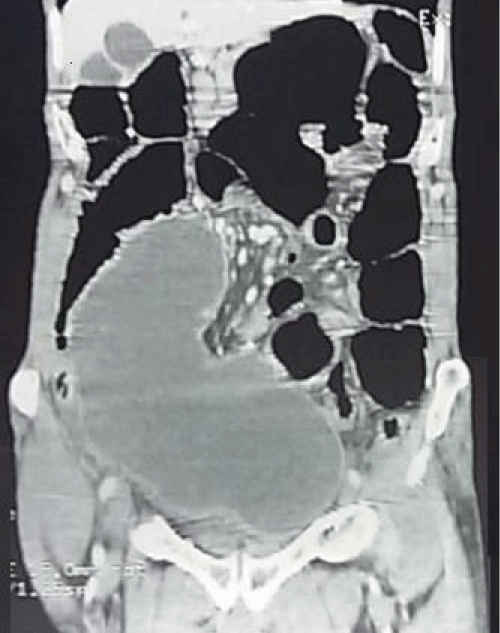
He is a 38-year-old patient followed for chronic dermatitis under corticotherapy and was admitted to the emergency room for occlusive syndrome with symptomatology evolving five days before the consultation with clinical examination: conscious patient stable on the hemodynamic and respiratory plan The examination noted a distended abdomen, hypertympanic the hernial orifices were free and the rectal ampulla was empty The biological workup revealed an HB 13 g/d; hyperleukocytosis with predominantly PNN at 15 000 elements/mm3, C RP was elevated to 150 mg/l, renal function was normal urea 6 mmol/L creatinemia 10 mg/l, unprepared abdomen in the standing position, which revealed a hydrophobic level of the greaves and abdominal CT which showed a colonic occlusion on volvulus of the colon with the collapse of the left and right colonic frame with highlighting of an aspect of whirl sign at the level of the right flank (Figures 1,2).
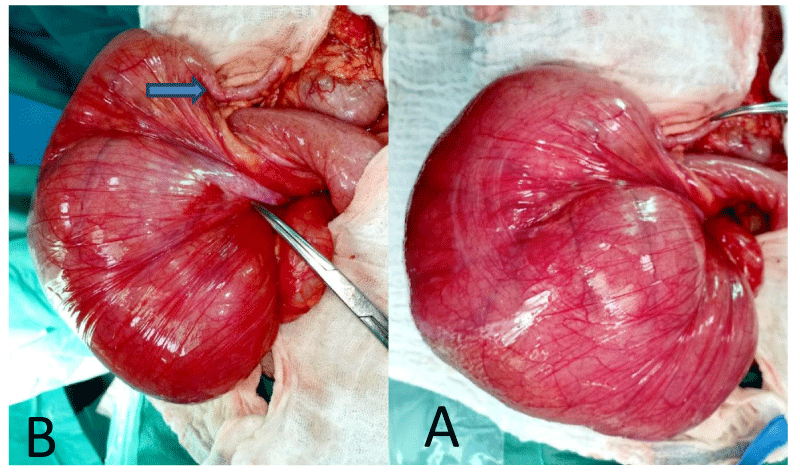
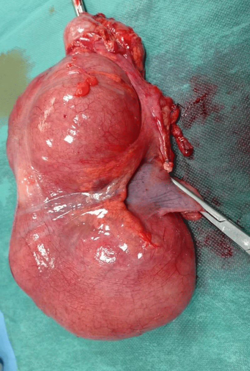
The patient was operated on in the emergency room, approached by laparotomy with the exploration we found a volvulated cecum realizing 1 turn of a spire in the anticlockwise direction (Figures 3,4), the patient had an ileocecal resection with manuaileocolicic terminal-lateral anastomosis.
Discussion
Caecal volvulus is a rarely diagnosed clinical situation for intestinal obstruction [7] and an uncommon etiology [7]. It is responsible for about 1 to 1.5% of all intestinal obstructions, while 20 to 40% of all colonic volvulus. Its incidence is 2.8-7.1 cases per million population per year [3].
The volvulus of the cecum is a torsion of the right colon around its mesenteric axis which is only possible if the proximal colon is mobile or if congenital anomalies in which the right colon does not fuse properly with the lateral peritoneum [8] Excessive mobility of the cecum is due to an incomplete embryological rotation of the intestine or to a defect in the attachment of the ascending colon to the posterior parietal peritoneum [9,10].
Caecal volvulus may occur as a result of adhesions due to abdominal surgery, chronic constipation, pregnancy, or prolonged immobility [11]. Two types of volvulus have been described: either by a rotation of the colon around its axis, with the cecum remaining in the right lower abdominal, or by a tilting of the cecum associated with a rotation of the colon which is then placed in the left upper abdominal quadrant of the abdomen [12-14]. The diagnosis of cecal volvulus is difficult because the clinical signs are not specific and the intensity of the pain is extremely variable [10].
Cugnenc et al. reported in a series a mean age of onset of cecal volvulus of 61.8 years and no gender-related predisposition was established [15].
The Mobile caecum syndrome has been reported to occur in nearly 50% of patients before the onset of acute volvulus [16].
Typically, patients present with generalized abdominal pain or predominantly lower quadrant abdominal pain with abdominal distension, and pain resolution after the passage of flatus, The physical findings in patients during symptomatic episodes may include high pitched bowel sounds and right lower quadrant abdominal tenderness. However, these abnormal physical findings generally disappear as the patients’ symptoms resolve [17].
In the acute volvulus stage, the patient typically presents with signs of acute bowel obstruction and it is difficult to differentiate caecal volvulus from other forms of small bowel obstruction. A tender and dilated cecum may be palpable in the patient with low BMI and may help to differentiate cecal volvulus from other forms of bowel obstruction [18].
In the advanced stage, patients typically present with generalized and severe abdominal pain, peritoneal irritation disturbances of consciousness, and hemodynamic instability [19].
In the advanced stage, patients typically present with generalized and severe abdominal pain, peritoneal irritation disturbances of consciousness, and hemodynamic instability [19].
However, up to 30% of patients do not have these radiographic features [20].
A barium enema is not recommended for patients with suspected perforation and gangrene [21,22].
CT scan replaced barium enema as the preferred imaging modality for the diagnosis of acute cecal volvulus [5]. The abdominal CT scan is a powerful diagnostic tool, it allows the diagnosis of an associated complication such as ischemia or perforation [13].
The three pathognomonic CT signs associated with acute cecal volvulus are: ‘‘whirl signs’’, ‘‘coffee bean’’ and ‘‘bird beak’’; In the setting of acute cecal volvulus, the whirl is composed of the spiraled loop of the collapsed caecum, with low attenuating fatty mesentery and engorged mesenteric vessels. Visualization of a gas-filled appendix is also a CT scan sign associated with cecal volvulus. The CT scan may show signs of intestinal necrosis or ischemia, which manifest as submucosal edema, diminished or non-enhancement of the intestinal wall, pneumatosis intestinalis, or signs of intestinal perforation such as pneumoperitoneum [5].
Several studies have reported a successful colonoscopic reduction of cecal volvulus. The success rate of colonoscopic reduction of cecal volvulus is only 30% with the risk of colonic perforation colonoscopy is not recommended for the treatment of cecal volvulus [23].
It is generally accepted that the only effective treatment for cecal volvulus is surgery.
Endoscopic detorsion is feasible in the absence of severe ischemia but carries a non-negligible risk of perforation [24]. Treatment has three goals: to remove the obstacle by detorsion, if possible, to treat progressive complications, and to prevent recurrence [25]. It remains a controversial subject. Right hemicolectomy with primary anastomosis is recommended by several teams even in the absence of colonic necrosis because it eliminates the risk of recurrence [26-28]. Caecostomy is effective for the prevention of recurrence but carries a high risk of wall infection and exposes the risk of digestive fistula requiring a closure procedure
Laparoscopic procedures are being increasingly used to manage cecal volvulus. Several reports of laparoscopic treatment of cecal volvulus were published [6] and infectious complications are less frequent with caecopexy but recurrences are more frequent [2].
Conclusion
The variability of the topography of the ileocecal region and its peritoneal attachments is important. Caecal volvulus should be considered in patients with acute abdominal pain, especially when there are suggestive radiological signs. The diagnosis is most often delayed due to non-specific clinical signs. Rapid and adapted management is necessary to decrease the risk of morbidity and mortality.
- Rokitansky C. Intestinal strangulation. Arch Gen Med. 1837; 14(1): 202–210.
- Rabinovici R, Simansky DA, Kaplan O. Cecal volvulus. Dis Colon Rectum. 1990; 33(9): 765–769.
- Pulvirenti E, Palmieri L, Toro A, Di Carlo I. Is laparotomy the unavoidable step to diagnose caecal volvulus? Ann R Coll Surg Engl. 2010 Jul;92(5):W27-9. doi: 10.1308/147870810X12699662980231. Epub 2010 Jun 7. PMID: 20529467; PMCID: PMC5696952.
- Bandurski R, Zareba K, Kedra B. Cecal volvulus is a rare cause of intestinal obstruction. Pol Przegl Chir. 2011; 83(9): 515–517.
- Tsushimi T, Kurazumi H, Takemoto Y, Oka K, Inokuchi T, Seyama A, Morita T. Laparoscopic cecopexy for mobile cecum syndrome manifesting as cecal volvulus: report of a case. Surg Today. 2008;38(4):359-62. doi: 10.1007/s00595-007-3620-7. Epub 2008 Mar 27. PMID: 18368329.
- Delabrousse E, Sarliève P, Sailley N, Aubry S, Kastler BA. Cecal volvulus: CT findings and correlation with pathophysiology. Emerg Radiol. 2007 Nov;14(6):411-5. doi: 10.1007/s10140-007-0647-4. Epub 2007 Jul 6. PMID: 17618472.
- Habre J, Sautot-Vial N, Marcotte C, Benchimol D. Caecal volvulus. Am J Surg. 2008; 196(5): 48-49.
- Friedman JD, Odland MD, Burbrick MP. Experience with colonic volvulus. Dis Colon Rectum. 1989; 32(1):409-416.
- Wolfer JA, Beaton LE, Anson BJ. Volvulus of the cecum. Anatomical factors in its etiology. Surg Gynecol Obstet. 1942; 882–894.
- Cugnenc PH, Gayral F, Larrieu H, Garbay M. Volvulus aigu du cæcum : à propos de 10 observations. Chirurgie. 1982; 108(3): 279–283.
- Howard RS, Catto J. Cecal volvulus. A case for nonresectional therapy. Arch Surg. 1980 Mar;115(3):273-7. doi: 10.1001/archsurg.1980.01380030025006. PMID: 7356382.
- Pirró N, Merad A, Sielezneff I, Sastre B, Di Marino V. Volvulus du cæcum, bases anatomiques et physiopathologie: à propos de 8 cas consécutifs. Morphologie. 2006; 90(1): 197- 20.
- Printen KJ. Mobile cecal syndrome in the adult. Am Surg. 1976 Mar;42(3):204-5. PMID: 1259253.
- Berger JA, Leersum MV, Plaisier PW. Cecal volvulus: Case report and overview of the literature. European Journal of Radiology Extra. 2005; 55(4): 101-103.
- O'Mara CS, Wilson TH Jr Stonesifer GL, Stonesifer L, Cameron JL. Cecal volvulus: analysis of 50 patients with long-term follow-up. Ann Surg. 1979; 189(6): 724- 731. PubMed | Google Scholar
- Moore CJ, Corl FM, Fishman EK. CT of cecal volvulus: unraveling the image. AJR Am J Roentgenol. 2001 Jul;177(1):95-8. doi: 10.2214/ajr.177.1.1770095. PMID: 11418405.
- Perret RS, Kunberger LE. Case 4: Cecal volvulus. AJR Am J Roentgenol. 1998 Sep; 171(3): 855-860.
- Anderson JR, Mills JO. Caecal volvulus: a frequently missed diagnosis? Clin Radiol. 1984 Jan;35(1):65-9. doi: 10.1016/s0009-9260(84)80240-8. PMID: 6690184.
- DONHAUSER JL, ATWELL S. Volvulus of the cecum with a review of 100 cases in the literature and a report of six new cases. Arch Surg (1920). 1949 Feb;58(2):129-48. PMID: 18111729.
- Madiba TE, Thomson SR. The management of cecal volvulus. Dis Colon Rectum. 2002 Feb;45(2):264-7. doi: 10.1007/s10350-004-6158-4. PMID: 11852342.
- Halabi WJ, Jafari MD, Kang CY, Nguyen VQ, Carmichael JC, Mills S, Pigazzi A, Stamos MJ. Colonic volvulus in the United States: trends, outcomes, and predictors of mortality. Ann Surg. 2014 Feb;259(2):293-301. doi: 10.1097/SLA.0b013e31828c88ac. PMID: 23511842.
- Young WS. Further radiological observations in caecal volvulus. Clin Radiol. 1980 Jul;31(4):479-83. doi: 10.1016/s0009-9260(80)80200-5. PMID: 7418350.
- Consorti ET, Liu TH. Diagnosis and treatment of caecal volvulus. Postgrad Med J. 2005 Dec;81(962):772-6. doi: 10.1136/pgmj.2005.035311. PMID: 16344301; PMCID: PMC1743408.
- Anderson JR, Welch GH. Acute volvulus of the right colon: an analysis of 69 patients. World J Surg. 1986 Apr;10(2):336-42. doi: 10.1007/BF01658160. PMID: 3705613.
- O'Mara CS, Wilson TH, Stonesifer GL, Cameron JL. Cecal volvulus: analysis of 50 patients with long-term followup. Ann Surg. 1979; 189: 724–731
- Breda R, Mathieu L, Mlynski A, Montagliani L, Duverger V. Volvulus du cæcum. J Chir (Paris). 2006; 143(1): 330-2.
- Sedik A, Bar EA, Ismail M. Cecal volvulus: Case report and eview of literature. Saudi Surg J. 2015; 3(2): 47-9.
- Meyers JR, Heifetz CJ, Baue AE. Cecal volvulus: a lesion requiring resection. Arch Surg. 1972 Apr;104(4):594-9. doi: 10.1001/archsurg.1972.04180040208035. PMID: 5013801.
Article Alerts
Subscribe to our articles alerts and stay tuned.
 This work is licensed under a Creative Commons Attribution 4.0 International License.

 Save to Mendeley
