Archives of Clinical Gastroenterology
Mucinous adenocarcinomas arising from a retro-rectal hamartoma: Unusual malignant transformation of a tailgut cyst
María Recuero Pradillo1*, Carlos Quimbayo-Arcila1, Beatriz Moreno Torres1, Angel Romo Navarro1, Juan-Sebastian Malo Corral2 and Juan Ruiz Martín1
2Department of Surgery, Hospital Virgen de la Salud, Toledo, Spain
Cite this as
Aijaz M, Hasan M, Alam F (2019) Mucinous adenocarcinomas arising from a retro-rectal hamartoma: Unusual malignant transformation of a tailgut cyst. Arch Clin Gastroenterol 5(2): 027-030. DOI: 10.17352/2455-2283.000065Introduction
Retrorectal cystic hamartomas, or tailgut cysts, are rare congenital lesions that are usually present as tumors with a considerable size in presacral area. Frequently, these lesions are asymptomatic so the diagnosis may be delayed.
It’s in the fourth week of pregnancy when the cloacal membrane comes to lie in a ventral position, enclosing the distal caudal portion into the eventual hindgut, receiving the name of tailgut. The tailgut ordinarily regresses in the sixth week of gestation. However, a tailgut cyst is thus created if the mucoussecreting remnants fail to regress [1].
Tailgut cysts are not related to any specific age-group, and may appear at moment, although it has been found a higher prevalence in women [2].
About five hundredth of the cases are discovered by chance; the other half of patients complain about symptoms that are related to a retrorectal space, such as discomfort when seated, rectal bleeding, or infections [3].
The diagnosis needs to be confirmed radiologically, through Magnetic Resonance Imaging (MRI) and Computed Tomography (CT).
Complete surgical resection is recommended in all cases, benign tumors included, as they can potentially become malignant. In spite of it, malignant change is extremely rare. The current article reviews scientific literature about cases with malignant transformations. In addition, it is also briefly discussed the differential diagnosis of retrorectal cystic lesions.
Material and Methods
The clinicopathological characteristics of a mucinous adenocarcinoma within a Retrorectal Cystic Hamartoma (RCH) in a 52-year-old woman is described. In this study, the anatomopathological examination is emphasized but Computed Tomography (CT) scans, Magnetic Resonance Imaging (MRI) are also included. A comprehensive review of the literature is also present.
The specimen was first preserved by fixing in formaldehyde. Next, sections was cut from tissue embedded in paraffin blocks. Finally, tissue sections (5µm) were stained with hematoxylin and eosin and analyzed with a optical microscope. Immunohistochemistry stains were performed on selected sections for CK7, CK20, MIB1, p53, CDX2, CD20, PAX8, Estrogen Receptors (ER), Progesterone Receptor (PR).
Case Report
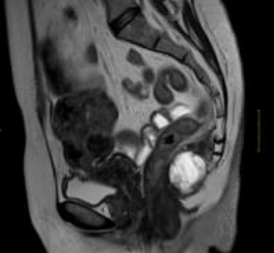
A 52-year-old female with a 6-month history of a low back pain and enlarging mass in the glutealarea is admitted in the service. On physical examination, a 40×30mm tumor was identified in the intergluteal crease. In the MRI, a presacral uniloculated cystic mass posterior to the rectum was detected, measuring 40×30×33mm. There was no evidence of invasion into surrounding structures (Figure 1). The mass is located in the retrorectal space without features of colon invasion. Subsequently, surgical resection was performed through posterior approach. The post-operative recovery of the patient was uneventful.
Pathological findings
The macroscopic specimen consisted of a 3.5cm×2.5cm cystic tumor with well-circumscribed margin. The cut surface revealed a unilocular cyst lesion, filled with a creamy-yellow substance. Additonally, contained multiple gelatinous masses, both floating and attached to the cyst wall. No solid areas were found.
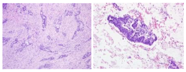
Microscopic pathological examination revealed an intestinal-type epithelium with variable degrees of nuclear and architectural atypia next to an associated desmoplastic hyaline stroma. There were also identified pools of extracellular mucin which contained floating malignant epithelial cells (Figure 2). Invasion of the cyst wall by the adenocarcinoma was not detected, nor lymphovascular invasion.
Immunohistochemical stains showed the tumor cells to be positive for CDX2. Immunohistochemical staining for CK7 showied focal cytoplasmic positivity and negative staining with CK20.
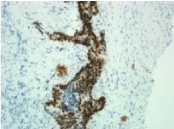
There was a marked overexpression of p53 in the mucinous adenocarcinoma arising in the retrorectal cystic hamartoma. Also, there was marked overexpression of p53 in areas of severe dysplasia (Figure 3). Moreover, a significantly higher expression of the Ki-67 antibody (MIB-1), indicating a high proliferation rate in malignant epithelium. Other immunohistochemical stains were performed to determine other possible primary origin areas, with negative results (PAX8, estrogen receptors, progesterone receptor, WT1 and p16).
The final pathology data revealed a mucinous adenocarcinoma in the retrorectal space.
Discussion
Retrorectal Cyst Hamartoma (RCH) or tailgut cyst is a rare malformation of the retrorectal space. RCHs are not related to any specific age-group, and may appear at moment. Most RCH occurs in women, as illustrated in the case presented. Approximately, half of the patients are symptomatic, being pain in the rectal area and lower back, or symptoms associated with the mass size their most common complaints [4].
The presacral space (also called retrorectal space) is a space posteriorly limited by the sacrum and anteriorly by the fascia of the rectum. Tumors occurring in the retrorectal space are typically benign, although a variety of neoplastic conditions could happen in this region [5].
The best therapeutic option is a complete excision of the tumor, even in asymptomatic patients. Surgical resection may be necessary for treating conditions such as potential growth or malignancy. Negative surgical margin is recommended in order to relieve pressure symptoms and to make a definitive diagnosis.
However, choosing the best surgical approach may be challenging. The most attractive option is the posterior surgical approach of presacral masses, but a laparoscopic approach should be regarded too, as it reduces the surgical trauma and postoperative complications [6]. Complications that are typically associated with surgery include urinary and fecal incontinence, bleeding, abscess or fistula formation, or nerve root injury leading to weakness. The regular postoperative follow-up consists of the clinical examination with anal endosonography. In our case, the tumor was excised successfully, with complete resection. The patient had an uncomplicated postoperative stay and a satisfactory recovery without morbidity. She was not needed additional treatments.
Analyzed raw, the lesion is typically composed of multilocular cyst ranging in size from one to fifteen cm and tends to present opaque mucoid content.
For diagnosing that the mucinous adenocarcinoma has developed from a congenital tailgut cyst, the patient history and its localization, morphology and immunohistochemical phenotype must be taken into account. In our case, the morphology and the immunohistological phenotype (positive for CDX2) were similar to those of colo-rectal carcinomas. Nonetheless, the retrorectal localization, female sex, the entire tumor-free colon and the finding of dysplastic columnar epithelium within the cysts were evidences that suggested that this lesion developed within a preexisting congenital tailgut cyst rather than extending from the intestinal tract.
Other differential diagnoses of tailgut cysts include epidermoid and dermoid cysts, rectal duplication cyst, and anal gland cyst. Epidermoid cysts are lined only by stratified squamous epithelium. Dermoid cysts contain, in addition, dermal appendages. Duplication cysts are usually unilocular and are lined by an intestinal or respiratory epithelium. Many of them are located prerectally, whereas congenital tailgut cysts are invariably retrorectal [7]. Finally, another differential diagnosis is with chordoma, which is the most common primary sacral lesion.
The positive nuclear staining for p53 in immunohistochemical stains suggests a mutation in the tumor-inhibitory gene (mutant p53). The moderated intensity of the tumor proliferative index marker Ki-67 (MIB-1) usually indicates a high capacity of the malignant neoplastic process. Thus, in our case, following the results from both immunohistochemical markers, the patient experienced a malignant transformation of the congenital retrorectal cystic hamartoma associated with the p53 gene mutation.
In a review of the scientific literature, malignant transformation within a retrorectal cyst hamartoma has only been documented in 33 cases (including the current report), carcinoid tumor and adenocarcinoma included in the search [8,9], while there have been found 29 cases of neuroendocrine tumours arising in tailgut cysts [10,11].
Conclusion
Retrorectal cystic hamartomas (tailgut cysts) are rare congenital lesions thought to arise from remnants of the embryonic postanal gut. They exists predominantly as asymptomatic retrorectal multicystic masses in women. Surgical resection provides a convenient specimen for histopathological examination, relieves symptoms caused by local compression, avoids subsequent infection, and rules out malignant transformation. Malignant transformation in the form of adenocarcinoma or carcinoma, although rare, may occur. Our case was unusual because, besides from presenting classic congenital tailgut cysts with malignant transformation, it also appeared in the form of a mucinous adenocarcinoma.
- Haydar M, Griepentrog K (2015) Tailgut cyst: A case report and literature review. Int J Surg Case Rep 10: 166–168. Link: http://bit.ly/2L9Nv3x
- Baek SK, Hwang GS, Vinci A, Jafari MD, Jafari F, et al. (2016) Retrorectal Tumors: A Comprehensive Literature Review. World J Surg 40: 2001–2015. Link: http://bit.ly/2qL3gGY
- Andea AA, Klimstra DS (2005) Adenocarcinoma arising in a tailgut cyst with prominent meningothelial proliferation and thyroid tissue: case report and review of the literature. Virchows Arch 446: 316–321. Link: http://bit.ly/2OjzeD6
- Hjermstad BM, Helwig EB (1988) Tail gut cysts: report of 53 cases. Am J Clin Pathol 89: 139-147. Link: http://bit.ly/2KT5bjJ
- Patel N, Maturen KE, Kaza RK, Gandikota G, Al-Hawary MM, et al. (2016) Imaging of presacral masses-a multidisciplinary approach. Br J Radiol. 89: 20150698. Link: http://bit.ly/2OiUSYe
- Saxena D, Pandey A, Bugalia RP, Kumar M, Kadam R, et al. (2015) Management of presacral tumors: Our experience with posterior approach. Int J Surg Case Rep 12: 37-40. Link: http://bit.ly/2KUfW5c
- Dahan H, Arrive L, Wendum D, Docou le Pointe H, Djouhri H, et al. (2001) Retrorectal developmental cysts in adults: clinical and radiologic–histopathologic review, differential diagnosis, and treatment. Radiographics 21: 575–584. Link: http://bit.ly/2OjxwBQ
- Kaistha S, Gangavatiker R, Harsoda R, Kinra P (2018) A case of adenocarcinoma in a tail gut cyst and review of literature. Med J Armed Forces India 74: 390-393. Link: http://bit.ly/2qKg00o
- Niicoll K, Bartrop C, Walsh S, Foster R, Duncan G, et al. (2019) Malignant transformation of tailgut cysts is significantly higher than previously reported: systematic review of cases in the literature. Colorectal Dis 21: 869-878. Link: http://bit.ly/2QQQyRC
- Al Khaldi M, Mesbah A, Dubé P, Isler M, Mitchell A, et al. (2018) Neuroendocrine carcinoma arising in a tailgut cyst. Int J Surg Case Rep 49: 91–95. Link: http://bit.ly/35yWUJn
- Lee A, Suhardja TS, Nguyen TC, Teoh WM (2019) Neuroendocrine tumour developing within a long-standing tailgut cyst: case report and review of the literature. Clin J Gastroenterol. Link: http://bit.ly/33ji3Wz
Article Alerts
Subscribe to our articles alerts and stay tuned.
 This work is licensed under a Creative Commons Attribution 4.0 International License.


 Save to Mendeley
