Archives of Clinical Gastroenterology
Isolated splenic metastasis: An unusual presentation of colonic adenocarcinoma
Mohsin Aijaz1*, Mahboob Hasan2 and Feroz Alam3
2Professor, Department of Pathology, Jawaharlal Nehru Medical College, Aligarh Muslim University, Aligarh 202002 U.P, India
3Assistant Professor, Department of Pathology, Jawaharlal Nehru Medical College, Aligarh Muslim University, Aligarh 202002 U.P, India
Cite this as
Aijaz M, Hasan M, Alam F (2019) Isolated splenic metastasis: An unusual presentation of colonic adenocarcinoma. Arch Clin Gastroenterol 5(2): 027-030. DOI: 10.17352/2455-2283.000064It is very uncommon situation in which primary colonic carcinoma is asymptomatic and presents as isolated splenic metastasis. Involvement of spleen by secondary tumors is usually seen in disseminated spread of tumor. However, isolated splenic involvement by tumor metastasis is an infrequent event, except in cases of lymphoid origin malignancy where spleen is commonly involved. We hereby report a case of 50 years old man who presented with gradually increasing pain abdomen for 3 months. USG report showed splenomegaly indicating either splenic abscess or hemangioma. Splenectomy was performed followed by pathological examination. Histopathological examination (HPE) revealed diffuse infiltration of spleen by sheets and nests of malignant cells, suggesting metastatic adenocarcinoma to spleen. Subsequently computed tomography was done to find out the site of primary tumor. Thus a cystic mass in left splenic flexure of colon was identified on CT scan. Biopsy was done that suggested colonic cancer. Hence a diagnosis of colonic mucinous adenocarcinoma with metastatic splenic involvement was made. Patient was operated for the same and managed accordingly. Based on this case, we concluded that surgeons should pay careful attention to splenic lesions as metastatic deposits can be there, especially in old aged patients having features that favor some ongoing malignant disease.
Introduction
Spleen is a very unusual site of metastatic tumor spread. This is based on various anatomical, histological and functional features of the spleen, that make it impervious for secondary tumors [1]. Majority of the cases are part of disseminated metastatic diseases and usually arise from breast, ovary, lung, gastric, colorectal cancer and skin melanoma [2]. Isolated metastasis to spleen is an infrequent event. Most cases are usually asymptomatic and diagnosis is based on correlation of clinicopathological findings and radiological imaging. Here we report a similar case where a middle aged male presented with left upper abdomen pain. Based on clinical examination and sonographic findings, a provisional diagnosis of splenic abscess or hemangioma was suggested. Hence subsequently splenectomy was done. Results of histopathological examination, however, surprisingly revealed sheets of malignant glands within the splenic parenchyma. Thus a diagnosis of metastatic adenocarcinoma to spleen was made. The interesting point of our report is that the spleen metastasis was detected in the absence of clinical suspicion of malignancy. Consequently, CT Scan was done post operatively that revealed growth in right colon. There are very few cases of colorectal carcinoma seen in literature that present as solitary splenic involvement.
Case Report
We report a rare case of 50 years old male patient who visited surgical outpatient department of JNMCH, AMU Aligarh with chief complaint of pain in left upper abdomen for 3 months. Pain was localized to left hypochondrium and was dull aching, non-colicky and non-radiating, with gradual increase in its intensity. For last 20 days, pain was so much severe that it hampered his routine activities. It was not associated with vomiting or diarrhea. No history of malena, hematemesis or hematuria was there. No history of alcohol intake was there but the patient was a chronic smoker. Patient also gave history of weight loss and decreased appetite for the same duration. Clinical examination revealed stable vitals. General and systemic examination were within normal limits except mild pallor and tenderness in left hypochondrium. With this clinical presentation he was admitted and full work up was done in order to make a definite diagnosis.
Routine investigations including complete blood count, renal function test, liver function test, blood sugar, serum electrolytes were advised. All the investigations revealed normal parameters except mild decrease in haemoglobin and slightly raised total leukocyte count. Serum amylase and serum lipase were also within normal limits. Montaux test and widal tests were negative. Quantitative buffy coat test (QBC) for malaria was done that also exhibited negative findings. Further ultrasonography of abdomen was done that revealed a well-defined heterogenous hypoechoic lesion in left supra renal fossa along with splenomegaly. Provisional diagnosis of splenic abscess or haemangioma was made. Subsequently splenectomy was done followed by histopathological examination.
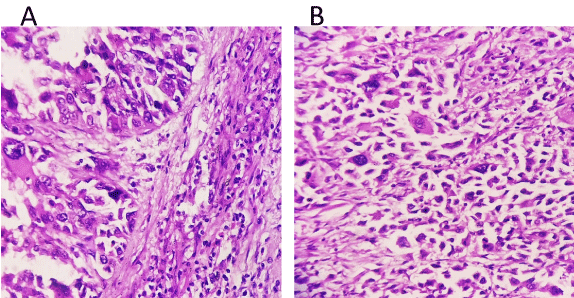
Gross examination showed splenic enlargement with a dimension of 14 x 10 x 4 cms. Splenic capsule showed strong adherence with perisplenic fats. Cut section showed white homogenous area with specks of haemorrhage (Figure 1). Results of histopathology were very surprising and showed sheets, nests and island of mucin containing malignant cells often forming glands, infiltrating throughout the spleen and also in the attached perisplenic fats (Figure 2). Thus the diagnosis of adenocarcinoma metastatic to spleen was made. Immunohistochemistry (IHC) was applied as a useful ancillary adjunct to morphologic examination for determining site of origin for adenocarcinoma. Cytokeratin staining is helpful in the diagnostic differentiation of metastatic lesions and assists in determining the site of origin from two common primaries (lung and colon). Hence Cytokeratin 7 and 20 was applied. CK7 was found to be negative in tumor cells (ruling out lung, upper gastrointestinal tract adenocarcinomas and many other adenocarcinomas). However, Cytokeratin 20 was found to be positive in tumor cells (seen in colonic adenocarcinoma). Keeping in view the suspected metastasis from colon, further CDX2 and p53 were applied that also showed strong positive nuclear staining in tumor cells, thus adhering us to diagnosis of metastasis from colonic carcinoma (Figure 3). Post operatively CT Scan was advised to confirm the primary site.
Result of computed tomography suggested colon cancer that further strengthened our diagnosis. Biopsy was taken that revealed similar morphology of tumor cells as seen in spleen. This favoured the diagnosis of primary colonic adenocarcinoma which has metastasized to spleen. Finally, hemicolectomy was done. Very few cases have been found in literature where colonic adenocarcinoma presented with solitary splenic metastasis. This case also has similar presentation and hence highlights the importance of spleen as one of the rare site of metastatic tumor, that too in the absence of clinical suspicion.
Discussion
Spleen is one of the such visceral organ where isolated metastasis is very rare. The anatomical, histological or physiological features of spleen makes it an unusual site for tumor secondaries [1]. A number of hypotheses have been laid down by various authors. According to Sappington, there is sharp angle of the splenic artery with celiac axis that is associated with low incidence of splenic metastasis [3]. whereas Kettle suggested the role of rhythmic contraction of the spleen that might prevent growth of tumor emboli there [4]. It has been seen that even if the neoplastic cells reach the spleen, growth of tumor cells is opposed by splenic microenvironment. This is partly attributed to production of a humoral factor, the splenic factor that avoids tumoral cells adhesion and trigger their cytolysis [5]. Being a reticuloendothelial organ, phagocytic activity has also been suggested as possible factors preventing malignant cells development in the spleen [6].
Both haematological and lymphatic pathways have been supposed to be the channels for metastatic spread. But unlike other visceral organs, the splenic parenchyma lacks afferent lymphatic vessels. However lymphatic channels present in capsular and subcapsular regions can convey subcapsular splenic metastasis [7]. Many authors have the view that vascular route is the major pathway because the metastasis is usually limited to splenic parenchyma.
Various studies show that nearly 7% of cancer autopsies have metastasis to the spleen [8]. This usually seen in disseminated cancers. Isolated splenic metastasis is a rare event. Lung, endometrium, ovary, cervix, stomach, colon, melanoma, breast and bladder are the most common solid tumors in which splenic metastasis occurs [2]. Few cases of splenic metastases have also been reported from prostate, salivary gland and esophageal carcinomas [8,9].
Colorectal carcinoma usually metastasizes to regional lymph node, peritoneum and liver [10]. Lungs and spleen are rare sites. According to previous studies isolated splenic metastasis is seen in only 4.4 % of colon cancers [2]. On reviewing literature, it was noted that cases which show isolated splenic metastasis, have either metachronous or synchronous splenic metastasis from colorectal carcinoma, and the left colon was the predominant site of the primary tumor [11]. In our own patient too, the primary tumor was located in the left splenic flexure, thus favoring the hypothesis of Indudhara. According to Indudhara et al., it is the retrograde flow of blood through the inferior mesenteric vein and splenic vein that carries neoplastic cells to spleen [12]. Many authors are of the opinion that metachronous splenic metastasis is more prevalent than synchronous. Splenectomy is usually performed in both types of involvement. Patients of metachronous involvement have better survival than synchronous. However, no clear explanation of this has been seen in literature. Our patient has also synchronous involvement. However, our case differs from previous cases seen in literature in that the patient has no symptoms of primary malignancy. He only had left hypochondrium pain and splenomegaly. A solitary splenic mass in a patient is usually suggestive of a primary splenic lesion such as lymphoma, hemangioma, abscess or infarction. Hence our patient underwent splenectomy. It is the microscopic examination that revealed deposits of metastatic adenocarcinoma. Hence this incidental finding prompted us for whole work up of patient to look for primary tumor.
Owing to the development of advance imaging and molecular testing, diagnosis of colorectal carcinoma and micrometastases to spleen is made at an earlier stage. Ultrasonography and computed tomography are helpful in diagnosing primary as well as metastatic lesions. PET scan is supportive in detecting recurrences. Review of literature showed the elevated serum CEA level (4.6-223 ng/mL) in colonic adenocarcinoma [13], as was also observed in our patient (184 ng/mL). CEA has been supposed to modulates the immune response and suppresses the humoral response as well as lymphocyte and NK cell activity [14]. It provides adhesion between cancer cells and macrophages, thus playing a significant role in the pathogenesis of isolated splenic tumor metastasis [15].
Surgery is the primary modality of treatment followed by chemotherapy with or without radiotherapy in metastatic colorectal carcinoma. Our patient also underwent left hemicolectomy followed by chemotherapy (5-FU, leucovorine and oxaliplatin). Majority of cases in literature show a disease free survival period of 3–144 months after the diagnosis of primary tumor [16,17]. Whereas survival after splenectomy in metachronous involvement varies between 6 months to 7 years [18]. So, prognosis of isolated splenic colorectal metastasis is more favorable, although these cases show distant metastatic spread.
Conclusion
Spleen is an uncommon site of metastatic tumor spread. Any splenic mass in an asymptomatic patient is usually primary splenic lesion such as lymphoma, hemangioma or abscess. However, clinician and surgeons must keep in mind the possibility of metastatic tumor deposits there, especially in chronically ill patient. This will be helpful in early diagnosis of primary malignancy. Thus improving the management and survival of patients with isolated splenic metastases from colorectal carcinoma as was recommended in similar publications.
Highlights
• Spleen is a very unusual site of metastatic tumor spread
• Various hypotheses have been laid down to justify the low incidence
o Rhythmic contraction of the spleen that might prevent growth of tumor emboli there.
o Growth of tumor cells is opposed by splenic microenvironment.
o Phagocytic activity has also been suggested as possible factors preventing malignant cells development in the spleen.
- Skandalakis LJ, Gray SW, Skandalakis JE (1990) Splenic realities and curiosities. Prob Gen Surg 7: 28-32.
- Berge T (1974) Splenic metastases. Frequencies and patterns. Acta Pathol Microbiol Scand A 82: 499-506. Link: http://bit.ly/2Hm5Acy
- Sappington SW (1922) Carcinoma of the spleen: its microscopic frequency; a possible etiologic factor. JAMA 78: 953-955. Link: http://bit.ly/33W47mM
- Kettle EH (1912) Carcinomatous metastases in the spleen. J Pathol Bacteriol 17: 40-46. Link: http://bit.ly/2KRBhMQ
- Place RJ (2001) Isolated colon cancer metastasis to the spleen. Am Surg 67: 454-457. Link: http://bit.ly/2MA2HZK
- Klein B, Stein M, Kuten A, Steiner M, Barshalom D, et al. (1987) Splenomegaly and solitary spleen metastasis in solid tumors. Cancer 60: 100-102. Link: http://bit.ly/2PcgFTT
- Cavallaro A, Modugno P, Specchia M, Potenza AE, Lo Schiavo V, et al. (2004) Isolated splenic metastasis from colon cancer. J Exp Clin Cancer Res 23: 143-146. Link: http://bit.ly/2NsgrFy
- Comperat E, Bardier-Dupas A, Camparo P, Capron F, Charlotte F (2007) Splenic metastases: clinicopathologic presentation, differential diagnosis, and pathogenesis. Arch Pathol Lab Med 131: 965-969. Link: http://bit.ly/2ziwfmc
- Inouye CM, Anagnostou V, Li QK (2017) Primary parotid adenocarcinoma metastasis to the spleen with PIK3CA mutation: cytological findings and review of the literature. Int J Clin Exp Pathol 10: 5999-6005. Link: http://bit.ly/2Z8Kcm5
- Genç V, Akbari M, Karaca AS, Çakmak A, Ekıncı C, et al. (2010) Why is isolated spleen metastasis a rare entity? Turk J Gastroenterol 21: 452-453. Link: http://bit.ly/2ZnInAT
- Pisanu A, Ravarino A, Nieddu R, Uccheddu A (2007) Synchronous isolated splenic metastasis from colon carcinoma and concomitant splenic abscess: a case report and review of the literature. World J Gastroenterol 13: 5516-5520. Link: http://bit.ly/2zfWGJl
- Indudhara R, Vogt D, Levin HS, Church J (1997) Isolated splenic metastases from colon cancer. South Med J 90: 633-636. Link: http://bit.ly/2ZowwhS
- Abdou J, Omor Y, Boutayeb S, Elkhannoussi B, Errihani H (2016) Isolated splenic metastasis from colon cancer: Case report. World J Gastroenterol 22: 4610-4614. Link: http://bit.ly/2KOLzNQ
- Prado IB, Laudanna AA, Carneiro CR (1995) Susceptibility of colorectal carcinoma cells to natural-killer mediated lysis: relationship to CEA expression and degree of differentiation. Int J Cancer 61: 854-860. Link: http://bit.ly/2NrPxhm
- Teixeira CR, Tanaka S, Haruma K, Yoshihara M, Sumii K, et al. (1994) Carcinoembryonic antigen staining patterns at the invasive tumor margin predict the malignant potential of colorectal carcinomas. Oncology 51: 228-233. Link: http://bit.ly/2L1Z8IO
- Genna M, Leopardi F, Valloncini E, Molfetta M, De Manzoni G, et al. (2003) Metachronous splenic metastasis of colon cancer. A case report. Minerva Chir 58: 811-814. Link: http://bit.ly/2P8SsOd
- Avesani EC, Cioffi U, De Simone M, Botti F, Carrara A, et al. Synchronous isolated splenic metastasis from colon carcinoma. Am J Clin Oncol 24: 311-312. Link: http://bit.ly/30t9urG
- Okuyama T, Oya M, Ishikawa H (2001) Isolated splenic metastasis of sigmoid colon cancer: a case report. Jpn J Clin Oncol 31: 341-345. Link: http://bit.ly/2ZmH8xG
Article Alerts
Subscribe to our articles alerts and stay tuned.
 This work is licensed under a Creative Commons Attribution 4.0 International License.


 Save to Mendeley
