Archives of Clinical Gastroenterology
Ciliated hepatic foregut cyst, about a case and review of imaging features
Mbengue A*, Ndiaye AR, Diallo M, Amar NI, Diack A, Ndao MD, Diop M, Fall Amath, Diouf CT, Soko TO and Diakhate IC
Cite this as
Mbengue A, Ndiaye AR, Diallo M, Amar NI, Diack A, et al. (2018) Ciliated hepatic foregut cyst, about a case and review of imaging features. Arch Clin Gastroenterol 4(3): 037-039. DOI: 10.17352/2455-2283.000058Ciliated hepatic foregut cyst is a rare benign liver lesion with about a hundred cases described in the literature. It is the hepatic equivalent of the bronchogenic cyst, of which it shares the same histological characters. One of these key features in imaging is its location in the anterior sub-capsular region of the IV segment of the liver. The treatment is surgical because of the risk of degeneration. We report a case of ciliated cyst examined in ultrasound, CT and MRI.
Observations
This is a woman of 56 years old who is hospitalized for a table of dermatopolymyositis evolving for 3 months with a good clinical evolution under corticotherapy.
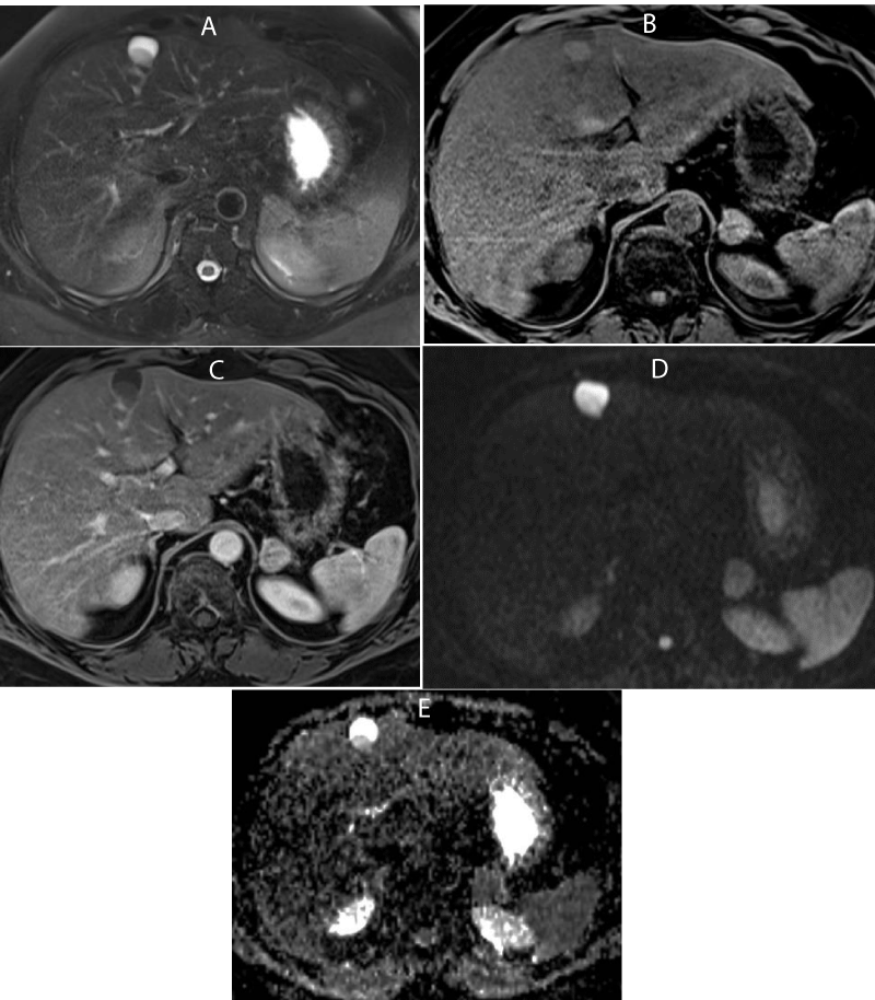
His biological assessment showed an increase in muscle enzymes (LDH) and mild hepatic cytolysis (ALAT, ASAT, Bilirubin). The rest of the biological assessment was normal. A thoraco-abdominopelvic CT scan performed as part of the etiological assessment, had returned normal except for a 1cm hepatic focal lesion, anterior subcapsular segment IV, hypodense at arterial and portal time without enhancement (Figure 1). The ultrasound performed as a supplement because of the superficial seat of the lesion showed a pseudo solid hypoechoic formation, with discreet posterior reinforcement (Figure 1). An MRI was performed on a Siemens Avento 1.5T machine with T2SE, Diffusion, IP / OP and EGT1 sequences without and with gadolinium injection. The focal lesion appeared in intense hypersignal T2 weighted imaging and hyposignal T1 weighted imaging. Its content was heterogeneous with declivitous sediment with T2 hyposignal, hypersignal T1. On diffusion weighted imaging (b800), the lesion appeared in hypersignal with diffusion restriction in sediment part. After Gadolinium injection, there was a fine peripheral contrast enhancement on the late phases (Figure 2). In front of this lesion of centimeter size, anterior subcapsular seat of segment IV, cystic type in MRI with sediment and peripheral enhancement, having a pseudo-solid aspect in ultrasound and CT, the diagnosis of ciliated hepatic foregut cyst has been retained, the patient refused surgery and biospy. The indication of a follow-up with annual control was retained.
Discussion
Ciliated hepatic foregut cyst is a rare cystic lesion that was first described in 1857 by Friederich. The term of ciliated hepatic foregut cyst was only introduced in 1984 by Wheeler and Edmondson [1]. The average age of discovery is around 50, but paediatric cases have been described [2,3]. It would be derived from an abnormal development of the anterior primitive intestine, during which a bronchiolar bud migrates abnormally into the peritoneal cavity [4,5].
In the majority of cases, it is an asymptomatic lesion of chance discovery. A few cases of symptomatic cysts have been reported by pain in the right hypochondrium [6,7]. A case of biliary compression and a case of portal compression have also been reported [2]. The diagnosis of certainty remains the anatomopathological examination which shows a characteristic architecture resembling the bronchogenic cyst at the thoracic level with four layers: a pseudo-stratified ciliated cylindrical epithelium associated with secretory mucus cells, a supportive connective tissue, a muscularis made of smooth muscle fibers and a thin peripheral fibrotic capsule [8].
Apart from the anatomopathological examination, the diagnosis can be asserted to the cytology of the suction fluid which can show hair cells. Other ectopic localizations of lesions presenting a ciliated type epithelium have been described at the retroperitoneal level, at the level of the pancreas and at the level of the tongue [8].
In imaging, Ciliated hepatic foregut cyst has three main characteristics, its preferential localization in the left liver, particularly the IV segment, the size of the lesion almost always less than 3 cm and the anterior subcapsular situation [1,9,10]. A review of the recent literature of 96 cases found a location at segment IV in 44 cases [10]. It is most often a unilocular cyst, only 7 out of 96 cases were multi-locular.
In sonography, ciliated hepatic foregut cyst appears most often hypoechoic with or without posterior reinforcement. The anechoic aspect is much rarer [8]. In CT, it is a hypodense lesion without enhancement after injection of contrast product. More rarely the lesion appears spontaneously hyperdense [11,12]. Calcium sediments have also been reported [8]. In MRI, ciliated hepatic foregut cyst has a variable signal in T1-weighted sequence, the lesion appears most often hyper-intense in more than 50% of cases, it can be iso or hypo-intense [11,12]. This variable aspect in T1-weighted sequence is a function of the mucus content and the presence of calcium or cholesterol crystals [11,12]. In T2-weighted sequences, the lesion appears most frequently in frank hypersignal. In our case, there is a sediment of intermediate signal in T2, in hypersignal T1 probably related to sedimented crystals [1,13]. The hypersignal diffusion of our case is very rarely reported. It is likely related to the content rich in mucus. The absence of enhancement after injection is the rule, unlike in our case where there is a fine and regular peripheral enhancement.
The particularities in our patient is the sediment in frank hypersignal T1, discreet hyposignal T2, the hypersignal diffusion at b 800 but especially the presence of a peripheral enhancement.
The appearance in imaging is often very evocative but the formal diagnosis is based on the anatomopathology obtained after surgical resection or after suction puncture. The latter has a positive predictive value of 73% [1]. The differential diagnosis for this lesion includes simple cyst, hydatid cyst, mesenchymal hamartoma and epidermoid cyt.
The evolutionary potential is poorly known, two cases of portal and biliary vascular compression have been reported, but especially three cases of degeneration in squamous cell carcinoma have been observed [1,3], which indicates the surgical resection of this type of lesions.
Conclusion
Ciliated hepatic foregut cyst is a rare hepatic benign lesion, with histology identical to that of the bronchogenic cyst at the thoracic level [2,10]. His diagnosis must be evoked by a thick-walled unilocular cystic lesion in segment IV in a middle-aged adult [10,12]. The management is surgical because of the risk of degeneration.
Informed consent statement
Patient gave informed consent to publication of case report.
Institutional review board statement
This study received the favourable opinion of the Institutional ethics committee.
- Benlolo D, Vilgrain V, Terris B, et al. (1996) Coll. Imagerie des kystes ciliés. À propos de 4 cas. Gast Clin Biol 20: 497-501.
- Ambe C, Gonzalez-Cuyar L, Farooqui S, Hanna N, Cunningham SC (2012) Ciliated Hepatic Foregut Cyst: 103 Cases in the World Literature. Open J Path 2: 45-49. Link: https://tinyurl.com/y7ps9txp
- Wilson JM, Groeschl R, George B, Turaga KK, Patel PJ, Saeianv K, et al. (2013) Ciliated hepatic cyst leading to squamous cell carcinoma of the liver –A case report and review of the literature. Int Journ Surg Case Reports 4: 972-975. Link: https://tinyurl.com/yb5wd7sr
- Wheeler DA, Edmondson HA (1984) ciliated hepatic foregut cyst. Am J Surg Pathol 8: 467-470.
- Vick DJ, Goodman ZD, Ishak KG (1999) Squamous cell carcinoma araising in a ciliated hepatic foregut cyst. Arch Pathol Lab Med 123: 1115-1117. Link: https://tinyurl.com/ycpf85ag
- Benlolo D, Vilgrain V, Terris B, et al. (1996) Coll. Imagerie des kystes ciliés. À propos de 4 cas. Gast. Clin Biol 20: 497-501.
- Cai XJ, Huang DY, Liang X, Yu H, Li W, Wang XF, et al. (2004) Ciliated hepatic foregut cyst: report of first case in China and review of littérature. J Zhejiang Univ Sci 5: 483-485. Link: https://tinyurl.com/ychaxdep
- Harty MP, Hebra A, Rucheli ED, Schnaufer L. (1998) Ciliated hepatic foregut cyst causing portal hypertension in an adolescent. Am J Roent 170: 688-690. Link: https://tinyurl.com/yblrevd6
- Catherine L, Dumas de la Roque A, Mabille M, Dagher I, Prevot S, Franco D, et al. (2009) Kyste hépatique à revêtement cilié : à propos d’un et revue de la littérature. J Radiol 90: 59-62. Link: https://tinyurl.com/ychohzob
- Sharma S, Dean AG, Corn A, Kohli V, Wright HI, Sebastian A, et al. (2008) Ciliated hepatic foregut cyst: an increasingly diagnosed condition. Hepatobiliary Pancreat Dis Int 7: 581-589. Link: https://tinyurl.com/yb35lnry
- Vilgrain V (2010) Lésions kystiques du foie. In Imagerie de l’abdomen. Paris: Lavoisier, Médecine sciences/Publications. 86-87.
- Ansari-Gilani K, Esfeh JM. (2017) Ciliated hepatic foregut cyst: report of three cases and review of imaging features. Gast Report 5: 75–78. Link: https://tinyurl.com/y99ajw93
- Chatelain D, Chailley Heu B, Terris B (2005) Thse ciliated heparic foregut cyst, an unusual bronchiolar foregut malformation: a histological, histochemical cyst. Hinyokika Kiyo 51: 25-29.
Article Alerts
Subscribe to our articles alerts and stay tuned.
 This work is licensed under a Creative Commons Attribution 4.0 International License.


 Save to Mendeley
