Annals of Circulation
A Suspected Complication of Radial Access Gone Good
Krzywicki P*, Wąsek W and Horszczaruk G
Cite this as
Krzywicki P, Wąsek W, Horszczaruk G (2017) A Suspected Complication of Radial Access Gone Good. Ann Circ 2(1): 001-002. DOI: 10.17352/ac.000003Case Report
A female patient, 43 years of age, was scheduled for coronary angiography due to suspicion of coronary heart disease on the basis of cardiovascular risk factors: obesity, arterial hypertension, hypercholesterolemia, CCS II angina and positive ECG exercise test.
Due to standard protocol, a radial 6F sheath (Balton, Warsaw, Poland) was introduced into right radial artery (RA), then 5000 U of unfractionated heparin and 200µg of gliceryl trinitrate were administered intraarterially.
Angiography of the right coronary artery was performed employing Judkins 4, 5F diagnostic catheter (Cordis, Miami, Fl).
Because of marked arterial spasm the catheter was slowly withdrawn with inadvertent removal of the guidewire from the RA.
Arterial spasm and considerable pain made re-introduction of diagnostic guidewire impossible. After intraarterial administration of additional 200µg of gliceryl trinitrate, 5mg of verapamil and 200mg of magnesium sulphate, angiography of the right arm’s vessels was performed.
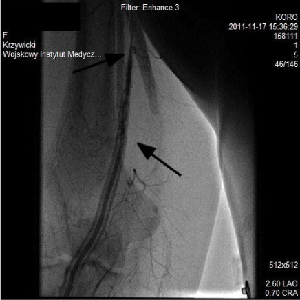
We found type 1. High bifurcation of brachial artery and angiographic signs of obstructive spasm of the RA. In the late phase of the same cine loop angiographic appearance was highly suggestive of RA dissection and contrast flow between RA and brachial vein (Figure 1). That raised suspicion of iatrogenic perforation with subsequent arterio-venous fistula. Intriguingly enough, there was no sign of contrast extravasation and the patient did not report any pain or swelling which also were not found in physical examination of the arm.
Because of this suspicion, angiography of the left coronary artery was performed via transfemoral approach and eventually both coronary arteries proved angiographically normal.
After that, we performed another angiogram of the right radial artery antegradely - via transfemoral approach – and retrogradely – via transradial approach. Both showed normal arterial flow without any signs of spasm, perforation, contrast extravasation or, indeed any other pathological flow or connection between arterial and venous vasculature.
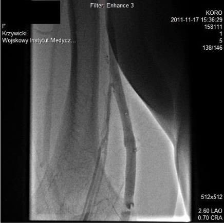
Quick resolve of what we had considered a serious complication prompted us to review the previous angiograms again. To our relief, in the very late phase of the incriminated cine loop (Figure 2) we found that what we considered to be dissection of the radial artery and AV fistula was in fact flow of contrast forced by high pressure from radial artery via arterio-venous anastomoses to concomitant veins and subsequently and naturally to the brachial vein – a proper venogram.
After the spasm abated, normal intraarterial pressures and hemodynamics were restored thus causing the closure of the AV anastomoses. That we could later appreciate as a normal angiography of the brachial arteries. During 24hr observation and in a follow up telephone call physical examination revealed no pathological findings and the patient did not report any problems.
To the best of our knowledge such apparent complication has not been described in the literature. As radial access for coronary diagnostic and therapeutic interventions becomes prevalent it is of increasing importance for medical personnel to be aware of possible real and apparent complications.
Presentation of our case report underlines the importance of thorough knowledge of anatomy and mindful review and interpretation of angiograms.
- Agostoni P, Biondi-Zoccai GG, de Benedictis ML, Rigattieri S, Turri M, et al. (2004) Radial versus femoral approach for percutaneous coronary diagnostic and interventional procedures: Systematic overview and meta-analysis of randomized trials J Am Coll Cardiol 44: 349-356. Link: https://goo.gl/RlzVBe
- Hamon M, Pristipino C, Mario CD, Nolan J, Ludwig J, et al. (2013) Consensus document on the radial approach in percutaneous cardiovascular interventions: position paper by the European Association of Percutaneous Cardiovascular Interventions and Working Groups on Acute Cardiac Care and Thrombosis of the European Society of Cardiology. EuroIntervention 8: 1242-1251. Link: https://goo.gl/pS84XG
Article Alerts
Subscribe to our articles alerts and stay tuned.
 This work is licensed under a Creative Commons Attribution 4.0 International License.


 Save to Mendeley
