Archive of Biomedical Science and Engineering
Rapid diagnosis in thyroid tumors by using touch cytology
Suzan Seno1*, Adnan Almarrawi2, Mohannad Nasani2 and Safa Alhalabi3
2Department of Biotechnology Engineering, Faculty of Technical Engineering, Aleppo University, Syria
3Specialist Pathology at Al Razi Hospital, Aleppo, Syria
Cite this as
Seno S, Almarrawi A, Nasani M, Alhalabi S (2022) Rapid diagnosis in thyroid tumors by using touch cytology. Arch Biomed Sci Eng 8(1): 001-004. DOI: 10.17352/abse.000027Copyright
© 2022 Seno S, et al. This is an open-access article distributed under the terms of the Creative Commons Attribution License, which permits unrestricted use, distribution, and reproduction in any medium, provided the original author and source are credited.The study aims to assess the role of imprint cytology in the diagnosis of thyroid nodules and compare it with paraffin Section in the diagnosis of thyroid lesions.
It included 40 patients who visited Private Hospitals in Aleppo during the period from April to December 2021. The results indicated that there were 19 patients with thyroid cancer, representing 47.5%, of whom 14 were diagnosed with Papillary Carcinoma, with a percentage of 35%, and 5 cases with Follicular Carcinoma, with a percentage of 12.5%, while the remaining cases included 21 patients with Benign Adenomas at a rate of 52.5%, including 11 cases of Hashimoto’s disease at a rate of 27.5% and 10 cases of Benign Follicular Tumors at a rate of 25%. These results were compared with Paraffin Sections and reached sensitivity, specificity, and accuracy in diagnosis for this technology, 94.73%, 95.23% and 95%, respectively, and the positive predictive value was 94.73%, while the negative predictive value was 95.23%. The results also indicated that this technology is fast in determining the Histologic Grading of Tumor Differentiation, but it does not determine the Histological Type. It also indicated that there was a significant association between gender and the incidence of thyroid tumors, while no significant statistical evidence was observed between age and the possibility of thyroid cancers.
Introduction
Thyroid cancer is the most common type of endocrine malignancy, and it is one of the few types of cancer whose incidence has increased in recent years and constitutes 3% of all diagnosed cancers worldwide [1,2]. Infection rates vary by geographic region, age, and gender. In 2021, more than 44,280 people (12,150 males and 32,130 females) were newly diagnosed with thyroid cancer in the United States alone.
As for Syria, according to the statistics of the World Health Organization (WHO), issued in 2020, the number of infected cases reached 2186 people.
The annual mortality rate for thyroid cancer cases is low, representing five deaths per million individuals annually and this is likely reflected due to the good prognosis of most thyroid cancers, which are considered to be a good prognosis. Age-specific mortality rates rise steadily from around age 40-44 and more steeply from around age 55-59. The highest rates are in the 90+ age group for both females and males.
Thyroid cancer affects all age groups, from children to the elderly, is more common in females than males, and ranks seventh among all malignant tumors that affect women. The disease affects more than 7 out of 10 people, all of whom are female [3].
It starts from abnormal growth of cells in the thyroid gland and then multiplies irregularly, which eventually leads to the formation of clumps of cells called tumors, and thus can invade other tissues in the neck and then spread to the surrounding lymph nodes, or through the bloodstream, It is then transmitted to other parts of the body [4]. This disease can take many forms depending on the type of cells affected in the gland, and genetic factors, age, sex, family history of infection, iodine deficiency, and radiation exposure play a decisive role in increasing the incidence [5].
Since the rapid and accurate diagnosis of thyroid tumors during surgical operations helps determine the treatment plan and spares patients the additional costs and undergoing further surgery, the use of Touch Cytology (TC), which is one of the most important rapid diagnostic technologies in developing countries.
In 1927, Dudgeon and Patrick in London developed this method for obtaining a rapid cytological diagnosis from freshly taken tissue samples, and they proved that the results they reached in the study of 200 samples are It was of high accuracy and reliability compared to the examination of frozen and paraffin-embedded tissues, and since then, this technology has been used during surgical excision of tumors to reach a more accurate diagnosis [6].
That It was able to determine the presence of tumor malignancy in the thyroid gland and determine the degree of histologic grading without specifying the histologic classification of it, thus preserving cellular material by reducing tissue damage and transferring the image of cells located from tissues to glass slides, i.e. corresponding tissues. It reveals details of chromatin condensation in the nucleus and abnormalities within the examined cells and thus shortens the time in diagnosis compared to Paraffin Section and Frozen Section. Thus, it allows surgeons to remove one lobe of the thyroid gland or to completely excise it according to the clinical situation, but patients are also subjected to paraffin sections to find out the histological pattern and confirm the degree and stage of the tumor [7].
Materials and methods
The patients
The study included 40 patients who attended private hospitals in Aleppo (Al-Issa Hospital, Al-Furqan Hospital, Al-Salam Hospital) and private histopathology laboratories in the city of Aleppo during the period from March to December 2021. They were diagnosed with the possibility of thyroid cancer through the appearance of enlargement in the nodes. lymph nodes in the neck, as well as pain in the frontal area of the neck, voice changes as a result of pressure on the fourth laryngeal nerve, difficulty breathing and swallowing, and they underwent imaging using ultrasound, which helps determine the number and size of nearby enlarged lymph nodes, which confirms the spread of gland cancer The patients were divided according to age into two age groups, and they underwent surgeries to remove the masses, and they underwent TC surgery to verify the presence of tumor malignancy by taking tissue samples from the newly excised masses, where a very small part of them was taken using a sterile scalpel. The sample was sharpened and then placed on sterile gauze to get rid of the excess blood, and using forceps, it was passed over several glass slides by light pressure to give the shape of an imprint of cells The cells were fixed with absolute alcohol and then treated with hematoxylin and eosin dyes, and examination using light microscopy to determine the presence of malignant neoplastic cells by observing dense chromatin and abnormal divisions within the cell nuclei [6,7]. Then the samples taken were treated with formalin to make paraffin tissue sections through fixation with gradient concentrations of alcohol (75%, 95%, 100%) and cleared using xylol and then incubated with paraffin and cut using a microtome, then stained the slides with hematoxylin and eosin and examining them Microscopically [8], in order to compare with it and make sure that the results match the diagnosis [10].
Statistical study
Statistical tests were carried out using IBM SPSS Statistics program version 24, qualitative variables were expressed using frequencies and percentages, descriptive stats were used to find out the dimensions of the size of tumor masses, and cross-tabulation and Chi-square were also used to study the relationship Between age, sex and the extent of malignancy.
The following statistical equations were used to calculate Sensitivity, Specificity, Accuracy, Positive Predictive Value (PPV) and Negative Predictive Value (NPV):
Sensitivity = true positive/ (true positive + false negative) x 100%.
Specificity = True Negative / (True Negative + False Positive) x 100%.
Positive predictive value = true positive / (true positive + false positive) x 100%.
Negative predictive value = true negative / (true negative + false negative) x 100%.
Accuracy = (true positive + true negative) / total number of cases x 100%.
Results
Distribution of patients according to gender and incidence of thyroid cancer
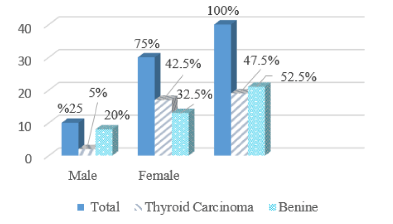
The study included 40 patients, 10 of whom were males with a percentage of 25%, of whom only two patients were found to have thyroid cancer with a percentage of 5%, and 8 patients with benign tumors of 20%, and the number of females was 30 by 75%, of whom 17 cases were diagnosed with thyroid cancer with a percentage of 42.5 %, and 13 cases with benign tumors at a rate of 32.5%, and we note that the distribution of males to females in this study is 3:1. (Figure 1) shows the distribution of patients according to gender and the incidence of thyroid cancer.
Distribution of patients according to the histological pattern of the tumor by sex and age group
The histological patterns of the tumor were determined using paraffin tissue sections because the cell fingerprinting technology does not enable the identification of any patterns compared to its weak effectiveness in other organs such as the breast, and these patterns were distributed according to sex as in (Table 1), as it was noted that the most common pattern in males is Hashimoto’s disease By 20%, while the most frequent malignant type in females was papillary adenocarcinoma with 32.5%, which corresponds to international statistics.
When studying the relationship between sex and the extent of malignant tumors, a significant correlation was observed, P = 0.04<0.05, which confirmed that women are more susceptible to infection compared to males, whose number was very few, due to the high levels of estrogen and progesterone hormones they have. This result did not match study [12].
The age range of patients ranged from 23 to 60 years, with an average age of 43 years, and the age groups were divided into two groups, the first group was less than or equal to 45 years old, and the second group was over 45 years old, and the histological patterns of the tumor were distributed according to age groups as shown in the (Table 2), as it was noted that the most common histological pattern in the first age group was papillary thyroid carcinoma by 45%, while the type in the second age group was Hashimoto’s disease by 33.8%.
When studying the relationship between age and the risk of thyroid cancer, it was found that there is no significant correlation between them, p = 0.5 > 0.05, meaning that the increase in the age of patients over 45 years does not increase the risk of malignancy, and the result is consistent with the study [13] while It did not match the study [14].
The distribution of patients according to the degree of differentiation of the tumor and the location of the tumor masses
The results of the current study indicated that there were 19 patients with thyroid cancer, 47.5%, and 21 patients with benign tumors, 52.5%, and the frequency of each degree of tumor differentiation was distributed for patients with cancer diagnosed using TC technology, as shown in (Table 3). The second degree is the most common with 68.42%.
As for the location of the tumor masses, it was found that 17 patients (42.5 percent) had masses in the right lobe, while 23 patients were diagnosed with their presence in the left lobe.
The dimensions of the tumor masses ranged from 2.5×3.5×5 cm to 6×6×7 cm, and the average size of the tumor was with dimensions of 2.5×4×5.5 cm.) 10 cases with a percentage of 52.63%, while the number of cancers with a diameter smaller than 2 cm (T1) was 7 cases with a percentage of 36.84%, and cancers with a diameter larger than 4 cm (T3) reached 4 cases with a percentage of 21.05%.
The efficiency of TC in diagnosing thyroid tumors
The extent of congruence between the results of cell fingerprint and paraffin tissue sections was studied, as the number of true positive cases was (18/40), while the number of false-positive cases was (1/40), while the number of true negative cases was (20/40), while the number of false-negative cases was (1/40). The sensitivity, specificity, and accuracy values of TC technology in diagnosing tumors were calculated for all clinical cases, which ranged, respectively: 94.73%, 95.23%, 95% and the positive predictive value reached 94.73%, while the negative predictive value was 95.23%, and the results were compared with some International reference studies, as it was noted that the results were similar, as shown in (Table 4) and the false-negative rate in this study was 2.5%.
In contrast to the effectiveness of this technology in breast tumors, it is not able to determine the histological patterns of the tumor, although its ability to do so is small [4].
Discussion
This study was conducted with the aim of assessing the role of Touch cytology in diagnosing thyroid lesions keeping Paraffin Sections as the gold standard.
It seems to be a useful, valuable, and inexpensive method in clinics, which do not have equipment for frozen sections. it increased the overall accuracy of diagnosis. Further studies are needed to validate the obtained results of this study it is rich in cells and thus has been assumed a more suitable cytological examination technique by pathologists, in spite of its limitation for detection of vascular and capsular invasion.
Conclusion
The study provides the possibility of rapid diagnosis of Lesions in the thyroid gland by using TC to check whether it is malignant or not, however, other studies with a higher clinical number of cases should be performed to more accurately determine the percentage of false negatives, and slow paraffin biopsy should be used to confirm the degree of tumor differentiation.
- Lim H, Devesa SS, Sosa JA, Check D, Kitahara CM. Trends in Thyroid Cancer Incidence and Mortality in the United States, 1974-2013. JAMA. 2017 Apr 4;317(13):1338-1348. doi: 10.1001/jama.2017.2719. PMID: 28362912; PMCID: PMC8216772.
- Siegel RL, Miller KD, Jemal A. Cancer statistics, 2020. CA Cancer J Clin. 2020 Jan;70(1):7-30. doi: 10.3322/caac.21590. Epub 2020 Jan 8. PMID: 31912902.
- Atlanta G. About Thyroid Cancer Overview and Types, American Cancer Society. 2021; 7-8.
- Alexander EK, Marqusee E, Orcutt J, Benson CB, Frates MC, Doubilet PM, Cibas ES, Atri A. Thyroid nodule shape and prediction of malignancy. Thyroid. 2004 Nov;14(11):953-8. doi: 10.1089/thy.2004.14.953. PMID: 15671774.
- Anand C. Role of Imprint Cytology in Diagnosis of Thyroid Lesions. World Journal of Endocrine Surgery, India. 2019; 11(2):49-52.
- Pantanowitz L, Xing J, Monaco S. Atlas of Touch Preparation Cytopathology, Demos Medical An Imprint of Springer Publishing, 2018, chapter 1,2.
- Alina R. Role of Touch Cytology in Diagnosis of Thyroid Tumors. 2017; 31(50): 11-15.
- Claudia D. Microtomy and Paraffin Section Preparation), Knowledge pathway. 2019; 1-22.
- Taneri F, Poyraz A, Salman B, Tekin E, Akyuerek N, Bayram O, Onuk E. Using imprint and frozen sections in determining the surgical strategies for thyroid pathologies. Endocr Regul. 2001 Jun;35(2):71-4. PMID: 11563934.
- Issam M. Role of Fine Needle Aspiration, Intraoperative Imprint Cytology and Frozen Section in Breast Lumps and Thyroid Lesions. Medical Principles and Practical. 8:173–182.
- Adhya AK, Mohanty R. Utility of touch imprint cytology in the preoperative diagnosis of malignancy in low resource setting. Diagn Cytopathol. 2017 Jun;45(6):507-512. doi: 10.1002/dc.23699. Epub 2017 Mar 7. PMID: 28267274.
- Yan HX, Pang P, Wang FL, Tian W, Luo YK, Huang W, Yang GQ, Jin N, Zang L, Du J, Ba JM, Dou JT, Mu YM, Lyu ZH. Dynamic profile of differentiated thyroid cancer in male and female patients with thyroidectomy during 2000-2013 in China: a retrospective study. Sci Rep. 2017 Nov 20;7(1):15832. doi: 10.1038/s41598-017-14963-z. PMID: 29158505; PMCID: PMC5696456.
- Kaliszewski K, Diakowska D, Nowak Ł, Wojtczak B, Rudnicki J. The age threshold of the 8th edition AJCC classification is useful for indicating patients with aggressive papillary thyroid cancer in clinical practice. BMC Cancer. 2020 Nov 30;20(1):1166. doi: 10.1186/s12885-020-07636-0. PMID: 33256657; PMCID: PMC7708212.
- Haymart MR. Understanding the relationship between age and thyroid cancer. Oncologist. 2009 Mar;14(3):216-21. doi: 10.1634/theoncologist.2008-0194. Epub 2009 Mar 6. PMID: 19270027.
Article Alerts
Subscribe to our articles alerts and stay tuned.
 This work is licensed under a Creative Commons Attribution 4.0 International License.


 Save to Mendeley
