Biomed Sci Eng
Myocardial Contrast and Stress Echocardiography: New Frontiers
Paulo Magno Martins Dourado1*, Jeane Mike Tsutsui1, Antonio Casella Filho2, Larissa de Almeida Dourado3, João Paulo de Almeida Dourado4, Leonardo Roever5 and Wilson Mathias Junior1
2Atherosclerosis Unity, Heart Institute (InCor), University of São Paulo Medical School
3Heart Hospital (HCor) , São Paulo
4University of the City of São Paulo (Unicid), São Paulo
5Federal University of Uberlândia, Department of Clinical Research, Uberlândia, Brazil
Cite this as
Martins Dourado PM, Tsutsui JM, Filho AC, de Almeida Dourado L, de Almeida Dourado JP, et al. (2017) Myocardial Contrast and Stress Echocardiography: New Frontiers. Biomed Sci Eng 3(1): 001-010. DOI: 10.17352/abse.000007Introduction
The assessment of myocardial perfusion by echocardiography following intravenous injection of ultrasound contrast agents has proved to be feasible [1]. Low-mechanical index imaging techniques that cause minimal microbubble destruction let simultaneous analysis of myocardial perfusion and function in real-time [2]. Real-time myocardial contrast echocardiography (RTMCE) increases the sensitivity of Dobutamine stress testing for the detection of CAD [3] and can distinguish between stunned and infarcted myocardium after acute ischemia [4].
The Dobutamine-Atropine Stress Echo (DASE) is a well-established method to noninvasive evaluation of the patients with known or suspected CAD. The ischemic myocardium evaluation is based on the detection of reduction of the systolic myocardium thickness by two-dimensional echocardiography. Those changes are induced by the balance between offer and consumption of oxygen during stress by Dobutamine. Meta-analysis studies have demonstrated that both DASE and myocardial contrast perfusion stress echocardiography are a clinical tool for the detection of CAD [5].
The importance of Dobutamine stress echocardiography (DSE) is well gathered in the risk stratification after acute myocardial infarct and also to evaluate myocardial viability in patients with ischemic cardiomiopathy. The contractile reserve detection can be utilized to predict myocardial regional function recovery in patients with chronic CAD. The method above is more specific to evaluate functional recovery after procedures of surgical revascularization than the methods which analyze the presence of viability by the metabolic integrity of myocardial cell, as scintigraphy by 201Talium and positron emission tomography. In patients with severe reduced ventricular function at rest, the demonstration of contractile reserve by DSE is associated with the reduction in the mortality level when the patients are subjected to bypass surgery.
One of the limitations of the DASE is the necessity of an adequate visualization and delineation of endocardium borders to detect transitory alterations and, sometimes, mild myocardial mobility. The inadequate delineation of the left ventricular endocardium border is a possible cause of false result, and improve the variability intra and inter observer in the exam analysis. New technological advances as tissue Doppler, second harmonic image and the utilization of contrast agents, associated with digital image development, has made stress echocardiography a method with high practicality and reproducibility to CAD evaluation in many situations [6, 7].
Pharmacological considerations and physiological basis to detection of ischemia by dobutamine-atropine stress echocardiography
Among the pharmacological agents used to induce stress, Dobutamine is the most utilized in the clinical practice to evaluate ischemia and myocardial viability. Dobutamine is a catecholamine with half-life between two and three minutes, that present good tolerance and, because its positive inotropism and chronotropic effect, may cause increment in the oxygen consumption by the myocardium [8].
Although cardiovascular stress testing does not define the degree of coronary stenosis, it determines the physiological importance of a particular blood flow obstruction. Reduction in the coronary flow cause myocardial ischemia because the imbalance between the consumption and supply of oxygen [9].
The myocardial ischemia occurring in areas addressed by an artery with significant narrowing degree obey a sequence of events known as ischemic cascade. The reduction or absence of myocardial thickening systolic expressed by segmental motility alterations are an early phenomenon sensitive and specific to ischemia [10]. The global and regional left ventricular function evaluation by echocardiography during stress allows not only the diagnosis, but also the determination of severity and extent of myocardial ischemia. Moreover, the echocardiography is able to identify, by analysis of the various myocardial segments, coronary artery ischemic area-related and new resources as Speckle tracking echocardiography (STE) that is a method of quantitative assessment of myocardial function complementary to ejection fraction (EF) and visual evaluation. Standard STE analysis, demands manual tracing of the myocardium whereas automated function imaging (AFI) offers more convenient (based on selection of three points) assessment of longitudinal strain. Both methods provided good and similar feasibility with only 1% segments excluded from analysis at peak stage of DSE with shorter time and lower coefficient of variance offered by AFI. Global and regional longitudinal strain achieved by faster and less operator-dependent AFI method correlate well with standard more time-consuming STE analysis during baseline and peak stage of DSE [11].
Evolution of the echocardiography protocols under stress by dobutamine-atropine
The conventional Protocol consists of intravenous infusion of D obutamine started in dose of 5 mg/kg/min and increased to 10, 20, 30 and 40 µg/Kg/min in three stages minutes [12]. Diagnostic endpoints of the test are: positive echocardiogram (new onset wall motion abnormalities or worsening of baseline dissinergy); achievement of 85% of maximal predicted heart rate (220 - age); severe chest pain and/or diagnostic ST-segment changes. The test is stopped without diagnostic endpoints for: Intolerable symptoms; hypertension; hypotension; supraventricular arrhythmias (supraventricular tachycardia or atrial fibrillation); or ventricular arrhythmias (ventricular tachycardia; frequent, polymorphous, premature ventricular beats) [13].
Over the years, some modifications have been made in DSE protocols. An early atropine administration during the DSE has been proposed to reduce the test duration [14]. Tsutsui et al. [15], evaluated the safety, efficacy and diagnostic accuracy of the early injection of atropine in a larger number of patients (n = 1664), compared with the conventional protocol (n = 3163). The early atropine protocol proposed consisted in beginning atropine injection in the dose of 20 µg/Kg/min of Dobutamine. The incidence of serious adverse effect was similar in both protocols (0.3% versus 0.4%; p = NS). The sensitivity, specificity and accuracy of the early atropine injection to CAD detection was 83%, 89%, and 86%, respectively, and conventional protocol were 85%, 77% and 82% (p = NS between the groups). The authors concluded that early atropine injection is a safe and effective alternative to conventional protocol, keeping similar diagnostic accuracy to CAD detection. The early atropine administration safety during DASE also was confirmed in a population of elderly patients (≥ 70 years old) [16].
Another modification in the conventional Dobutamine-atropine protocol proposed by Mathias et al. [17], is metoprolol injection in bolus at stress peak, to increase diagnostic accuracy of the exam. This modification consists in the injection of 5 mg of metoprolol in a 1 minute at the stress peak. When the heart rate is below 100 beats per minute or at most within 3 minutes after an injection of metoprolol the post-metolol images are acquired.
Technical aspects and interpretation of the images
As criteria of myocardial ischemia and viability are based in the detection of myocardial segmental alterations, a complete visualization of all the walls of the left ventricle is necessary to document or delete reliable abnormalities.
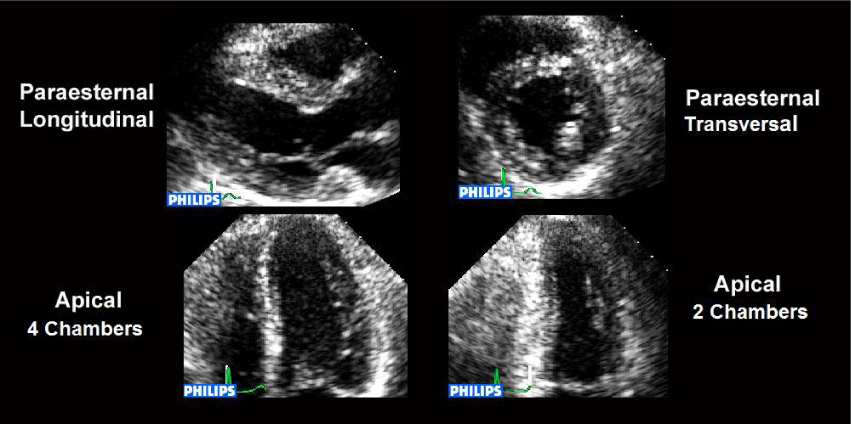
By use potentially ischemia stimuli, the tests should be performed in environments specially adapted for a possible procedure of cardio-respiratory reanimation and others possible complications. The patients should be fasting for a period of four hours. Generally are acquired and stored in quadruple screen format in the axis paraesternal longitudinal, paraesternal transverse, apical four chambers and apical two chambers (Figure 1). The images are acquired in basal state, with low dose of Dobutamine, stress peak, and at the stage of recovery. In the presence of alterations in segmental motility the images must be registered in order to allow the documentation of the presence, location, extent and duration of dysinergy. A 12 lead electrocardiogram must be done in basal state and in the end of each stage. A lead of the electrocardiogram should be monitored continuously by echocardiography apparatus to provide a constant reference to the operator to observe ST segment changes and presence of arrhythmias. The blood pressure is measured by sphygmomanometer in rest conditions and end of each stage.
By the years, different schemes of left ventricle segmentation were proposed to represent the myocardium distribution of major coronary artery territories. The most used so far in the literature was recommended by the American Society of Echocardiography in 1989 [18], defining 16 myocardial segments. However, a new segmentation including 17 segments were proposed, due to the advent of contrast echocardiography and the need for standardization of segmentation not only by echocardiography, but also for other methods of image, such as myocardium scintigraphy and positron emission tomography [19]. For semi quantitative assessment of segmental motility, scores are given to each of the left ventricle segments, based on evaluation of the endocardium thickening and in the degree of wall contraction, being the value 1 given to normal segments, value 2 to hypokinetic, value 3 to akinetic and value 4 to dyskinetics, in all stages of the test. The score index of segmental motility is obtained by the sum of the scores given to every one of left ventricle segments divided by the number of segments analyzed.
The test must be interpreted as positive whenever the presence of ischemia appears, in the form of new alteration during stress in the left ventricle segmental motility, which are expressed by augment in the score of more than one segment of the LV at least a point. When changes already exists in segmental motility at rest the test, it is considered positive if there are deterioration of motility compared to the pre-existing, or when changes of the left ventricle presenting at rest improves with low doses drug and later worsens with high doses, indicating viability and myocardial ischemia, respectively. Viability assessment is made by demonstrating improvement in the myocardium motility with low doses of Dobutamine, and this response can occur at higher doses, especially in patients using beta-blockers.
Diagnostic value and prognosis of dobutamine-atropine stress echocardiography
When compared the physical stress testing with DASE, the later has increased sensitivity and shown greater specificity for the diagnosis of CAD.
Many studies in literature have compared the diagnostic accuracy of different types of stress echocardiography [20,21].
While the DSE and by physical stress presents similar diagnostic accuracy, the dipyridamole stress echocardiography seems to present a somewhat diagnostic smaller, and this difference can be attributed a lower sensitivity of dipyridamole in the identification of uni arterial disease (38% to dipyridamole, 70% for physical test and 61% to Dobutamine).
The addition of atropine to DSE increases the diagnostic accuracy and reduces the percentage of ineffective tests, especially in patients using beta-blockers [22]. These findings were also supported by Pingitore et al., in international multicenter studies EPIC (Echo Presenting International Cooperative Study) [23] and EDIC (Echo Dobutamine International Cooperative Study) [24]. Mathias et al. [25], showed that DASE is a secure and accurate method to diagnostic CAD in patients with or without previous alterations of ventricular motility.
Tsutui et al. [26] showed that although a new protocol of dobutamine stress echocardiography with the early injection of atropine (EA-DSE) has been demonstrated to be useful in reducing adverse effects and increasing the number of effective tests and to have similar accuracy for detecting coronary artery disease (CAD) compared with conventional protocols, no data exist regarding its ability to predict long-term events. The aim of this study was to determine the prognostic value of EA-DSE and the effects of the long-term use of beta blockers on it. A retrospective evaluation of 844 patients who underwent EA-DSE for known or suspected CAD was performed; 309 (37%) were receiving beta blockers. During a median follow-up period of 24 months, 102 events (12%) occurred. On univariate analysis, predictors of events were the ejection fraction (p <0.001), male gender (p <0.001), previous myocardial infarction (p <0.001), angiotensin-converting enzyme inhibitor therapy (p = 0.021), calcium channel blocker therapy (p = 0.034), and abnormal results on EA-DSE (p <0.001). On multivariate analysis, the independent predictors of events were male gender (relative risk [RR] 1.78, 95% confidence interval [CI] 1.13 to 2.81, p = 0.013) and abnormal results on EA-DSE (RR 4.45, 95% CI 2.84 to 7.01, p <0.0001). Normal results on EA-DSE with beta blockers were associated with a nonsignificant higher incidence of events than normal results on EA-DSE without beta blockers (RR 1.29, 95% CI 0.58 to 2.87, p = 0.54). Abnormal results on EA-DSE with beta blockers had an RR of 4.97 (95% CI 2.79 to 8.87, p <0.001) compared with normal results, while abnormal results on EA-DSE without beta blockers had an RR of 5.96 (95% CI 3.41 to 10.44, p <0.001) for events, with no difference between groups (p = 0.36). In conclusion, the detection of fixed or inducible wall motion abnormalities during EA-DSE was an independent predictor of long-term events in patients with known or suspected CAD. The prognostic value of EA-DSE was not affected by the long-term use of beta blockers.
Myocardial deformation index is considered a sensitive marker of ischemia and may be useful in the quantification of hemodynamic significance of CAD. Some studies determined the diagnostic value of speckle-tracking echocardiography in derived myocardial deformation parameters at rest and during stress, can evaluate hemodynamically significance in coronary artery stenosis on those patients with moderate and high probability of CAD. Also, it showed that left ventricular strain and strain rate analyses during DSE can be used in the assessment of hemodynamic significance in coronary artery stenosis, with patients that have moderate and high risk for CAD, with good accuracy [27,28].
One the strategies proposed by Mathias et al. [17], to improve DASE sensitivity was the rapid injection of metoprolol in the stress peak with acquisition of post-metoprolol images in maximum range of three minutes. After metoprolol injection, occurred improvement in both sensitivity and accuracy.
Multiple factors can lead to a decrease in the definition of the endocardium borders mainly in the stress peak. An important technological advanced introduced was tissue harmonic image (THI) [29], that brings significant benefits of reproducibility and exequibility of the stress echocardiography [30]. Harmonic imaging, in which a pulse is transmitted at one frequency and received at twice the transmitted frequency, was originally developed to detect the nonlinear vibrations of ultrasound contrast microbubbles. Early in contrast imaging, however, researchers noticed that the images acquired prior to the arrival of the contrast had better image quality than those made at the fundamental, even at the higher frequency. It had long been know that sound propagating through tissue created harmonics of its center frequency. Tissue is generally considered to be incompressible and the speed of sound independent of pressure. However, at high acoustic amplitudes, sound speed is slightly higher than at ambient pressure and the peak of the sound wave actually travels faster than the local sound speed. Conversely, in the trough, the speed of sound is slower. This leads to peaking of the sound wave, and the creation of harmonics [31]. The other aspect of THI that has an even more dramatic effect on image quality is its ability to reduce artefacts from shallow structures. Acquisition of ultrasound images are often between ribs, or through surface fat layers. Ribs and fat layers produce distortions, reflections and reverberations that can lead to a near-field haze often seen in fundamental imaging. THI reduces this problem because the imaging beam is not created at the skin surface but by the propagating wave, and therefore does not even exist in the first couple of centimetres [32]. Another important contribution to improve the quality of images during stress echocardiography was the use of contrast agents based in microbubbles.
Diagnostic value and prognosis of dipyridamole stress echocardiography
The addition of myocardial perfusion (MP) imaging during dipyridamole real time contrast echocardiography (DipRCE) improves the sensitivity to detect coronary artery disease, but its prognostic value to predict hard cardiac events in large numbers of patients with known or suspected CAD remains unknown.
Gaibazzi et al. [33], studied 1252 patients with DipRCE and followed them for a median of 25 months. The prognostic value of MP imaging regarding death and nonfatal myocardial infarction was determined and related to wall motion (WM), clinical risk factors and rest ejection fraction using Cox proportional-hazards models, C index and risk reclassification analysis. A total of 59 hard events (4.7%) occurred during the follow up (24 deaths, 35 myocardial infarctions). The 2-year event-free survival was 97.9% in patients with normal MP and WM, independent, known or suspected CAD. Patients with normal MP responses have better outcome than patients with normal WM; patients with both reversible WM and MP abnormalities have the worst outcome.
Physical stress echocardiography
Physical stress echocardiography (PSE) has high sensitivity and specificity, and is more accurate than ECG, Treadmil Test and resting echocardiography in the detection of CAD [34]. LV WMA detected by PSE occur earlier than angina or ST-segment abnormalities [34]. Additionally, PSE also has an additional value in the location and quantification of myocardial ischemia as well as in the prediction of adverse events in patients with established CAD [34]. PSE presents high sensitivity and specificity to detect myocardial ischemia [35] both in patients without a history of prior intervention and in those previously submitted to percutaneous coronary intervention or coronary artery bypass graft surgery [35]. Detection of ischemia with exercise electrocardiographic testing is limited in patients with left bundle bunch block. PSE also face some limitations in these patients because of the paradoxical motion of the inter-ventricular septum [36]. The prognostic accuracy of myocardial perfusion and stress echocardiography appeared similar. PSE provides significant information for predicting outcome in patients with LVSD and known/suspected coronary artery disease [37]. PSE proved to be a safe modality of stress, with non-fatal complications only. Advanced age and enlargement of the left atrium are predictive of cardiac arrhythmias [38]. Another advantage of PSE is the radiation-free nature.
Role of the stress echocardiography in risk stratification after acute myocardial Infarct
The echocardiography at rest and during stress provides a variety of information’s about the left ventricular function, myocardial viability and presence of ischemia, with important therapeutic implications and prognostic after myocardial acute infarct. The GISSI study [39] showed that left ventricle function is one of the main predictors of cardiac mortality after myocardial acute infarction, with larger increments mortality associated with the progressive reduction of ventricular ejection fraction. News parameters reflecting left ventricular (LV) function as strain and strain rate provide strong prognostic information in patients after AMI. These parameters were superior to EF and WM in the risk stratification for long-term outcome [40].
There are landmarks studies in literature demonstrating the effectiveness of prognostic stratification with stress echocardiography by dipyridamole and Dobutamine in different subsets of patients after acute myocardial infarct as the EPIC and EDIC study [41].
Echocardiography evaluation of myocardial viability
The contractile improvement that occurs during stress reflects the functional cellular contractile mechanism. A simple echocardiographic analysis at rest provides information about the myocardial viability. Pierard, et al. [42]. Showed the identification of viable myocardium by echocardiography with low doses of Dobutamine in patients in the post-acute myocardial infarct subjected to thrombolytic therapy, showing good correlation with the positron emission tomography. MCE was a powerful predictor of mortality in patients with heart failure to evaluate patients with depressed ventricular function at rest, myocardium viability, ischemia and coronary flow reserve [43].
The Dobutamine inotropic response expresses the quantity of myocardial viable, and usually correlates with the recovery of function after restoration of blood flow by coronary revascularization. The inotropic positive response to viable myocardium generally occurs with doses of 5 to 10 g.Kg-1.min-1 of Dobutamine, however, the administration of higher doses are recommended, since the test being well tolerated by the patient.
The echocardiography has an important role in the evaluation of surgical indication in patients with chronic CAD and myocardial dysfunction, providing not only prognostic data based in the analysis of left ventricular function at rest, but also to evaluate the capacity of functional recovery detecting the presence of viability and myocardial ischemia.
Comparation between echocardiography versus cardiac magnetic resonance
To compare the value of Dobutamine stress echocardiography (DSE) with that provided by Dobutamine Cardiac Magnetic Resonance (DCMR) for the non-invasive risk stratification of patients with suspected or known coronary artery disease (CAD) Bikiri et al. [44], included in a study patients with suspected or known CAD underwent either DSE or DCMR using the same standardized protocol. Patient matching was then performed retrospectively for age, gender and risk factors. Outcome data including cardiac death and non-fatal myocardial infarction (defined as hard cardiac events) and ‘late’ revascularization performed >90 days after the diagnostic procedures were collected during at least 6 months. Follow-up data were available in 1852 patients who completed either DSE (n = 884) or DCMR (n = 884) during a mean follow-up duration of 4.1 ± 2.4 and 3.9 ± 1.9 years, respectively (p = NS). Matched patients exhibited an overall high risk profile (69 ± 9 years; 69% male, 70% history of CAD and 26% diabetes mellitus in both groups). Using multivariable analysis, both modalities successfully identified patients with inducible ischemia at higher risk for subsequent hard cardiac events, surpassing the value of conventional risk factors like age, male gender and diabetes (HR = 9.2; 95%CI = 5.6–14.9 for DCMR versus 2.5; 95%CI = 1.7–3.7 for DSE). By testing for interaction the predictive capacity of DCMR was higher than that provided by DSE (p = 0.02). Patients with negative DCMR exhibited lower event rates compared to those with negative DSE (annual hard cardiac event rate of 0.8% versus 3.2%, p = 0.002). DSE & DCMR aid the risk stratification of CAD patients. However, inducible WMA during DCMR are associated with a higher risk for subsequent cardiac events. Patients with negative DCMR on the other hand, exhibited a lower event rate compared to those with negative DSE.
New advances in DASE and contrast echocardiography
The tissue Doppler is better for quantification of the myocardium flow at rest and during stress, with most studies suggesting an increase in sensitivity in detecting ischemia when compared to visual analysis [45]. However, its routine use in Echocardiographic Laboratories is still limited, and studies involving more number of patients are still required to consolidate the technique.
The contrast echocardiography is gaining space. The contrast agents are solutions that contains gas microbubbles with the blood cells size, whose interface with the liquid ambient is highly refractional, improving the echocardiographic signal where they are [46].
The microbubbles more utilized are formed by perfluorocarbons and have enough stability to when injected by peripheral intravenous vein cross the pulmonary barriers and contrast left cardiac cavities and coronary circulation [47].
The echocardiographic contrast let a better definition of the endocardium borders, allowing an assessment more adequate of the parietal myocardial thickness and the global and segmental left ventricle contractile function, at rest and under stress.
The microbubbles have the feature to expand and collapse in a non-uniform way in the ultrasound presence, generating signals that contain multiple frequencies of the original frequencies (harmonics). The probes with second harmonic, transmit the echocardiographic signal in one frequency and receive signals with the frequency emitted and signals with second harmonic, produced by microbubbles. This technique improves the definition of the endocardial borders, especially in cases with inadequate acoustic windows, even without the use of echocardiographic contrast. The association of the contrast agents has additional benefits to the use of second harmonic image during the stress echocardiograph.
The use of contrast agents during pharmacological stress echocardiography showed benefits to improve endocardial delineation utilizing only fundamental image or when associated to harmonic image. The American Society of Echocardiography recommends the use of contrast agents during stress echocardiography when at least two myocardial segments are not adequate visualized in any apical axis [48].
In patients with multivessel disease and balanced ischemia, the addition of contrast with myocardial perfusion assessment, is not only able to overcome the limitation of false negative rate on a per-patient basis, but may also depict multivessel myocardial perfusion defects more efficiently than others techniques, thanks to high spatial resolution. Myocardial perfusion assessment during MCE, although not always technically feasible, has a very high spatial and temporal resolution which can easily demonstrate multivessel sub endocardial perfusion defects during maximal vasodilation [49]. Porter et al [2]. Showed that the analysis of myocardial perfusion in real-time associated with DSE improved the sensitivity to CAD detection. In this study, the overall concordance between the analysis of myocardial perfusion and quantitative angiography was of 83% (k = 0.65), meanwhile the concordance between the analysis of segmental motility and quantitative angiography was of 72% (p = 0.07).
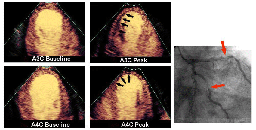
Tsutsui et al., evaluated the exequibility, safety and diagnostic accuracy of the DASE with simultaneous analysis of the segmental motility and myocardial perfusion utilizing real-time image, in a large number of patients [50]. The authors showed that the use of echocardiographic contrast allow an adequate assessment of the myocardial perfusion without any increase in the incidence of side effects when compared to the conventional protocol. The myocardial perfusion analysis presented good accuracy for CAD detection (Figure 2) and showed an independent predictor to cardiac events.
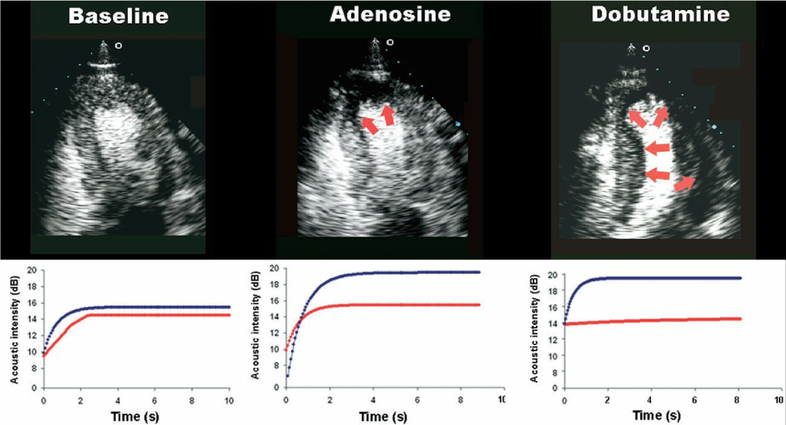
The dynamic of reperfusion of the myocardial contrast in real-time allow the quantification of the myocardial flow, when used contrast continuous infusion. The myocardial flow can be quantified using mathematical models that utilizes parameters of the plateau intensity and the mean velocity of the microcirculation replenishment by the microbubbles [1]. The replenishment of the microbubbles in the myocardium related to time can be measured by the augment in the acoustic intensity to each frame of the image sequence after the complete destruction of the microbubbles in the myocardium by a pulse of high ultrasonic energy (flash echocardiographic), as exampled in Figure 3. The use of specific computer programs to quantify myocardial contrast allows the analysis of the image sequences and quantification of the regional myocardial flow, such as at rest how after the cardiovascular stress induction, thus providing the myocardial flow reserve quantification.
Kowatsch I et al. [51], have studied the value of quantitative analysis of the myocardium perfusion to CAD detection during DASE and by adenosine. The authors showed that the reserve of velocity of the myocardial flow obtained by DASE real-time perfusion image presented similar accuracy to adenosine stress to CAD detection (accuracy of 80% to both pharmacies). Interestingly, the quantitative analysis of the myocardial perfusion during DASE presented additional value to CAD in relation to the examination of the 12 leads electrocardiogram, the segmental motility analysis and qualitative myocardial perfusion analysis (Figure 3). While the microbubbles improve definition of the endocardial borders, mostly contribution to stress echocardiography is the potential to allow detecting changes of myocardial perfusion. The development of contrasts containing microbubbles of smaller diameter and greater stability associated with technological advances such as STE may accurately identify early deformation impairment, while also being predictive of LV remodeling during follow-up. Some studies suggests that STE is associated with changes in LV volume or function regardless of underlying mechanisms and deformation direction. Meta-regression demonstrates a strong association between peak longitudinal systolic strain and adverse remodeling [52].
Echocardiographic strain and strain rate imaging
Echocardiographic strain and strain rate imaging is a technology enabling more reliable and comprehensive assessment of myocardial function. The spectrum of potential clinical applications is very wide due to its ability to differentiate between active and passive movement of myocardial segments, to quantify intraventricular dyssynchrony and to evaluate components of myocardial function, such as longitudinal myocardial shortening, that are not visually assessable. The high sensitivity of both Tissue Doppler Imaging (TDI) and two-dimensional speckle tracking derived strain and strain rate data for the early detection of myocardial dysfunction recommend these non-invasive diagnostic methods for routine clinical use. In addition to early detection of myocardial dysfunction of different etiologies, assessment of myocardial viability, detection of acute allograft rejection after heart transplantation and early detection of patients with transplant coronary artery disease, strain and strain rate measurements are helpful in the selection of different therapies and follow-up evaluations of myocardial function after different medical and surgical treatment. Strain and strain rate data also provide important prognostic information [53].
Tissue doppler-derived strain and strain-rate Imaging
TDI is accepted as a sensitive and sufficiently accurate echocardiographic tool for quantitative assessment of cardiac function [54,55]. Several tissue Doppler velocity parameters appeared to be useful for the diagnosis and prediction of long-term prognosis in major cardiac diseases [56,57]. Myocardial time-velocity curves can be obtained either online as spectral pulsed TDI, known as pulsed wave TDI (PW-TDI), or reconstructed offline from two-dimensional color coded TDI images, known as color TDI loops. In addition to velocity and displacement (tissue tracking) measurements, due to the relationship between velocity and strain rate, TDI also allows the reconstruction of strain curves and color coded images. Thus, the transmural velocity gradient is equal to the transmural strain rate, whereas the longitudinal velocity gradient over a segment with a fixed distance is a measure of longitudinal strain rate. Before the development of color-coded TDI, it was difficult to distinguish myocardial contraction from translational motion of the heart. Thus, during systole, in addition to radial wall thickening and longitudinal wall shortening, the left ventricle also rotates about its long axis and translates anteriorly. Calculation of myocardial velocity gradients allows the assessment of wall motion independently from the translational motion of the heart [54,58].
Taking into account all the aspects mentioned above it is not surprising that TDI-derived strain and strain rate (SR) measurements are not highly reproducible (more than 10-15% interobserver variability). However, in the hands of very experienced and highly trained operators this method can be a valuable non-invasive tool for routine clinical use to evaluate the myocardial contractile function. Despite all limitations this technique has been initially validated with sonomicrometry and also with magnetic resonance imaging [59,60].
Non-doppler speckle-tracking derived 2d-strain imaging
Non-Doppler two dimensional (2D)-strain imaging derived from speckle tracking is an echocardiographic technique for obtaining strain and SR measurements [60,61]. It analyzes motion by tracking speckles in the 2D ultrasonic image. These acoustic markers are statistically equally distributed throughout the myocardium and their size is about 20 to 40 pixels. These markers within the ultrasonic image are tracked from frame to frame. Special software allows spatial and temporal image processing with recognition and selection of such elements on ultrasound images. The geometric shift of each speckle represents local tissue movement. When frame rate is known, the change in speckle position allows determination of its velocity. Thus, the motion pattern of myocardial tissue is reflected by the motion pattern of speckles. By tracking these speckles, strain and strain rate can be calculated. The advantage of this method is that it tracks in two dimensions, along the direction of the wall, not along the ultrasound beam, and thus is angle independent [61]. The 2D echocardiographic loops obtained from paraesternal and apical views are processed offline. This requires only one cardiac cycle to be acquired but strain and SR data can be obtained only with high-resolution image quality at high frame rate [61]. The necessity of high image quality is a major limitation for routine clinical applicability in all patients. At present, the optimal frame rate for speckle-tracking seems to be 50-70 frames per second (FPS), which is lower compared to TDI (>180 FPS). This could result in under-sampling, especially in patients with tachycardia and during strain and SR measurements performed throughout stress echocardiography. Moreover, rapid events during the cardiac cycle such as isovolumetric phases may not appear on images and peak SR values may be reduced due to under-sampling, in isovolumetric phases and in early diastole. Using higher frame rates could reduce the under-sampling problem, but this will result in a reduction of spatial resolution and consequently less than optimal region of interest (ROI) tracking [62], Low frame rate increases the spatial resolution, but because speckle-tracking software uses a frame-by-frame approach to follow the myocardial movement and searches each consecutive frame for a speckle pattern closely resembling and in close proximity to the reference frame, with too low a frame rate the speckle pattern could be outside the search area, again resulting in poor tracking [63].
It is also important to know that different tracking algorithms potentially produce different results and therefore it should be kept in mind that a periodical update of the software package conceivably influences reference values.
Although speckle-tracking derived 2D-strain and TDI-derived strain calculations do not give the same values (2D-strain imaging gives lower SR values), strain and SR measurements obtained by these two different imaging techniques correlate well [61]. For the LV, the reproducibility of 2D-strain measurements is better than that of TDI-derived strain measurements. The intraobserver and inter observer variability for 2D-strain and SR measurements were found to be low: 3.6% to 5.3% and 7% to 11.8%, respectively [61] Ingul et al., found lower interobserver variability for non-Doppler 2D-strain measurements in comparison to TDI-derived strain measurements and automated non-Doppler 2D-strain measurements also appeared significantly less time consuming [64]. The lack of angle dependency is a great advantage of non-Doppler 2D-strain imaging in comparison to TDI-derived strain data.
Real time 3-dimensional dobutamine stress echocardiography
Two dimensional contrast-enhanced DSE is used clinically to diagnose stress-induced wall-motion abnormality (WMA). Takeushi et al. [65], hypothesized that contrast-enhanced real-time 3-dimensional (RT-3DE) DSE could improve the detection rate of WMA, because from a single full-volume acquisition, multiple segments can be visualized. They acquired both 2D and 3D DSE in 78 patients with known or suggested CAD (mean age: 65 years; 44 men). Dobutamine was infused using a standard protocol, and atropine added, if required. For 2D DSE, the intravenous contrast agent was injected at each stage and images displayed in a quad screen format. For RT-3DE DSE, contrast harmonic 3D data sets (full volumes) were acquired at baseline and peak stress. Using custom software, 3 short-axis views (from apex to base) were created, and wall motion scored using a wall-motion score index using a 16-segment model. A positive stress test was defined as new or worsened WMA or fixed abnormality during stress. In their results Heart rate increased from 72 +/- 13 to 133 +/- 15/min (86 +/- 11% of age-predicted). A total of 1248 segments were analyzed at each stage for both modalities. A single segment at baseline and 5 segments at peak stress could not be assessed with contrast 2D DSE. In contrast, 88 segments at baseline and 39 segments at peak stress could not be assessed with contrast RT-3DE DSE. With RT-3DE DSE, the majority of uninterpretable segments were in the anterior and lateral walls. Significant correlations were noted between wall-motion score index by 2D and 3D DSE at baseline (r = 0.78) and peak stress (r = 0.83). The concordance rate (positive/negative findings) between modalities was 69% (54/78) on a patient basis and 88% (206/234) on a perfusion territory basis. When using 2D DSE results as the gold standard, sensitivity and specificity for detecting WMA by 3D DSE was 58% and 75%, respectively. Sensitivity and specificity values were 67% and 94% for the right coronary artery, 53% and 81% for the left anterior descending coronary artery, and 88% and 100% for the left circumflex coronary artery territory, respectively. The authors concluded that Contrast-enhanced 3D DSE was feasible in the majority of patients. However, the moderate concordance between both modalities was a result of: (1) difficulties in visualizing the anterolateral segments because of the relatively large imprint of the transducer; and (2) lower frame rates with 3D DSE resulting in the erroneous diagnosis of dyssynchrony.
Aggeli et al. [66], in a study with 56 patients referred for coronary angiography, were examined by 2DE and RT-3DE during the same Dobutamine stress protocol showed that RT-3DE identifies wallmotion abnormalities more readily in the apical region than 2DE, which may explain the tendency towards higher sensitivity in the left anterior descending artery territory. RT-3DE results were validated using angiography as reference and findings indicate diagnostic equivalence to 2DE, with the advantage of considerable shorter acquisition times .
Conclusions
Due to the level of sensitivity, specificity, positive predictive value, negative predictive value and accuracy, stress echocardiography perhaps has the highest overall utility and is the most preferred and prescribed modality for the assessment of CAD. Pharmacologic stress echocardiography and MCE turn out to be the most cost-effective and risk-effective modality, without significant environmental/radiation/bio hazard, with the added advantage of repeatability, portability, an acceptable learning curve and high level of safety. Strain, Strain Rate, TDI, RT-3DE, Speckle tracking, combined MCE and RT-3DE, combined Strain rate and TDI add newer dimensions and may unleash the full potential of SE. The use of contrast has broadened the indications for transthoracic echocardiography and has increased the accuracy of stress echocardiography, while reducing the number of patients who cannot be scanned because of a limited acoustic window. Finally, echocardiography will be seen in the future not only as a diagnostic tool in those affected by cardiovascular disease but also as a method for prediction of risk and perhaps activation of targeted treatment.
- Wei K, Jayaweera AR, Firoozan S, Linka A, Skyba DM, et al. (1998) Quantification of myocardial blood flow with ultrasound-induced destruction of microbubbles administered as a constant venous infusion. Circulation 97: 473-483. Link: https://goo.gl/CSd84A
- Porter TR, Xie F, Silver M, Kricsfeld D, Oleary E, et al. (2001) Real-time perfusion imaging with low mechanical index pulse inversion Doppler imaging. J Am Coll Cardiol 37: 748-753. Link: https://goo.gl/QM7pG4
- Plana JC, Mikati IA, Dokainish H, Lakkis N, Abukhalil J, et al. (2008) A randomized cross-over study for evaluation of the effect of image optimization with contrast on the diagnostic accuracy of Dobutamine echocardiography in coronary artery disease The OPTIMIZE Trial. JACC Cardiovasc Imaging 2: 145-152. Link: https://goo.gl/8YUxak
- Dourado PM, Tsutsui JM, Mathias Jr W, Andrade JL, da Luz PL, et al. (2003) Evaluation of stunned and infarcted canine myocardium by real-time myocardial contrast echocardiography. Braz J Med Biol Res 36: 1501-1509. Link: https://goo.gl/u821px
- Senior R, Becher H, Monaghan M, Agati L, Zamorano J, et al. (2009) Contrast echocardiography: evidence-based recommendations by European Association of Echocardiography. Eur J Echocardiogr 2: 194-212. Link: https://goo.gl/pAAAyT
- Kaul S (2008) Myocardial contrast echocardiography: a 25-year retrospective. Circulation 3: 291-308. Link: https://goo.gl/L5VVPp
- Gaibazzi N, Lorenzoni V, Reverberi C, Wu J, Xie F, et al. (2016) Effect of Pharmacologic Stress Test Results on Outcomes in Obese versus Nonobese Subjects Referred for Stress Perfusion Echocardiography. J Am Soc Echocardiogr 29: 899-906. Link: https://goo.gl/yhkVZl
- Mahoney L, Shah G, Crook D, Rojas-Anaya H, Rabe H, et al. (2016) A Literature Review of the Pharmacokinetics and Pharmacodynamics of Dobutamine in Neonates. Pediatr Cardiol 37: 14-23. Link: https://goo.gl/IAZI1l
- Martínez GJ, Yong AS, Fearon WF, Ng MK (2015) The index of microcirculatory resistance in the physiologic assessment of the coronary microcirculation. Coron Artery Dis. 26 Suppl 1: e15-e26. Link: https://goo.gl/vmTLb1
- Nesto RW, Kowalchuk GJ (1987) The ischemic cascade: temporal sequence of hemodynamic, electrocardiographic and symptomatic expressions of ischemia. Am J Cardiol 9: 23C-30C. Link: https://goo.gl/13e5nW
- Wierzbowska-Drabik K, Hamala P, Roszczyk N, Lipiec P, Plewka M, et al. (2014) Feasibility and correlation of standard 2D speckle trackingechocardiography and automated function imaging derived parameters of left ventricular function during Dobutamine stress test. Int J Cardiovasc Imaging 30: 729-737. Link: https://goo.gl/mce5Gh
- Obokata M, Kane GC, Reddy YN, Thomas Olson P, Barry Borlaug A, et al. (2017) Role of Diastolic Stress Testing in the Evaluation for Heart Failure With Preserved Ejection Fraction: A Simultaneous Invasive-Echocardiographic Study. Circulation. 28: 825-838. Link: https://goo.gl/vCyixt
- Picano E, Mathias W Jr, Pingitore A, Bigi R, Previtali M, et al. (1994) Safety and tolerability of Dobutamine-atropine stress echocardiography: a prospective, multicentre study. Echo Dobutamine International Cooperative Study Group. Lancet 29: 1190-1192. Link: https://goo.gl/zrY3ll
- Lewandowski TJ, Armstrong WF, Bach DS (1998) Reduced test time by early identification of patients requiring atropine during Dobutamine stress echocardiography. J Am Soc Echocardiogr 11: 236-242. Link: https://goo.gl/ApxBQn
- Tsutsui JM, Osorio AF, Lario FA, Fernandes DR, Sodre G, et al. (2004) Comparison of safety and efficacy of the early injection of atropine during Dobutamine stress echocardiography with the conventional protocol. Am J Cardiol 1: 1367-1372. Link: https://goo.gl/5LOm9f
- Tsutsui JM, Lario FC, Fernandes DR, Kowatsch I, Sbano JC, et al. (2005) Safety and cardiac chronotropic responsiveness to the early injection of atropine during Dobutamine stress echocardiography in the elderly. Heart 29: 1563–1567. Link: https://goo.gl/A7QWdB
- Mathias W Jr, Tsutsui JM, Andrade JL, Kowatsch I, Lemos PA, et al. (2003) Value of rapid beta-blocker injection at peak Dobutamine-atropine stress echocardiography for detection of coronary artery disease. J Am Coll Cardiol 41: 1583-1589. Link: https://goo.gl/e2qbk1
- Schiller NB, Shah PM, Crawford M, DeMaria A, Devereux R, et al. (1989) Recommendations for quantitation of the left ventricle by two-dimensional echocardiography. American Society of Echocardiography Committee on Standards, Subcommittee on Quantitation of Two-Dimensional Echocardiograms. J Am Soc Echocardiogr 2: 358-367. Link: https://goo.gl/qEdg8X
- Cerqueira MD, Weissman NJ, Dilsizian V, Jacobs AK, Kaul S, et al. (2002) Standardized myocardial segmentation and nomenclature for tomographic imaging of the heart: a statement for healthcare professionals from the Cardiac Imaging Committee of the Council on Clinical Cardiology of the American Heart Association. Circulation 29: 539-542. Link: https://goo.gl/lZ9k3K
- Beleslin BD, Ostojic M, Stepanovic J, Djordjevic-Dikic A, Stojkovic S, et al. (1994) Stress echocardiography in the detection of myocardial ischemia. Head-to-head comparison of exercise, Dobutamine, and dipyridamole tests. Circulation 90: 1168-1176. Link: https://goo.gl/Tk0zK3
- Dagianti A, Penco M, Agati L, Sciomer S, Dagianti A, et al. (1995) Stress echocardiography: comparison of exercise, dipyridamole and Dobutamine in detecting and predicting the extent of coronary artery disease. J Am Coll Cardiol 26: 18-25. Link: https://goo.gl/djLu25
- Quiñones A, Benenstein R, Saric M (2013) New-onset seizure after perflutren microbubble injection during Dobutamine stress echocardiography. Echocardiography. 30: E95-E97. Link: https://goo.gl/SYF9CG
- Picano E, Landi P, Bolognese L, Chiaranda G, Chiarella F, et al. (1993) Prognostic value of dipyridamole echocardiography early after uncomplicated myocardial infarction: a large-scale, multicenter trial. The EPIC Study Group. Am J Med 95: 608-618. Link: https://goo.gl/nJgQSB
- Pingitore A, Picano E, Colosso MQ, Reisenhofer B, Gigli G, et al. (1996) The atropine factor in pharmacologic stress echocardiography. Echo Persantine (EPIC) and Echo Dobutamine International Cooperative (EDIC) Study Groups. J Am Coll Cardiol 27: 1164-1170. Link: https://goo.gl/2BLau8
- Mathias JW, Doya EH, Ribeiro EE, Silva LA, Gasques A, et al. (1993) [Detection of myocardial ischemia during Dobutamine stress echocardiography. Correlation with coronary cineangiography]. Arq Bras Cardiol 60: 229-234. Link: https://goo.gl/joWt5O
- Tsutsui JM, Dourado PM, Falcão SN, Figueiredo M, Guerra VC, et al. (2008) Prognostic value of dobutamine stress echocardiography with early injection of atropine with versus without chronic beta-blocker therapy in patients with known or suspected coronary heart disease. Am J Cardiol 102: 1291-1295. Link: https://goo.gl/nXbIAQ
- Rumbinait? E, Žaliaduonyt?-Pekšien? D, Vieželis M? eponien? I, Lapinskas T, et al. (2016) Dobutamine-stress echocardiography speckle-tracking imaging in the assessment of hemodynamic significance of coronary artery stenosis in patients with moderate and high probability of coronary artery disease. Medicina (Kaunas). 52: 331-339. Link: https://goo.gl/fjkuL3
- Uusitalo V, Luotolahti M, Pietilä M, Wendelin-Saarenhovi M, Hartiala J, et al. (2016) Two-Dimensional Speckle-Tracking during Dobutamine Stress Echocardiography in the Detection of Myocardial Ischemia in Patients with Suspected Coronary Artery Disease. J Am Soc Echocardiogr. 29: 470-479.e3. Link: https://goo.gl/9c3alt
- Caidahl K, Kazzam E, Lidberg J, Neumann AG, Nordanstig J, et al. (1998) New concept in echocardiography: harmonic imaging of tissue without use of contrast agent. Lancet 17: 1264-1270. Link: https://goo.gl/oa7e5I
- Pasovic M, Danilouchkine M, Faez T, van Neer PL, Cachard C, et al. (2011) Second harmonic inversion for ultrasound contrast harmonic imaging. Phys Med Biol. 7: 3163-3180. Link: https://goo.gl/P7Yk7m
- Powers J and Kremkau F (2011) Medical ultrasound systems. Interface Focus. 1: 477-489. Link: https://goo.gl/ctbJij
- Desser TS, Jeffrey RB (2001) Tissue harmonic imaging techniques: physical principles and clinical applications. Semin. Ultrasound CT MR 22: 1-10. Link: https://goo.gl/6qhXU8
- Gaibazzi N, Reverberi C, Lorenzoni V, Molinari R, Porter TR, et al. (2012) Prognostic value of high-dose dipyridamole stress myocardial perfusion contrast echocardiography. Circulation. 4: 1217-1224. Link: https://goo.gl/l3DOXe
- Cheitlin MD, Armstrong WF, Aurigemma GP, et al. (2003) ACC/AHA/ASE 2003 Guideline Update for the Clinical Application of Echocardiography: summary article. A report of the American College of Cardiology/American Heart Association Task Force on Practice Guidelines (ACC/AHA/ASE Committee to Update the 1997 Guidelines for the Clinical Application of Echocardiography). J Am Soc Echocardiogr 16: 1091-1110.
- Picano E, Ciampi Q, Citro R, D’Andrea A, Scali MC, et al. (2017) Echo 2020: the international stress echo study in ischemic and non-ischemic heart disease. Cardiovasc Ultrasound 15: 3. Link: https://goo.gl/cQ0tfv
- Bouzas-Mosquera A, Peteiro J, Alvarez-García N, Broullón FJ, García-Bueno L, et al. (2009) Prognostic value of exercise echocardiography in patients with left bundle branch block. JACC Cardiovasc Imaging 2: 251-259. Link: https://goo.gl/SY5JUd
- Peteiro J, Bouzas-Mosquera A, Pazos P, Pazos P, Castro-Beiras A (2010) Prognostic value of exercise echocardiography in patients with left ventricular systolic dysfunction and known or suspected coronary artery disease. Am Heart J 160: 301-307. Link: https://goo.gl/6DcImJ
- Andrade SM, Telino CJ, Sousa AC, Melo EV, Teixeira CC, et al. (2016) Low Prevalance of Major Events Adverse to Exercise Stress Echocardiography. Arq Bras Cardiol 107: 116-123. Link: https://goo.gl/arnoHH
- Volpi A, De VC, Franzosi MG, Geraci E, Maggioni AP, et al. (1993) Determinants of 6-month mortality in survivors of myocardial infarction after thrombolysis. Results of the GISSI-2 data base. The Ad hoc Working Group of the Gruppo Italiano per lo Studio della Sopravvivenza nell'Infarto Miocardico (GISSI)-2 Data Base. Circulation 88: 416-429. Link: https://goo.gl/jSOZr1
- Antoni ML, Mollema SA, Delgado V, Atary JZ, Borleffs CJ, et al. (2010) Prognostic importance of strain and strain rate after acute myocardial infarction. Eur Heart J. 31: 1640-1647. Link: https://goo.gl/Xi1s5q
- Varga A, Picano E, Cortigiani L, Petix N, Margaria F, et al. (1996) Does stress echocardiography predict the site of future myocardial infarction? A large-scale multicenter study. EPIC (Echo Persantine International Cooperative) and EDIC (Echo Dobutamine International Cooperative) study groups. J Am Coll Cardiol 28: 45-51. Link: https://goo.gl/UFxTiJ
- Pierard LA, De Landsheere CM, Berthe C, Rigo P, Kulbertus HE, et al. (1990) Identification of viable myocardium by echocardiography during Dobutamine infusion in patients with myocardial infarction after thrombolytic therapy: comparison with positron emission tomography. J Am Coll Cardiol 15: 1021-1031. Link: https://goo.gl/sH5AON
- Anantharam B, Janardhanan R, Hayat S, Hickman M, Chahal N, et al. (2011) Coronary flow reserve assessed by myocardial contrast echocardiography predicts mortality in patients with heart failure. Eur J Echocardiogr 12: 69-75. Link: https://goo.gl/WQnglG
- Bikiri E, Mereles D, Voss A, Greiner S, Hess A, et al. (2014) Dobutamine stress cardiac magnetic resonance versus echocardiography for the assessment of outcome in patients with suspected or known coronary artery disease. Are the two imaging modalities comparable? Int J Cardiol 171: 153-160. Link: https://goo.gl/b23v7h
- Pasquet A, Yamada E, Armstrong G, Beachler L, Marwick TH (1999) Influence of Dobutamine or exercise stress on the results of pulsed-wave Doppler assessment of myocardial velocity. Am Heart J 138: 753-758. Link: https://goo.gl/9lJr9o
- Crouse LJ, Cheirif J, Hanly DE, Kisslo JA, Labovitz AJ, et al. (1993) Opacification and border delineation improvement in patients with suboptimal endocardial border definition in routine echocardiography: results of the Phase III Albunex Multicenter Trial. J Am Coll Cardiol 22: 1494-1500. Link: https://goo.gl/DYXp8Z
- Porter TR, Xie F (1995) Transient myocardial contrast after initial exposure to diagnostic ultrasound pressures with minute doses of intravenously injected microbubbles. Demonstration and potential mechanisms. Circulation 92: 2391-2395. Link: https://goo.gl/k9rX33
- Mulvagh SL, DeMaria AN, Feinstein SB, Burns PN, Kaul S, et al. (2000) Contrast echocardiography: current and future applications. J Am Soc Echocardiogr 13: 331-342. Link: https://goo.gl/OQMoks
- Donataccio MP, Reverberi C, Gaibazzi N (2014) The dilemma of ischemia testing with different methods. Echo Res Pract 1: K1-4. Link: https://goo.gl/PHyRM9
- Tsutsui JM, Elhendy A, Xie F, O'Leary E, McGrain AC, et al. (2005) Safety of Dobutamine stress real-time myocardial contrast echocardiography. J Am Coll Cardiol 45: 1235-1242. Link: https://goo.gl/bfhZEU
- Kowastch I, Tsutsui JM, Osorio AFF, Uchida AH, Machiori GGA, et al. (2007) Head-to-Head Comparison of Dobutamine and Adenosine Stress Real-time Myocardial Perfusion Echocardiography for the detection of Coronary Artery Disease. J Am Soc Echocardiograph 20: 1109-1117. Link: https://goo.gl/Vuga0j
- Huttin O, Coiro S, Selton-Suty C, Juillière Y, Donal E, et al. (2016) Prediction of Left Ventricular Remodeling after a Myocardial Infarction: Role of Myocardial Deformation: A Systematic Review and Meta-Analysis. PLoS One 11: e0168349. Link: https://goo.gl/3nugU4
- Dandel, Hetzer R (2009) Echocardiographic strain and and strain rate imaging - new aplications. Int. J Cardiol 132: 11-24. Link: https://goo.gl/OQE2JX
- Sutherland G, Steward M, Groundstroen K, Moran CM, Fleming A, et al. (1994) Color Doppler myocardial imaging: a new technique for the assessment of myocardial function. J Am Soc Echocardiogr 7: 441-458. Link: https://goo.gl/noyaRf
- Stouylen A, Heimdal A, Bjornstad K, Torp HG, Techn, et al. (1999) Strain rate imaging by ultrasound in the diagnosis of regional dysfunction of the left ventricle. Echocardiography 16: 321-329. Link: https://goo.gl/w8RpWC
- Yu CM, Sanderson JE, Marwick TH, Oh JK (2007) Tissue Doppler imaging a new prognosticator for cardiovascular diseases. J Am Coll Cardiol 20: 234-243. Link: https://goo.gl/WDTN2C
- Waggoner AD, Bierig SM (2001) Tissue Doppler imaging: a useful echocardiographic method for the cardiac sonographer to assess systolic and diastolic ventricular function. J Am Soc Echocardiogr 14: 1143-1152. Link: https://goo.gl/8vueUw
- Uematsu M, Miyatake K, Tanaka N, Matsuda H, Sano A, et al. (1995) Myocardial velocity gradient as a new indicator of regional left ventricular contraction: detection by two-dimensional tissue Doppler imaging technique. J Am Coll Cardiol 26: 217-223. Link: https://goo.gl/102ZtE
- Urheim S, Edvardsen T, Torp H, Angelsen B, Smiseth OA (2000) Myocardial strain by Doppler echocardiography: validation of a new method to quantify regional myocardial function. Circulation 102: 1158-1164. Link: https://goo.gl/KU5Dal
- Edvardsen T, Gerber BL, Garot J, Bluemke DA, Lima JAC, et al. (2002) Qualitative assessment of intrinsic regional myocardial deformation by Doppler strain rate echocardiography in humans: Validation against three-dimensional tagged magnetic resonance imaging. Circulation 106: 50-56. Link: https://goo.gl/NygTec
- Perk G, Tunick PA, Kronzon I (2007) Non-Doppler two-dimensional strain imaging by echocardiography - from technical considerations to clinical applications. J Am Soc Echocardiogr 49: 1903-1914. Link: https://goo.gl/6NIQVa
- Teske AJ, De Boeck BWL, Olimulder M, Prakken NH, Doevendans PAF, et al. (2007) Echocardiographic assessment of regional right ventricular function. A head to head comparison between 2D-strain and tissue Doppler derived strain analysis. J Am Soc Echocardiogr 21: 275-283. Link: https://goo.gl/5yRhy2
- Korinek J, Kjaergaard J, Sengupta PP, Yoshifuku S, McMahon EM, et al. (2007) High spatial resolution speckle tracking improves accuracy of 2-dimensional strain measurements. J Am Soc Echocardiogr 20: 165-170. Link: https://goo.gl/a0p3t8
- Ingul CB, Torp H, Aase SA (2005) Automated analyses of strain rate and strain: feasibility and clinical implications. J Am Soc Echocardiogr 18: 411-418. Link: https://goo.gl/zojP4D
- Takeuchi M, Otani S, Weinert L, Spencer KT, Lang RM (2006) Comparison of contrast-enhanced realtime live 3-dimensional Dobutamine stress echocardiography with contrast 2-dimensional echocardiography for detecting stress-induced wall-motion abnormalities. J Am Soc Echocardiogr 19: 294-299. Link: https://goo.gl/CNJZRh
- Aggeli C, Giannopoulos G, Misovoulos P, Roussakis G, Christoforatou E, et al. (2007) Real-time three-dimensional Dobutamine stress echocardiography for coronary artery disease diagnosis: Validation with coronary angiography. Heart 93: 672-675. Link: https://goo.gl/QLMmRj
Article Alerts
Subscribe to our articles alerts and stay tuned.
 This work is licensed under a Creative Commons Attribution 4.0 International License.


 Save to Mendeley
